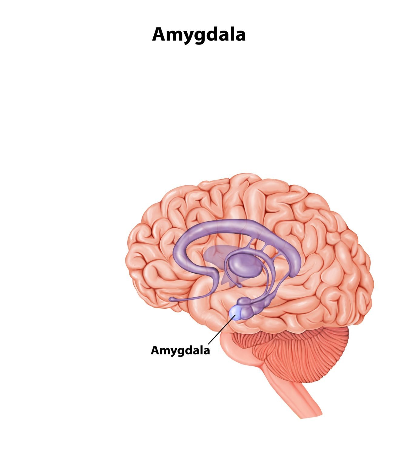
The amygdala is a small, almond-shaped structure buried deep in each temporal lobe of the brain. It scans everything a person senses for emotional and biological significance, especially threat, and triggers the fear, memory, and bodily reactions that follow.
Key Takeaways
- Location: Two almond-shaped clusters sit deep in the temporal lobes, roughly level with the eyes, one in each hemisphere.
- Beyond Fear: The amygdala flags anything emotionally significant, not just fear; modern theory treats it as one hub in a wider network, not a single emotion organ.
- Fear Conditioning: It links a neutral cue to danger via a fast subcortical path and a slower cortical path that can override it.
- Memory: Working with the hippocampus, it tags emotional experiences so they are remembered more vividly than ordinary ones.
- Clinical Role: Over-reactivity is linked to anxiety, PTSD, and depression, while under-reactivity is linked to psychopathy.
- Damage Effects: Losing both amygdalae, as in Klüver-Bucy syndrome, blunts fear and aggression and disrupts reading others’ emotions.
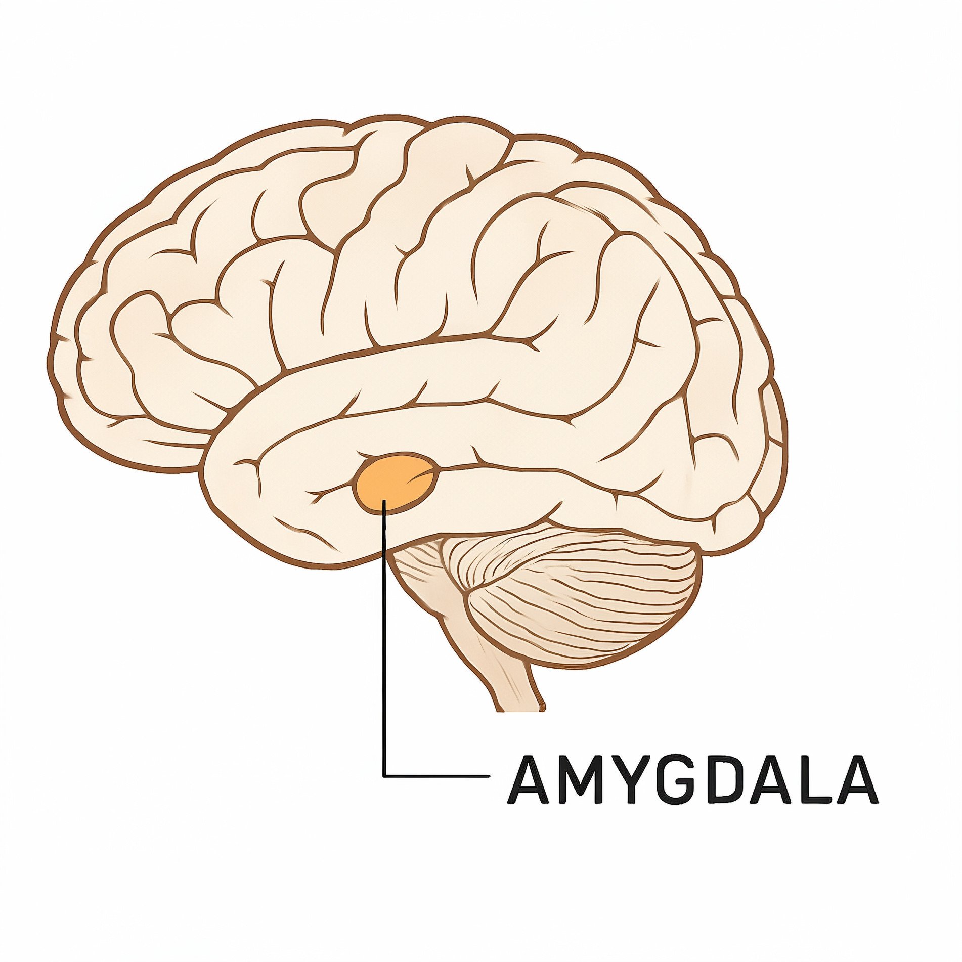
Where is the amygdala located?
The amygdala consists of two almond-shaped clusters tucked deep inside the temporal lobes (the area of the brain near your temples).
You have one on each side, sitting roughly level with your eyes.
Picture pointing one finger into your ear and another straight through your eye. Those two lines would meet roughly where the amygdala sits.
It is a core part of the limbic system, which is the brain’s “emotional headquarters.” Its specific location is key to how it functions:
- Next to the Hippocampus: Positioned right in front of the brain’s memory center (the hippocampus), it allows the amygdala to “label” memories with strong emotions like fear or joy.
- Near the Hypothalamus: Being close to this regulatory center allows the amygdala to quickly trigger physical reactions, like a racing heart, when it senses danger.
- Deep and Protected: Because it is located beneath the outer layers of the brain, it can process survival instincts much faster than our conscious, “thinking” brain.

What Does The Amygdala Do?
The amygdala is essential for processing emotional reactions and attaching emotional significance to experiences, playing a central role in various aspects of behavior and cognition.

1. Emotional Regulation
The amygdala is the primary hub for processing emotions like fear, aggression, and happiness.
It is essential for fear conditioning, a form of associative learning where a neutral stimulus (like a sound) is linked to an aversive event.
This creates a conditioned response, allowing the organism to anticipate danger.
Dual-Path Processing (The LeDoux Pathways)
Research by Joseph LeDoux identified two pathways the amygdala uses to process fear stimuli.
A “fast path” runs directly from the thalamus for immediate reaction, while a “slow path” through the cortex allows more detailed processing:
- Fast Path (Subcortical): A rapid, direct pathway from the thalamus to the amygdala. It bypasses the conscious mind for nearly instantaneous “fight-or-flight” activation.
- Slow Path (Cortical): A slower pathway where information travels through the sensory cortex for detailed analysis. This allows the Prefrontal Cortex (PFC) to “brake” or dampen the amygdala’s response if the threat is deemed false.
These two pathways converge on the same neurons in the lateral amygdala. Repeated pairing of the tone and shock strengthens that shared synapse, a process called long-term potentiation (LTP). LTP was first identified in the hippocampus, and it is thought to be the cellular basis of the fear memory.
Rogan, Stäubli and LeDoux (1997) confirmed this directly. After pairing a tone with a foot-shock in rats, the tone alone triggered a larger electrical response in the lateral amygdala than before training. This closed the loop between the learned behaviour and its synaptic basis.
2. Memory Consolidation
The amygdala does not store most memories itself. Instead, it makes emotional experiences stick in the hippocampus.
Emotional arousal releases stress hormones, including adrenaline and cortisol. These act on stress-hormone receptors in the basolateral amygdala, strengthening how vividly the hippocampus stores the memory (McGaugh, 2004).
This is why high-stakes events, like trauma or joy, are remembered more vividly than an ordinary day.
This tagging process is not the same as fear conditioning. Tagging controls how strongly a memory is stored; fear conditioning creates the learned association in the first place. On a cellular level, the memory itself likely involves lasting changes in synaptic plasticity within the amygdala’s own circuits.
3. Modulation of Aggression
The amygdala acts as a biological “trigger” for aggressive behavior.
Research shows that stimulating this region increases hostile responses, while damage (lesions) leads to a marked decrease in aggression, highlighting its role in social survival instincts.
4. The Stress Response (Fight or Flight)
The amygdala is sensitive to both acute and chronic stressors.
When a threat is detected, the amygdala signals the hypothalamus, which activates the sympathetic nervous system.
This results in the physiological fight or flight response: increased heart rate, adrenaline release, and heightened alertness.
Exposure to stress affects neuronal activity and synaptic plasticity within the amygdala, indicating its pivotal role in processing stress responses.
5. Social & Addictive Behaviors
The amygdala also influences social interactions and relationships.
It modulates social behaviors through bidirectional connections with the hippocampus.
The amygdala helps interpret facial expressions and social cues. Interestingly, neuroimaging shows that it also plays a role in culturally learned social biases
- Social Networks: Larger amygdala volume is positively correlated with more complex social networks, suggesting it helps process social cues and bond formation.
- Addiction: The basolateral amygdala links drug-related environmental cues with stress responses, often driving the “craving” cycle and increasing the risk of relapse.
6. Clinical Significance & Mental Health
Dysfunction in the amygdala is a hallmark of several conditions:
- Hyperactivity: Hypervigilance, panic attacks, and an overactive fight-or-flight response. Linked to PTSD and Social Anxiety (over-responsiveness to perceived threats).
- Volume Changes: Reduced amygdala volume (up to 15-20% in some studies) has been observed in patients with chronic Bipolar Disorder or long-term Depression.
- Hyporeactivity: In individuals with psychopathic tendencies, a lack of amygdala activation often correlates with an inability to learn from punishment (impaired fear conditioning).
- Structural Changes: Persistent negative emotional states due to a lack of “top-down” regulation from the Prefrontal Cortex.
- Autism: Atypical amygdala structure, especially a reduced response to faces and eye contact, features in theories of the social-communication difficulties seen in autism.
7. Respiratory Control
Recent evidence suggests the amygdala influences the medulla oblongata to regulate breathing.
During seizures, over-activation of the amygdala can cause apnea (temporary cessation of breathing). Patients often show no air hunger or alarm during these episodes, revealing that breathing control here runs separately from the felt urge to breathe (Harmata et al., 2023).

Amygdala Hijack
Amygdala hijack refers to an immediate, overwhelming emotional response that bypasses the brain’s rational processing centers.
This occurs when the amygdala, the brain’s emotional center, reacts to a stimulus before the cerebral cortex (the thinking brain) has a chance to evaluate it.
Essentially, the amygdala overrides the frontal lobes to hijack stress response control.
The Neural Mechanism: Two Pathways to Emotion
Research by neuroscientist Joseph LeDoux identifies two distinct pathways through which the brain processes fear and emotion.
An amygdala hijack effectively represents the dominance of the “fast path” over the “slow path”:
- The Fast Path (The Hijack): Sensory information travels directly from the thalamus to the amygdala. This transmission is extremely rapid and allows for an almost instantaneous response to a threat. Because this path bypasses the cortex, the emotional reaction occurs separately from, or prior to, cognitive interpretation.
- The Slow Path (The Rational Response): The same cortical route described above, which lets the Prefrontal Cortex weigh in and override a false alarm before it escalates.
Vulnerability Factors
Certain conditions can make the brain more susceptible to this hijacking by increasing amygdala sensitivity or decreasing cortical control:
- Sleep Deprivation: Poor sleep makes the amygdala more reactive. Brain scans show it responding more strongly to fearful faces after a bad night, which helps explain irritability when tired.
- Stress and Trauma: Chronic stress sensitizes the brain’s threat-response systems, making them over-react. In depression, the amygdala shows heightened activity even to stimuli presented outside conscious awareness.
Amygdala’s Connections and Neuronal Circuits
The amygdala is not a uniform mass; it is a complex, modular structure of roughly a dozen nuclei, grouped into three functionally distinct divisions.
The amygdala is modular, with different “subnuclei” handling specific stages of information processing:
1. Functional Anatomy: The Three Subnuclei Groups
The amygdala’s internal architecture allows it to specialize in different types of biological signaling:
- Basolateral Group: Sitting below and to the side, this is the primary input hub. It links sensory stimuli to emotional significance and works with the hippocampus to form emotional memories.
- Central and Anterior Group: Sitting at the front and centre, this is the primary output hub. It connects to the brainstem and hypothalamus, triggering the autonomic fight-or-flight response once a threat is detected.
- Medial Group: This group sits in the middle and connects heavily to the olfactory bulb. It processes scent to influence social and reproductive behaviour, reflecting the ancient link between smell and emotion.
2. The Connectivity Map: Integrating the System
To coordinate survival, the amygdala acts as a “relay station” between different parts of the Central Nervous System (CNS):
These connections are mostly bidirectional, running both ways between the amygdala and the rest of the brain. That two-way traffic is what lets the brain “self-regulate”: the amygdala alerts the cortex to a threat, and the cortex later signals back for the amygdala to stand down.
A. Sensory Input (The Intake)
- Source: Sensory thalamus and sensory cortices (visual, auditory, somatosensory).
- Result: Provides the amygdala with raw data about the environment (e.g., seeing a snake or hearing a loud bang).
B. Regulation and Decision-Making (The Filter)
- Connection: Bidirectional link with the Prefrontal Cortex (PFC).
- Result: Integrates “cold” logic with “hot” emotion, allowing the PFC to dampen amygdala activity once a threat is resolved.
C. Memory and Learning (The Record)
- Connection: Bidirectional link with the Hippocampus.
- Result: Ensures that emotionally significant events are prioritized for long-term storage, helping the organism learn to avoid future dangers.
D. Physiological Output (The Action)
- Connection: Output to the Hypothalamus and Brainstem.
- Result: Activates the sympathetic nervous system for the fight-or-flight response.
E. Reward and Motivation (The Drive)
- Connection: Output to the Nucleus Accumbens and Ventral Striatum.
- Result: Involved in reward-seeking behavior and addiction, linking positive emotions to specific actions.
What Happens If The Amygdala Is Damaged?
Damage to the amygdala primarily results in profound deficits in emotional processing, particularly regarding fear, aggression, and the ability to attach emotional significance to memories.
Because the amygdala acts as a central hub for integrating sensory information with emotional and autonomic responses, its destruction disrupts survival instincts and social behavior.
1. Disruption of Emotional Processing
The most significant impact of amygdala damage is the blunting of fear and aggression.
-
Loss of Fear Conditioning: Since the basolateral complex is the site of associative learning, damage here prevents an organism from linking a neutral stimulus (like a warning sound) with an aversive event (like a shock).
-
Deficits in Social Perception: Humans with amygdala damage often cannot recognize fear in the facial expressions of others. They lose the “emotional instinct” required to navigate social hierarchies.
-
The “Taming” Effect: In animal studies, damage to the amygdala can turn naturally aggressive or wild animals into docile ones, as they lose the ability to perceive a threat or mount a defensive response.
Key case: patient S.M. The clearest human evidence comes from patient S.M. A rare condition called Urbach-Wiethe disease almost completely destroyed her amygdala on both sides, while leaving nearby brain tissue intact.
- Aim: Feinstein et al. (2011) tested whether S.M. could still feel fear when confronted with real threats, not just photographs of frightened faces.
- Method: Researchers exposed her to live snakes and spiders, a tour of a reputedly haunted house, and a montage of frightening films.
- Results: S.M. showed essentially no fear in any of these situations. She reported curiosity, and even tried to touch the snakes and spiders.
- Conclusion: The amygdala appears necessary for triggering the felt state of fear in response to external threats.
2. Klüver–Bucy Syndrome
A landmark area of study in neurobiology is Klüver–Bucy Syndrome, which occurs when the temporal lobes (including the amygdala) are removed or damaged.
It is characterized by:
-
Docility: A complete lack of fear or anger.
-
Hyperphagia: An indiscriminate drive to eat, often attempting to consume non-food objects.
-
Hyperorality: A tendency to examine objects with the mouth.
-
Hypersexuality: An excessive and indiscriminate sex drive.
3. Impact on Memory: Loss of “Emotional Boost”
While the hippocampus handles the “what, where, and when” of a memory, the amygdala handles the “emotional intensity.”
-
Memory Consolidation: Normally, stress hormones (like adrenaline) activate the amygdala to strengthen memory storage in the hippocampus.
-
The Result of Damage: Patients with bilateral amygdala damage can remember facts about a traumatic event but do not experience the vivid, “highlighted” memory that healthy individuals do.
Case Study: Henry Molaison (H.M.) H.M. had his hippocampus and amygdala removed to treat epilepsy. This resulted in anterograde amnesia (the inability to form new facts) but also a striking inability to recognize the emotional significance of new people or experiences.
How to Soothe Your Amygdala
Soothing the amygdala is biologically defined as re-engaging the Cortical (Slow) Path.
This involves inhibiting the amygdala’s output by strengthening the Prefrontal Cortex (PFC) and shifting the Autonomic Nervous System from a Sympathetic (Fight-or-Flight) state to a Parasympathetic (Rest-and-Digest) state.
Calming the amygdala combines mental health strategies with everyday lifestyle changes. The techniques below target different parts of that process.
Practical Techniques to Ease Panic
-
- Breathing Exercises: Practice deep breathing to regulate your nervous system. Techniques like the 4-7-8 breathing method can be particularly effective during stressful moments.
-
- Grounding Exercises: Use grounding techniques, such as focusing on your five senses, to anchor yourself in the present and diminish overwhelming emotions.
- Affect Labeling: Simply naming an emotion (“I feel anxious”) measurably changes brain activity. It engages the prefrontal cortex, which helps dampen the emotional response, similar to cognitive reappraisal.
Stress and Anxiety Management
-
- Adopt a Mindful Practice: Meditation and yoga are excellent for reducing stress levels and relaxing your amygdala. Apps like Headspace or Calm offer guided practices to get you started.
-
- Stay Active: Regular physical activity releases endorphins, which naturally combat anxiety. Whether it’s a brisk walk or a heart-pumping workout, integrate movement into your daily routine.
-
- Pursue Hobbies: Engage in activities you enjoy, whether it’s painting, gardening, or playing an instrument. Hobbies provide a productive distraction from anxiety and can be an outlet for expression.
- Social Support: The presence of a supportive person can directly affect brain processing; for instance, holding someone’s hand can reduce the activation of stress-related brain areas.
Titration: Moving In and Out of Emotion
Trying to suppress the amygdala’s response entirely is often counterproductive. A more effective approach is titration, or dosing the emotional experience.
In chemistry, titration is the slow addition of one solution to another to reach a reaction point without overshooting.
In neurobiology, it refers to “dosing” a stressful memory or emotion so the amygdala stays within its “Window of Tolerance.”
1. Pendulation: Managing Neural Arousal
Pendulation is the process of swinging the focus between a “resourced” state (feeling safe/calm) and a “stressed” state (the emotional memory).
-
The Mechanism: By moving “in and out” of the stress response, you prevent the amygdala from reaching the threshold of a hijack.
-
The Goal: This allows the Prefrontal Cortex to stay “online” and continue processing the information, rather than being shut down by an overwhelming fear signal.
2. The Failure of Suppression
Trying to “block” or “ignore” an emotion is often counterproductive.
-
Rebound Effect: Suppression requires high levels of metabolic energy from the Prefrontal Cortex. When the PFC tires, the amygdala often reacts with even greater intensity (hyperactivity).
-
Avoidance Learning: Avoiding a thought reinforces the amygdala’s belief that the thought is a genuine threat, strengthening the synaptic pathways of fear.
3. Acceptance and Habituation
Acceptance is a biological strategy to induce habituation – the diminishing of a physiological response to a frequently repeated stimulus.
-
Adaptive Processing: Facing a negative emotion in small, manageable amounts (e.g., one minute) allows the brain to realize the “threat” does not result in actual physical harm.
-
Synaptic Changes: Over time, this “exposure” weakens the excitatory connections between the thalamus and the amygdala, effectively “de-sensitizing” the alarm system.
Critical Evaluation
The amygdala is often introduced as the brain’s “fear centre,” but that label oversimplifies the evidence. Three findings complicate the simple story:
- Not really a “fear centre”: the researcher who built the amygdala’s fear reputation now argues against the label.
- No dedicated “emotion” region: the amygdala activates across many emotional and even positive states, not just fear.
- Fear can happen without it: patients missing both amygdalae can still panic when their body signals suffocation.
Not Really a “Fear Centre”
Joseph LeDoux is the neuroscientist whose own research built the amygdala’s reputation as the brain’s fear centre. LeDoux himself now disagrees. That label, he argues, is a mistake (LeDoux, 2012, 2020). On his current view, the amygdala runs a non-conscious “defensive survival circuit” that detects and reacts to threat automatically.
The feeling of fear is different, LeDoux argues. It is assembled separately, in cortical circuits, not in the amygdala itself.
Treating amygdala activity as identical to felt fear is a category error, according to LeDoux and Pine’s (2016) two-system framework. That confusion, they argue, has complicated both emotion theory and the design of anti-anxiety drugs.
This matters clinically. If a drug or therapy only dampens amygdala activity, LeDoux warns, it may reduce the automatic defensive reaction without touching the conscious dread a person still reports.
No Single “Emotion Centre”
Psychologist Lisa Feldman Barrett goes further. According to Barrett’s (2017) theory of constructed emotion, there is no dedicated brain region for any single emotion.
Fear is not special. The amygdala responds to salience, meaning how much a stimulus stands out or matters, rather than to fear specifically. Instead, Barrett argues, it computes a general signal of how significant a situation is, leaving the cortex to turn that signal into a specific, labelled feeling.
Brain-scan studies show the amygdala activating during surprise, arousal, and even positive events, not just threat. This is a problem for amygdala-based fear research. Seeing the amygdala light up does not prove someone feels afraid.
Psychologists call this mistake the reverse-inference fallacy. It undermines a great deal of amygdala brain-imaging research.
Fear Can Happen Without the Amygdala
The clearest challenge comes from patient S.M. She lost both amygdalae to a rare condition called Urbach-Wiethe disease.
Feinstein et al. (2013) had her and two other patients with bilateral amygdala damage breathe air enriched with 35% carbon dioxide. That dose reliably creates a strong sensation of suffocation.
This was surprising. Without an amygdala, these patients should, in theory, have been the last people to panic.
All three patients panicked. S.M. panicked even more intensely than healthy volunteers tested the same way. Fear triggered by an internal, bodily threat can bypass the amygdala completely.
This does not overturn the rest of the evidence.
It shows the amygdala is one important node in a wider threat-detection network, rather than fear’s only gateway, and that internal bodily signals can trigger panic on their own.
Contemporary Research
Since 2015, the field has changed its central question. Researchers no longer just ask which brain “blob” lights up. They ask how specific circuits behave, using tools precise enough to test that directly.
Optogenetics is the key new tool. It uses light to switch genetically defined neurons on or off, letting researchers trigger or silence specific amygdala populations directly. Janak and Tye (2015), reviewing this fast-growing literature, describe the shift from correlation to causal, cell-by-cell circuit mapping.
It works within larger networks too. Zheng et al. (2023) found disrupted connectivity between the amygdala and other brain regions in people with schizophrenia.
Findings like this fit a broader pattern. Altered amygdala networks, not the amygdala in isolation, track a range of clinical conditions.
Circuit-specific work also separates positive from negative valence. Felix-Ortiz and Tye (2014) showed that basolateral-amygdala inputs to the ventral hippocampus bidirectionally modulate social behaviour, pointing to distinct microcircuits for approach versus avoidance within the same structure.
A dopamine signal adds another layer. Tang et al. (2020) identified a projection from the ventral tegmental area to the basal amygdala that flags salient events during fear learning. Older lesion studies could only implicate such circuitry as a whole, not pin it down this precisely.
References
Arehart-Treichel, J. (2014). Changes in Children’s Amygdala Seen After Anxiety Treatment.
Barrett, L. F. (2017). The theory of constructed emotion: An active inference account of interoception and categorization. Social Cognitive and Affective Neuroscience, 12(1), 1-23.
Bickart, K. C., Wright, C. I., Dautoff, R. J., Dickerson, B. C., & Barrett, L. F. (2011). Amygdala volume and social network size in humans. Nature Neuroscience, 14(2), 163-164.
Blumberg, H., Kaufman, J., & Martin, A. (2005). Amygdala and Hippocampal Volumes in Adolescents and Adults With Bipolar Disorder. Year Book of Psychiatry & Applied Mental Health, 2005, 31-32.
Carlson, N. R. (2012). Physiology of behavior. Pearson Higher Ed.
Cheng, H., & Liu, J. (2020). Alterations in amygdala connectivity in internet addiction disorder. Scientific Reports, 10(1), 1-10.
Correll, C. M., Rosenkranz, J. A., & Grace, A. A. (2005). Chronic cold stress alters prefrontal cortical modulation of amygdala neuronal activity in rats. Biological Psychiatry, 58(5), 382-391.
LeDoux, J. E. (1996). The emotional brain: The mysterious underpinnings of emotional life. Simon and Schuster.
LeDoux, J. (2012). Rethinking the emotional brain. Neuron, 73(4), 653-676.
Feinstein, J. S., Adolphs, R., Damasio, A., & Tranel, D. (2011). The human amygdala and the induction and experience of fear. Current Biology, 21(1), 34-38.
Feinstein, J. S., Buzza, C., Hurlemann, R., Follmer, R. L., Dahdaleh, N. S., Coryell, W. H., Welsh, M. J., Tranel, D., & Wemmie, J. A. (2013). Fear and panic in humans with bilateral amygdala damage. Nature Neuroscience, 16(3), 270-272.
Felix-Ortiz, A. C., & Tye, K. M. (2014). Amygdala inputs to the ventral hippocampus bidirectionally modulate social behavior. Journal of Neuroscience, 34(2), 586-595.
Harmata, G. I., Rhone, A. E., Kovach, C. K., Kumar, S., Mowla, M. R., Sainju, R. K., … & Dlouhy, B. J. (2023). Failure to breathe persists without air hunger or alarm following amygdala seizures. JCI insight, 8(3).
Janak, P. H., & Tye, K. M. (2015). From circuits to behaviour in the amygdala. Nature, 517(7534), 284-292.
Kiehl, K. A. (2006). A cognitive neuroscience perspective on psychopathy: Evidence for paralimbic system dysfunction. Psychiatry Research, 142(2-3), 107-128.
Klüver, H., & Bucy, P. C. (1939). Preliminary analysis of functions of the temporal lobes in monkeys. Archives of Neurology and Psychiatry, 42(6), 979-1000.
LeDoux, J. E. (2020). Thoughtful feelings. Current Biology, 30(11), R619-R623.
LeDoux, J. E., & Pine, D. S. (2016). Using neuroscience to help understand fear and anxiety: A two-system framework. American Journal of Psychiatry, 173(11), 1083-1093.
Lilienfeld, S. O. (1998). Methodological advances and developments in the assessment of psychopathy. Behaviour Research & Therapy, 36, 99–125.
Maren, S. (2001). Neurobiology of Pavlovian fear conditioning. Annual review of neuroscience, 24(1), 897-931.
McGaugh, J. L. (2004). The amygdala modulates the consolidation of memories of emotionally arousing experiences. Annual Review of Neuroscience, 27, 1-28.
Phan, K. L., Fitzgerald, D. A., Nathan, P. J., & Tancer, M. E. (2006). Association between amygdala hyperactivity to harsh faces and severity of social anxiety in generalized social phobia. Biological Psychiatry, 59(5), 424-429.
Rogan, M. T., Stäubli, U. V., & LeDoux, J. E. (1997). Fear conditioning induces associative long-term potentiation in the amygdala. Nature, 390(6660), 604-607.
Salzman, C. Daniel (2019, February 27). Amygdala. Encyclopedia Britannica. https://www.britannica.com/science/amygdala
Sethi, A., McCrory, E., Puetz, V., Hoffmann, F., Knodt, A. R., Radtke, S. R., … & Viding, E. (2018). Primary and secondary variants of psychopathy in a volunteer sample are associated with different neurocognitive mechanisms. Biological Psychiatry: Cognitive Neuroscience and Neuroimaging, 3(12), 1013-1021.
Sheline, Y. I., Barch, D. M., Donnelly, J. M., Ollinger, J. M., Snyder, A. Z., & Mintun, M. A. (2001). Increased amygdala response to masked emotional faces in depressed subjects resolves with antidepressant treatment: an fMRI study. Biological Psychiatry, 50(9), 651-658.
Swaab, D. F. (2008). Sexual orientation and its basis in brain structure and function. Proceedings of the National Academy of Sciences, 105(30), 10273-10274.
Tang, W., Kochubey, O., Kintscher, M., & Schneggenburger, R. (2020). A VTA to basal amygdala dopamine projection contributes to signal salient somatosensory events during fear learning. Journal of Neuroscience, 40(20), 3969-3980.
Vouimba, R. M., Yaniv, D., Diamond, D., & Richter‐Levin, G. (2004). Effects of inescapable stress on LTP in the amygdala versus the dentate gyrus of freely behaving rats. European Journal of Neuroscience, 19(7), 1887-1894.
Wang, X. Y., Zhao, M., Ghitza, U. E., Li, Y. Q., & Lu, L. (2008). Stress impairs reconsolidation of drug memory via glucocorticoid receptors in the basolateral amygdala. Journal of Neuroscience, 28(21), 5602-5610.
Zheng, G., Zhou, Y., Zhou, J., Liang, S., Li, X., Xu, C., Xie, G., & Liang, J. (2023). Abnormalities of the Amygdala in schizophrenia: a real world study. BMC psychiatry, 23(1), 1-9.
Further Reading