Henry Gustav Molaison, known as Patient H.M., is a landmark case study in psychology. After brain surgery in 1953 to treat severe epilepsy, he lost the ability to form new long-term memories. His case revealed that the hippocampus is crucial for turning short-term experiences into lasting memories, transforming our understanding of how memory works and shaping decades of brain research.
Key Takeaways
- Who: Henry Gustav Molaison, known as H.M., is psychology’s most famous case of anterograde and retrograde amnesia.
- The Surgery: In 1953, surgeons removed his hippocampus and amygdala to control his seizures.
- Anterograde Amnesia: He could no longer form new long-term memories, so new facts and faces never stuck.
- Retrograde Amnesia: He lost the 11 years before surgery but still recalled his childhood. Later work links part of that loss to his seizures and medication.
- Memory Spared: His short-term memory and his skill learning stayed intact. Memory is therefore not one single system.
- Legacy: Fifty years of testing showed the hippocampus is essential for making new long-term memories.
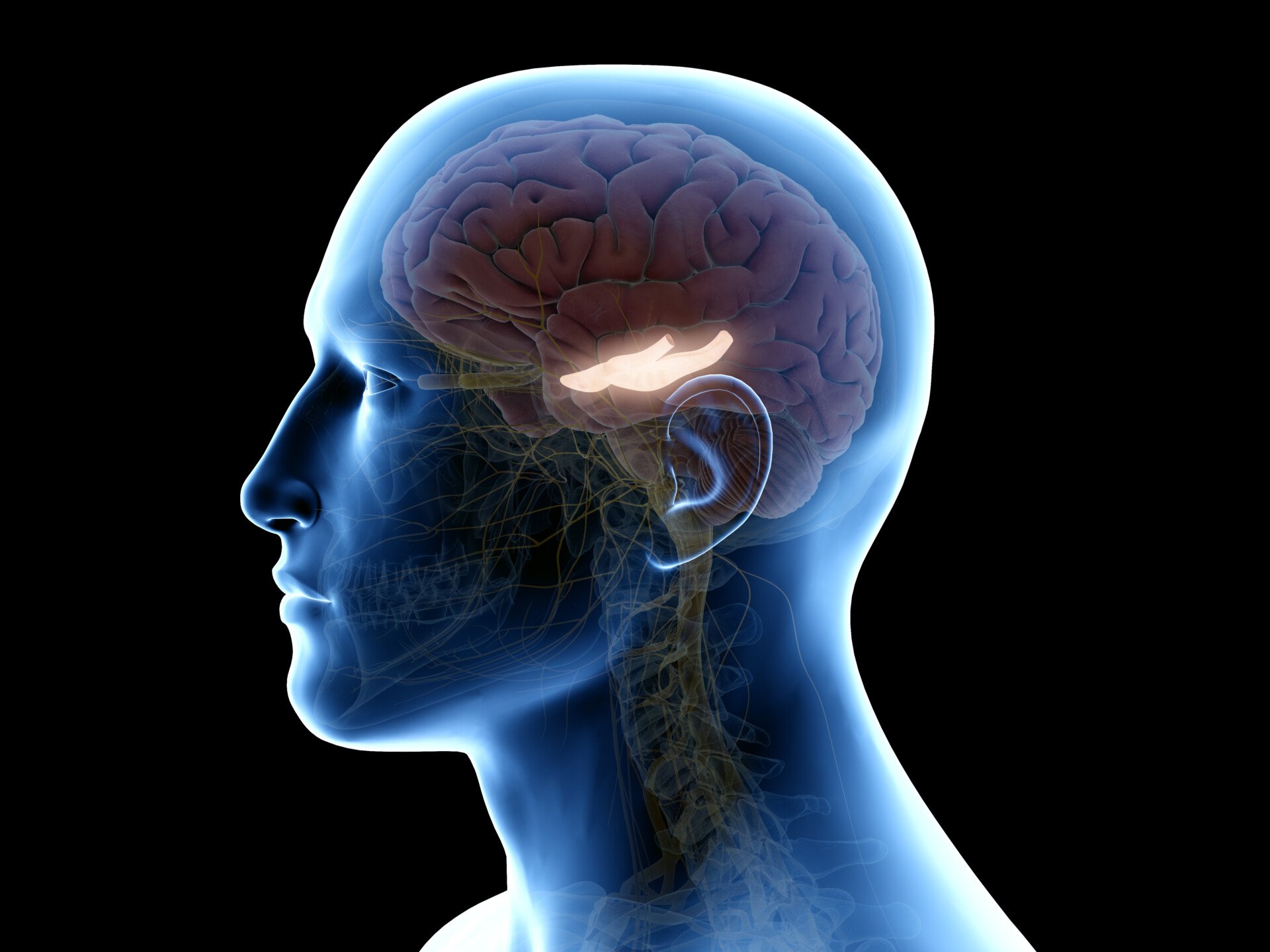
Who is H.M.?
Henry Gustav Molaison lost his memory on an operating table in 1953. Psychology and neuroscience textbooks know him simply as “H.M.”
H.M.’s Epilepsy
For years before his neurosurgery, H.M. suffered from epileptic seizures believed to be caused by a bicycle accident that occurred in his childhood.
The seizures started out as minor at age ten, but they developed in severity when H.M. was a teenager.
Continuing to worsen in severity throughout his young adulthood, H.M. was eventually too disabled to work.
Throughout this period, treatments continued to turn out unsuccessful, and epilepsy proved a major handicap and strain on H.M.’s quality of life.
At age 27, H.M. agreed to undergo radical surgery. The operation would remove the hippocampus, the region his doctors believed was the source of his seizures (Squire, 2009).
What Is Brain Resection Surgery?
For epilepsy patients, brain resection surgery refers to removing small portions of brain tissue responsible for causing seizures.
Although resection is still a surgical procedure used today to treat epilepsy, the use of lasers and detailed brain scans help ensure valuable brain regions are not impacted.
H.M.’s neurosurgeon had none of these tools in 1953. Nobody in the scientific or medical community yet understood what the hippocampus does, or how central it is to memory.
Life After the Surgery
In one regard, the surgery worked. H.M. did experience fewer seizures.
However, family and doctors soon noticed he also suffered from severe amnesia, which persisted well past when he should have recovered.
H.M. struggled to remember the years before his surgery. His memory had gaps across the 11 years prior.
Furthermore, he lacked the ability to form new memories — causing him to perpetually live an existence of moment-to-moment forgetfulness for decades to come.
He once described his state as “like waking from a dream…. every day is alone in itself” (Squire, 2009).
H.M. soon became a major case study. Psychologists and neuroscientists studied his memory deficits and cognitive abilities to understand the hippocampus.
When H.M. died on December 2, 2008, at the age of 82, he left behind a lifelong legacy of scientific contribution.
H.M.’s 1953 Surgery
Neurosurgeon William Beecher Scoville performed H.M.’s surgery in Hartford, Connecticut, in August 1953 when H.M. was 27 years old.
During the procedure, Scoville removed parts of H.M.’s temporal lobe. This is the region of the brain that sits behind each ear. It handles hearing and memory processing.
More specifically, the surgery was what Scoville and Milner (1957) called a “bilateral medial temporal lobe resection”: Scoville operated on both sides of the brain, not just one.
In this resection, Scoville reported removing 8 cm of tissue from the hippocampus, a seahorse-shaped structure deep in the temporal lobe.
That figure came from his surgical notes. Decades later, MRI scans of H.M.’s living brain put the lesion nearer 5 cm (Corkin et al., 1997).
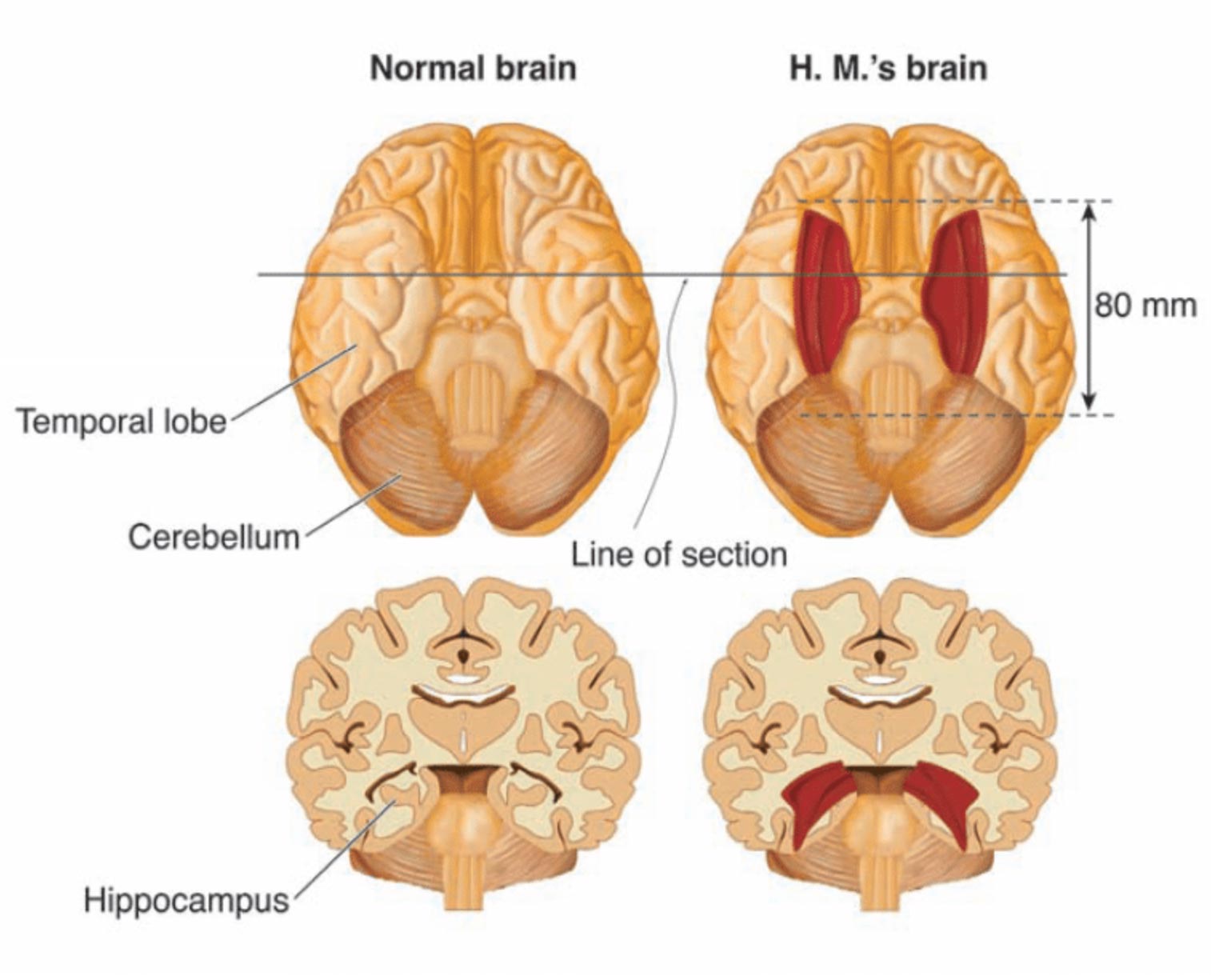
Bilateral resection of the anterior temporal lobe in patient HM.
Later research showed that Scoville probably destroyed two further structures. One was the “uncus,” thought to contribute to the sense of smell and to forming new memories.
The other was the “amygdala.” This structure helps control emotional responses such as fear and sadness.
The removal surgery reduced H.M.’s seizures, but it also cost him the ability to form new memories.
Why H.M. Was a Unique Patient
At the time, Scoville’s experimental procedure had previously only been performed on patients with psychosis, so H.M. was the first epileptic patient and showed no sign of mental illness.
In the original case study of H.M., which is discussed in further detail below, nine of Scoville’s patients from this experimental surgery were described.
However, because these patients had disorders such as schizophrenia, their symptoms were not removed after surgery.
In this regard, H.M. was the only patient with “clean” amnesia along with no other apparent mental problems.
H.M.’s Amnesia
The Original Study: Scoville and Milner (1957)
Aim: Scoville operated to control H.M.’s epilepsy. The research questions came only after the memory loss appeared.
Method: In August 1953, Scoville removed the medial temporal lobe on both sides, taking most of the hippocampus and amygdala (Scoville & Milner, 1957). Milner tested H.M. repeatedly over the decades that followed.
Results: Seizures eased, but H.M. could no longer form new long-term memories. His intelligence, language and immediate memory span stayed normal.
Conclusion: The medial temporal lobe is essential for turning experience into lasting memory. Old memories survived, so it is not the storehouse.
Evaluation: The lesion was surgically documented and H.M.’s intelligence was intact, so his deficits could be traced to specific structures. Still, conclusions drawn from one unusual brain must be treated with caution.
Retrograde and Anterograde Amnesia
H.M.’s amnesia took two forms, retrograde and anterograde. He had retrograde amnesia for the 11-year period before his surgery.
Retrograde amnesia means losing memories formed before the event.
The cause is disputed. Researchers now attribute much of H.M.’s retrograde loss to years of frequent seizures and antiepileptic medication, not to the surgery (Squire, 2009).
The loss was also uneven: childhood memories survived well, but memories from just before surgery were hit hardest. Researchers call this temporally graded retrograde amnesia (Squire, 2009).
This gradient fits how memory is thought to consolidate: new memories first depend on the hippocampus, a fast-learning index that ties together the scattered traces of an experience.
Over months and years those traces are gradually strengthened elsewhere in the cortex, so the memory can eventually be retrieved without the hippocampus. Old, fully consolidated memories survive losing the hippocampus. Memories still “in transit” do not (Squire, 2009).
In contrast, H.M.’s inability to form new memories after his operation, known as anterograde amnesia, was the result of the loss of the hippocampus.
This meant that H.M. could not learn new words, facts, or faces after his surgery, and he would even forget who he was talking to the moment he walked away.
Suzanne Corkin tested H.M. for decades. According to her account, the amnesia shaped every ordinary hour of his day.
He reread the same magazine without recognising it. He could not say the time unless he had just checked a clock, and he never retained the news (Corkin, 2014).
However, H.M. could perform tasks, and he could even perform those tasks easier after practice.
Explicit and Implicit Memory
This finding was a major scientific discovery. It separated two kinds of long-term memory, explicit and implicit.
The memory H.M. had lost covered facts, life events, and other experiences.
This type of long-term memory is referred to as “explicit” or “declarative” memories and they require conscious thinking.
H.M. still improved at tasks with practice. He could not recall practising, yet his “implicit” or “procedural” memory was intact (Scoville & Milner, 1957).
This type of long-term memory is unconscious. Examples include riding a bike, brushing your teeth, or typing on a keyboard.
Most importantly, losing his hippocampus cost H.M. his explicit memory but not his implicit memory. That dissociation matters. Implicit memory must depend on some other brain area.
Brenda Milner’s Testing of H.M.
The severity of H.M.’s side effects soon became clear. Doctors referred him to the Montreal Neurological Institute.
Two people there would define the case. They were the neurosurgeon Dr. Wilder Penfield and the neuropsychologist Dr. Brenda Milner.
H.M. was not the only patient to undergo this experimental surgery. He was, however, the only non-psychotic one. His memory impairment was also uniquely severe.
That made him a major focus for Milner and the wider scientific community.
Penfield and Milner were already running memory experiments on other patients. They saw H.M.’s potential at once.
His “dense amnesia, intact intelligence, and precise neurosurgical lesions made him a perfect experimental subject” (Shrader, 2012).
Milner tested H.M. through the 1950s and 1960s. From the 1960s onward, Suzanne Corkin and colleagues continued the testing for the rest of H.M.’s life, primarily at the Massachusetts Institute of Technology (MIT).
Their combined effort spanned roughly fifty years (Milner et al., 1968). Their longitudinal case study of H.M.’s amnesia became a sensation and remains one of the most widely-cited studies in psychology.
She protected Henry’s identity in print. She referred to him only as the patient H.M. (Shrader, 2012).
The Star Tracing Task
In the famous “star tracing task,” Milner tested if H.M.’s procedural memory was affected by the removal of the hippocampus during surgery.
In this task, H.M. had to trace an outline of a star, but he could only trace the star based on the mirrored reflection. H.M. then repeated this task once a day over a period of multiple days.
H.M. got faster. Over these days, Milner observed that he made fewer errors, and his learning curve looked normal.
Yet each time, he had no memory of ever having done the task before (Milner et al., 1968).
The task showed the split cleanly. H.M. had lost declarative memory but kept procedural memory, so researchers concluded that the hippocampus supports one and not the other.
Therefore, procedural memory must be localized somewhere else in the brain and not in the hippocampus.
Intact Short-Term and Working Memory
H.M. did not lose every kind of memory. His immediate memory span was normal, and he could hold information for about 15 seconds without rehearsal (Scoville & Milner, 1957).
With continuous rehearsal he could hold it much longer. The moment his attention was diverted, the material vanished.
This is the pattern predicted if short-term and long-term memory are separate stores, as the multi-store model claims. H.M. behaved as though the gateway between the two had been destroyed while both stores survived.
His working memory was also preserved. He could hold a conversation, follow instructions, and reason about whatever was in mind at the time.
His IQ stayed normal too. It rose slightly after surgery as the seizures eased, and his vocabulary and grammar were unimpaired (Kensinger et al., 2001).
H.M.’s Legacy
H.M. reshaped what science knows about memory. Milner and hundreds of other researchers used his case to establish how memory works and how the brain organises it.
H.M. volunteered his mind to science for fifty years. Without that contribution, our understanding of how memory functions separate in the brain would be far weaker.
Before H.M., nobody knew the hippocampus was essential for memory. Lose it, and you are confined to the moment-to-moment span of short-term memory.
Once this became clear, researchers publicised the findings about H.M. widely. Surgeons abandoned the operation (Squire, 2009).
H.M.’s case belongs to an earlier era of neuroscience. Researchers then learned about the brain mainly through dissection, lesioning, and watching how experimental procedures affected individual patients.
Patients like H.M. deserve recognition. They underwent dangerous operations in the mid-twentieth century, then allowed researchers to study them for the rest of their lives.
H.M.’s Brain After His Death
H.M. donated his brain to science. Researchers froze it and, in a 53-hour procedure, sliced it into 2,401 sections.
Each section was photographed and assembled into a three-dimensional digital map. That archive preserves his brain for future researchers (Corkin, 2014).
Suzanne Corkin, who tested H.M. for nearly five decades, put it best:
“H.M. was a pleasant, engaging, docile man with a keen sense of humor, who knew he had a poor memory but accepted his fate. There was a man behind the data. Henry often told me that he hoped that research into his condition would help others live better lives. He would have been proud to know how much his tragedy has benefitted science and medicine” (Corkin, 2014).
Critical Evaluation
H.M. is the flagship example of the case study method, an in-depth investigation of a single individual, applied to the brain. It shows what one patient can prove. It also shows where that evidence runs out.
Strengths of the H.M. Case Study
For a single patient, H.M. is unusually strong evidence. The surgery was documented, his other faculties were intact, and the testing ran for half a century.
- Precise lesion: Scoville documented the resection surgically and MRI later confirmed it, so H.M.’s deficits could be tied to specific structures (Corkin et al., 1997).
- Longitudinal depth: Fifty years of repeated testing produced converging evidence that no laboratory experiment on healthy volunteers could match (Milner et al., 1968).
- Falsifying power: His spared skill learning overturned the idea of a single unitary memory and forced the multiple-systems view.
- Clinical dividend: Recognising his deficit ended bilateral medial temporal surgery for epilepsy, protecting later patients (Squire, 2009).
- Converging cases: Patients such as Clive Wearing show the same dissociations from entirely different causes, easing the worry about a sample of one.
Limitations of the H.M. Case Study
A single case cannot settle everything. H.M.’s has clear limits, and several of them are still argued over today.
- Generalisability: Conclusions rest on one unusual brain, and his extensive bilateral lesion cannot ethically be reproduced.
- Confounding factors: Years of seizures and medication may have damaged his memory independently of the surgery (Squire, 2009).
- No baseline: H.M.’s pre-operative memory was never formally measured, so some comparisons are reconstructive.
- Researcher bias: A small group of investigators selected and interpreted the observations across decades, which invites confirmation bias.
- Revision over time: Key facts, including the size of the lesion and the extent of his retrograde amnesia, were revised as methods improved.
- Consent: H.M. could not remember agreeing to be tested, so he had to be re-consented at every session through a guardian.
Comparing H.M. With Other Amnesia Cases
H.M.’s case does not stand alone. Comparing him with another well-studied amnesic patient shows what his evidence does, and does not, establish.
Patient K.C. sustained severe brain damage in a motorcycle accident. Like H.M., he showed dense difficulty forming new memories alongside preserved short-term and skill memory.
Unlike H.M., his damage reached beyond the medial temporal lobes into wider cortex. He lost almost all memory of his own past experiences.
Yet he could still learn new facts about the world, slowly and with effort (Rosenbaum et al., 2005).
K.C. confirms H.M.’s central lesson: conscious memory for facts and events is a separate system from short-term and skill memory. He also refines it.
With more widespread damage, memory for personal experiences and memory for general facts can split apart in a way H.M.’s more limited lesion never revealed on its own.
The two cases also test a question H.M. leaves open. The standard view holds that the hippocampus hands memories to the cortex for permanent storage, so old memories no longer need it.
A rival theory, multiple trace theory, argues the hippocampus stays involved every time a detailed personal memory is replayed, however old it is (Nadel & Moscovitch, 1997).
H.M. never resolved this cleanly. He could recall the bare facts of his distant past, which fits the standard view. But he struggled to vividly relive even his oldest personal memories, which fits multiple trace theory instead.
Contemporary Research
Modern imaging has refined the classic picture. Corkin et al. (1997) scanned H.M.’s living brain with high-resolution MRI and mapped the lesion for the first time.
It was smaller than Scoville had estimated. The damage covered the amygdala, most of the entorhinal cortex and the front of the hippocampus, and the spared rear portion was cut off from its cortical inputs.
Later autobiographical-memory testing complicated the story. Steinvorth et al. (2005) probed remote personal memories in H.M. and a second amnesic patient. Re-experiencing detailed episodes was impaired, even for events from long before surgery.
The medial temporal lobe may therefore support vivid episodic recall indefinitely. It is not needed only to form a memory.
Sagar et al. (1985) had already shown that his retrograde loss was temporally graded. Childhood memories survived; the years just before surgery did not.
Real-World Event Memory: Dede et al. (2016)
A 2016 study tested H.M.’s pattern under real-world conditions. Five patients with hippocampal or wider medial-temporal-lobe damage, plus two groups of healthy volunteers, took a guided 25-minute walk. Eleven planned events happened along the way.
Aim: to test whether patients with this kind of damage show the same memory impairment for a realistic, extended experience. It also asked whether the effect is general, or specific to memory for event order.
Method: Researchers tested free recall, prompted recall, and forced-choice questions about each event’s detail, order, and location. One group was tested immediately, the other a month later.
Results: The patients recalled fewer details than volunteers tested immediately. They performed more like volunteers tested a month later. Their sense of event order broke down completely, unrelated to the true sequence, even though their memory for spatial and perceptual detail was only mildly affected.
Conclusion: The finding sharpens the classic picture. The hippocampus and medial temporal lobe seem especially important for binding separate experiences into the correct time order, not just for forming memories in general (Dede et al., 2016).
Applications of H.M.’s Case
H.M.’s case did not stay inside academic psychology. It reshaped clinical practice, surgery, and research ethics.
Memory Assessment and Rehabilitation
H.M.’s pattern, intact short-term memory but lost long-term learning, and preserved skills but lost conscious memory, shaped how clinicians test memory today.
Modern assessment batteries test immediate versus delayed recall separately. They also test implicit learning apart from explicit recall, since H.M. showed either can be lost or spared on its own (Squire, 2009).
His preserved skill learning also shaped treatment. Amnesic patients can still learn habits through repeated practice, even without remembering the practice itself.
Rehabilitation programmes for memory loss after stroke, brain injury, or early dementia use a technique called errorless learning. A patient is stopped from making mistakes during practice, so no wrong response ever gets implicitly reinforced.
Trial-and-error correction does not work here. Someone who cannot remember an error cannot learn from correcting it (Baddeley & Wilson, 1994).
Changing Neurosurgery and Research Ethics
H.M.’s outcome ended bilateral medial temporal lobe surgery as a treatment for epilepsy and psychosis. It also changed how surgeons prepare for operations near the hippocampus.
Surgeons test each temporal lobe’s contribution to memory before operating. For example, they may numb one side at a time to confirm the other side can support memory alone (Scoville & Milner, 1957; Squire, 2009).
This test is now routine before any one-sided temporal lobe operation, directly because of what H.M.’s case revealed.
The testing also raised a hard ethical problem. H.M. could never remember agreeing to take part in a study.
He had to be re-consented at every single session, through a guardian. His case helped researchers develop surrogate-consent and re-consent procedures now used more widely in research with patients who have dementia or other memory-impairing conditions.
References
Baddeley, A., & Wilson, B. A. (1994). When implicit learning fails: Amnesia and the problem of error elimination. Neuropsychologia, 32(1), 53–68. https://doi.org/10.1016/0028-3932(94)90068-X
Corkin, S. (2014). Permanent present tense: The man with no memory and what he taught the world. Penguin Books.
Corkin, S., Amaral, D. G., González, R. G., Johnson, K. A., & Hyman, B. T. (1997). H. M.’s medial temporal lobe lesion: Findings from magnetic resonance imaging. Journal of Neuroscience, 17(10), 3964–3979.
Dede, A. J., Frascino, J. C., Wixted, J. T., & Squire, L. R. (2016). Learning and remembering real-world events after medial temporal lobe damage. Proceedings of the National Academy of Sciences, 113(47), 13480–13485. https://doi.org/10.1073/pnas.1617025113
Hardt, O., Einarsson, E. Ö., & Nader, K. (2010). A bridge over troubled water: Reconsolidation as a link between cognitive and neuroscientific memory research traditions. Annual Review of Psychology, 61, 141–167.
Kensinger, E. A., Ullman, M. T., & Corkin, S. (2001). Bilateral medial temporal lobe damage does not affect lexical or grammatical processing: Evidence from amnesic patient H. M. Hippocampus, 11(4), 347–360. https://doi.org/10.1002/hipo.1049
Milner, B., Corkin, S., & Teuber, H.-L. (1968). Further analysis of the hippocampal amnesic syndrome: 14-year follow-up study of H. M. Neuropsychologia, 6(3), 215–234. https://doi.org/10.1016/0028-3932(68)90021-3
Nadel, L., & Moscovitch, M. (1997). Memory consolidation, retrograde amnesia and the hippocampal complex. Current Opinion in Neurobiology, 7(2), 217–227. https://doi.org/10.1016/S0959-4388(97)80010-4
Rosenbaum, R. S., Köhler, S., Schacter, D. L., Moscovitch, M., Westmacott, R., Black, S. E., Gao, F., & Tulving, E. (2005). The case of K.C.: Contributions of a memory-impaired person to memory theory. Neuropsychologia, 43(7), 989–1021. https://doi.org/10.1016/j.neuropsychologia.2004.10.007
Sagar, H. J., Cohen, N. J., Corkin, S., & Growdon, J. H. (1985). Dissociations among processes in remote memory. Annals of the New York Academy of Sciences, 444(1), 533–535. https://doi.org/10.1111/j.1749-6632.1985.tb37637.x
Scoville, W. B., & Milner, B. (1957). Loss of recent memory after bilateral hippocampal lesions. Journal of Neurology, Neurosurgery, and Psychiatry, 20(1), 11–21.
Shrader, J. (2012, January). HM, the man with no memory. Psychology Today. https://www.psychologytoday.com/us/blog/trouble-in-mind/201201/hm-the-man-no-memory
Squire, L. R. (2009). The legacy of patient H. M. for neuroscience. Neuron, 61, 6–9.
Steinvorth, S., Levine, B., & Corkin, S. (2005). Medial temporal lobe structures are needed to re-experience remote autobiographical memories: Evidence from H. M. and W. R. Neuropsychologia, 43(4), 479–496. https://doi.org/10.1016/j.neuropsychologia.2005.01.001


