Phineas Gage was a 25-year-old railroad foreman who survived an iron rod being driven through his skull and left frontal lobe in 1848.
His reported personality change afterward, from capable and even-tempered to impulsive and unreliable, made this one of psychology’s most famous case studies.
Key Takeaways
- Survival: In 1848, Phineas Gage lived through a shocking accident: an iron rod shot through his skull and brain.
- Personality Change: The rod destroyed parts of his frontal lobe, and early reports described major shifts in his behavior and temperament.
- Landmark Case: Gage’s story became crucial for linking brain regions to personality, decision-making, and social functioning.
- Modern Reassessment: Skull-based reconstructions since 1994 disagree on whether the damage was left-sided or bilateral, and Gage may have recovered better than assumed.
- Disputed History: Dr. Harlow’s original account was later embellished. Researchers now warn against overinterpreting Gage’s personality changes or idealizing his pre-injury self.
- Verdict: Supported with qualifications. Group studies link frontal damage to altered social decision-making, but they do not support a wrecked character or a single frontal centre for personality.

What Happened To Phineas Gage?
On September 13, 1848, Phineas Gage, a 25-year-old railroad construction foreman, was preparing explosives in Cavendish, Vermont.
As he tamped down blasting powder with a 43-inch, 13-pound iron rod, the charge exploded unexpectedly.
The tamping iron shot through his left cheek, behind his eye, and out through the top of his skull. It destroyed much of his left frontal lobe.
Amazingly, Gage survived. He convulsed briefly, then spoke within minutes. He walked with little assistance and sat upright in an ox-cart that carried him to his lodgings.
Dr. Edward H. Williams noted that Gage’s brain was visibly pulsing through the wound. Gage, showing grim composure, greeted him with words to the effect of “here is business enough for you.”
Fact Check: Did Phineas Gage Vomit Up His Brain?
One of the more gruesome claims about Gage’s injury is that he vomited out part of his brain. This detail comes from Dr. Edward Williams. He reported that during episodes of vomiting, “additional small amounts of Gage’s brain matter [were] expelled onto the floor through the frontal exit wound.”
While this account sounds shocking, it’s not entirely implausible. The explosion had forced a significant opening in his skull, and damaged tissue may have become dislodged.
However, experts caution that the extent of expelled brain matter is unknown. Such claims deserve skepticism, given the limited medical knowledge and observational tools of the time.
In short, yes, historical reports mention it. Modern interpretations, though, urge caution about taking it too literally.
Recovery and the First Clues of Change
Dr. John Martyn Harlow later took over Gage’s care.
The exit wound at the crown of the skull was large enough for him to introduce his index finger into it (Harlow, 1848). Gage endured weeks of infection and a semi-comatose spell, but he steadily recovered.
By October, he was walking and talking again. Physically, he appeared to regain his strength. Mentally and emotionally, however, something had changed.
Before the accident, Gage was described as energetic, responsible, and a favorite among his crew. Afterward, reports painted him as impulsive, rude, and unreliable.
Harlow wrote that “the equilibrium or balance… between his intellectual faculties and animal propensities, seems to have been destroyed.” He added:
“He is fitful, irreverent, indulging at times in the grossest profanity (which was not previously his custom)… at times pertinaciously obstinate, yet capricious and vacillating… devising many plans… which are no sooner arranged than they are abandoned… A child in his intellectual capacity and manifestations, he has the animal passions of a strong man”.
His friends famously said he was “no longer Gage.”
How Much Did Gage Really Change?
Though Harlow’s accounts are the primary source of information, later writers often embellished Gage’s personality both before and after the accident.
Some described him as unusually polite and mild-mannered pre-accident, and almost monstrously unstable afterward.
Accounts often idealise Gage’s character before the accident. The post-injury version is then exaggerated into near caricature, unlike Harlow’s own measured clinical account.
This embellishment likely reflects enthusiasm for making Gage’s case fit localization theories. Macmillan (2000) warns that such dramatised descriptions need cautious handling.
Harlow also recorded that Gage later held a demanding job as a stagecoach driver in Chile. Such sustained, responsible work suits no severely incapacitated man.
The photographic record backs this up. Daguerreotype portraits of Gage were only identified as genuine in the twenty-first century. They show him well-dressed and composed, holding his inscribed tamping iron, not the dishevelled wreck of popular legend (Macmillan & Lena, 2010).
One daguerreotype was identified in 2008. Jack and Beverly Wilgus had owned it for 30 years, believing it showed an anonymous whaler with a harpoon.
An online commenter suggested the “harpoon” might be Gage’s inscribed tamping iron (Wilgus & Wilgus, 2009). The collectors set out to test that.
They compared the image with Gage’s life mask and tamping iron at Harvard’s Warren Anatomical Museum. The forehead scar, the shape of the mouth and the depression beside the right eye matched. Writing on the rod lined up with the inscription on the surviving iron.
Severity of Gage’s Brain Damage
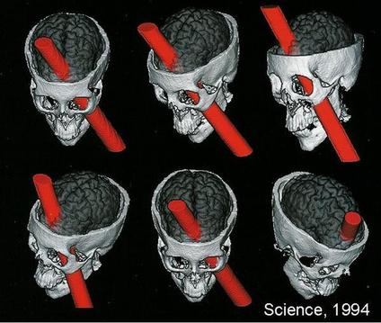
Researchers disagree about where the rod did its damage. Since 1994, they have reconstructed Gage’s skull using measurements, computer modelling and CT scans to estimate the rod’s trajectory.
Later work refined this picture.
Hanna Damasio and colleagues (1994) tackled this by photographing and measuring Gage’s skull, then using computer modelling to simulate the rod’s likely trajectory. They concluded that the damage reached the ventromedial prefrontal cortex in both hemispheres.
This region lies on the underside and inner surface of the prefrontal cortex, the front-most part of the frontal lobe. It is involved in emotional processing and real-world decision-making. A second team reached a different verdict.
Ratiu et al. (2004) used thin-slice computed tomography (CT) of the skull to build three-dimensional reconstructions of the injury. The picture looked different this time.
They found that the damage was limited to the left frontal lobe (see Fig. 2) and did not cross into the right hemisphere.
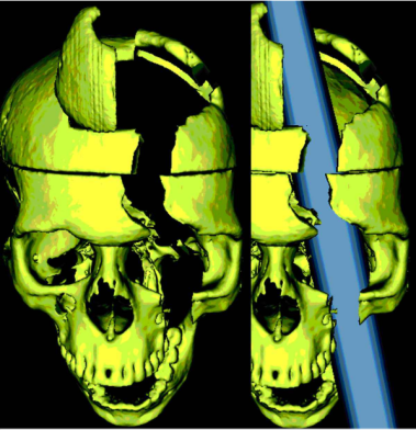
Van Horn et al. (2012) agreed with Ratiu et al. (2004) that the rod damaged only the left frontal lobe, not the right.
Van Horn’s team also found that Gage likely lost 11% of his white matter and 4% of his grey matter. This was enough to affect thinking and behavior, but not to prevent functional recovery.
A different question emerged.
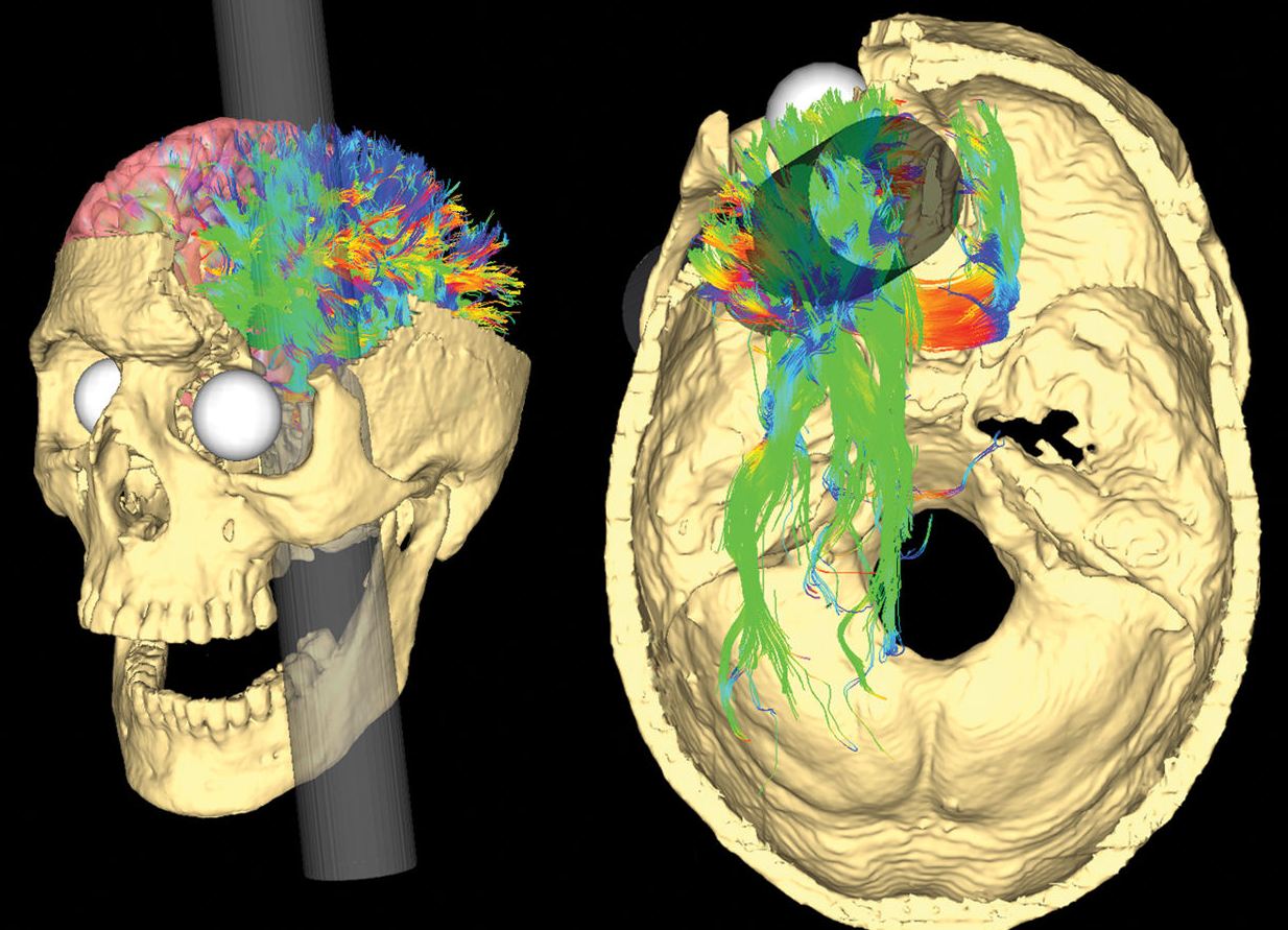
Some researchers speculated that Gage’s behavioral changes might resemble early-onset Alzheimer’s, since both involve white matter degradation.
However, Harlow only reported on Gage shortly after the accident, and there’s no concrete evidence he developed the disease. This remains purely speculative.
Why the Exact Damage Remains Unknown
Every study of Gage’s brain damage involves speculation. We cannot know for certain how much damage the accident itself caused.
We know that some brain tissue was destroyed, but any infections Gage suffered afterward may have destroyed further tissue.
We also cannot pinpoint exactly where the rod entered. Brain structure varies from person to person, so researchers can never know for certain which areas of Gage’s skull were destroyed.
What Happened to Phineas Gage After the Brain Damage?
Gage eventually moved to Chile and worked as a stagecoach driver. In 1860, he returned to the U.S. and began experiencing epileptic seizures.
He died in San Francisco in 1860. That was around twelve years after the accident, from complications linked to epilepsy.
In 1867, Harlow arranged to exhume Gage’s body and recovered his skull and tamping iron. Both are now held at Harvard’s Warren Anatomical Museum.
Why Is Phineas Gage Important to Psychology?
Launching Neuropsychology and Localisation of Function
Gage’s story helped launch neuropsychology, the study of normal brain function through patients with brain damage.
It provided early, powerful evidence that the frontal lobes are essential for regulating behaviour, personality, and social functioning in everyday life.
It also introduced the idea of localization of brain function: that specific parts of the brain serve specific roles. That idea reshaped psychology.
Here is how the inference runs, step by step:
- The dissociation: Some functions were lost while others were spared. After the rod, intellect, memory, language and movement stayed intact, but planning, emotional control and social conduct suffered.
- The lesion logic: If destroying one region removes a function and spares others, that region is probably necessary for that function.
- The localisation conclusion: A selective deficit means the brain is not an equipotential mass, one where any part can do any job. Different regions make different contributions.
- The region’s job: The prefrontal cortex supports executive functions, such as holding goals in mind, inhibiting impulses and foreseeing consequences. It also supports the social self.
- The limit: All of this rests on one patient, a retrospective record and a lesion known only from a skull. The inference is suggestive, not a controlled demonstration.
Gage arrived at exactly the right moment for this idea to take hold. Paul Broca’s speech-area findings came roughly a decade later. At the time, the debate between localisation and an undifferentiated, whole-brain view of the mind was still wide open.
His selective pattern of loss fits neither an all-purpose brain nor old-style faculty psychology. That is why the case still comes up wherever localisation of function is taught. Students often meet it first.
Ferrier’s Monkeys: An Experimental Parallel
Experimental support arrived within thirty years of the accident. In his 1878 Goulstonian Lectures, the physiologist David Ferrier reported that removing or cauterising the front of the frontal lobes in monkeys caused no obvious paralysis (Ferrier, 1878).
Yet he saw “a very decided alteration in the animal’s character and behaviour.”
The monkeys were not deprived of intelligence. They seemed to have lost the ability to observe attentively and intelligently.
Ferrier then traced the tamping iron’s probable path through Gage’s skull. He judged that it involved the prefrontal region. He concluded that “the absence of paralysis in this case is quite in harmony with the results of experimental physiology” (Ferrier, 1878).
That matters because it shows a changed self without paralysis was already treated as a signature of frontal damage, on independent animal evidence, in Harlow’s lifetime. The pattern was not invented later to fit twentieth-century theory.
Evidence of Recovery and Neuroplasticity
Yet Gage’s case is not just a tale of loss. His return to functional life suggests the brain’s remarkable capacity for neuroplasticity.
As Macmillan (2002) argues, Gage may have relearned lost abilities over time. He adapted. This is an early, striking example of resilience after brain injury.
This fits his later life: years of demanding, routine-based work as a stagecoach driver in Chile (see How Much Did Gage Really Change?, above). Routine, structured demands may have supported relearning, giving the brain repeated practice at planning and self-regulation.
That is a modest but real form of recovery.
Frontal-Lobe Syndrome and the Somatic Marker Hypothesis
Gage still matters to modern neuroscience. The case gave clinicians a name for what is now called frontal-lobe or dysexecutive syndrome, a pattern that follows damage to the ventromedial prefrontal cortex:
- Preserved: Intelligence and memory stay intact.
- Planning: Poor planning and foresight.
- Emotion: Blunted or volatile.
- Social conduct: Disinhibition and socially inappropriate behaviour.
This connects directly to modern research.
Antonio Damasio’s somatic marker hypothesis proposes that the ventromedial prefrontal cortex binds emotional signals to reasoning, tested using tasks such as the Iowa Gambling Task. Gage is considered the historical starting point for this entire line of research linking emotion to rational decision-making.
Gage remains a teaching touchstone for the prefrontal cortex’s role in personality and decision-making. It is a story everyone in psychology eventually meets.
He is also a byword for both the power and the pitfalls of case-study evidence. It is one of the most recognisable stories in the public understanding of neuroscience.
Evidence Check: Does Gage’s Case Hold Up?
Verdict: Supported with qualifications.
The broad claim stands. Damage to the ventral and medial frontal cortex can change social conduct and everyday decision-making. Group lesion studies that stand apart from Gage show this.
The narrower claims are not supported. The case is often used to argue that Gage’s own character was wrecked and that a single frontal “centre” governs personality. Both rest on a thin record.
Frontal Damage and Social Decision-Making: Real but Narrower
Level of evidence: Meta-analysis or systematic review; partly replicated.
The claim is that the frontal cortex, and especially its ventromedial part (the lower, inner surface), is necessary for regulating social conduct and making sound real-world decisions. On this view, destroying it leaves intellect intact but changes personality.
The evidence runs from systematic reviews down to one historical case.
- Yu et al. (2020): A systematic review of 184 papers on damage to the human ventromedial frontal lobe. It reports deficits in memory, executive function, emotion, social cognition and valuation.
- Messimeris et al. review: A 2023 review of lesion studies on value-based and social decision-making. Learning and updating reward value is consistently affected, but decision-making itself is often only moderately or context-specifically affected.
- Bechara et al. (1999): Patients with bilateral amygdala damage and patients with bilateral ventromedial damage were compared on a gambling task. Both groups were impaired, but only the ventromedial group still responded to reward and punishment.
- Anderson et al. (1999): Two adults (N = 2) had prefrontal damage before 16 months of age. Unlike adult-onset patients such as Gage, they also had defective social and moral reasoning.
- Gage himself: The evidence is observational: Harlow’s (1848, 1868) accounts, Bigelow’s (1850) report and skull-based reconstructions (Damasio et al., 1994; Ratiu et al., 2004). Gage cannot be replicated.
Yu et al. are the most trustworthy single item, because the literature was searched by a stated method. Their limits are real, though. A qualitative synthesis weighs no studies by quality and yields no pooled effect.
The Gage record is weakest.
It rests on one patient, no pre-injury baseline, a retrospective and partly second-hand description, and a dispute over which structures were destroyed (Macmillan & Lena, 2010).
Thiebaut de Schotten et al. (2015) use a sound anatomical method. It estimates the severed tracts from atlases of healthy adults (129 people), not Gage’s own brain. It supports a plausible mechanism and does not prove one.
So the strongest evidence qualifies the claim rather than overturning it. Yu et al. show that ventromedial damage disturbs several domains, including social cognition and valuation. The direction of Gage’s case holds up.
Messimeris et al. show the disturbance to decision-making is partial and task-dependent. Anderson et al. add that timing matters.
Early damage produces a different, more pervasive profile than the adult damage Gage suffered.
The finding is real but smaller and more variable than the popular story. The weakest element is the explanation: a localised moral faculty.
A fair current account treats Gage as a suggestive, poorly documented single observation. It fits the group-study finding that ventromedial frontal damage disrupts social and value-based decision-making in a partial, context-dependent way. It cannot establish that finding.
Where his lesion lay is disputed. Tractography models suggest the rod cut long-range white-matter connections too, so the change may reflect network disconnection as well as local cortical loss. On Harlow’s retrospective account, his later years show substantial functional recovery.
Critical Evaluation of the Phineas Gage Case Study
Gage’s case is both foundational and famously fragile as evidence. AQA and other exam boards often ask students to evaluate it directly, so the strengths and weaknesses below are worth knowing by name.
- No Control Group: As a single case study, Gage offers no comparison group, so the link between his injury and his behaviour can only be inferred, not proven.
- A Thin, Retrospective Record: The fullest account of his personality change was written twenty years after the accident, drawing on other people’s memories rather than contemporaneous notes.
- No Imaging in Gage’s Lifetime: Every modern claim about which brain regions were destroyed rests on a 19th-century skull, not a scan taken while Gage was alive.
- Genuine, Lasting Influence: Despite these flaws, the case genuinely launched the study of brain-behaviour links and still converges with modern research on emotion and decision-making.
- A Contested Explanation: Even Harlow’s own account may not need a neurological explanation at all; one historian argues the change reflects Gage’s reaction to disfigurement, not brain damage.
Why One Case Can’t Prove Causation
Some limitations cut deeper than others.
Gage was a natural experiment, an event that occurs naturally rather than one a researcher deliberately creates, not a designed one. No matched colleague stayed uninjured as a comparison.
Without a control group or any experimental manipulation, the link between his wound and his behaviour can only be inferred, never proven.
Case studies like this one are excellent for generating ideas but weak for testing them. Brain organisation differs from person to person. A change seen in one patient might reflect that patient’s own biology, not a general rule about the frontal lobes.
No two brains are organised identically.
This is why later researchers, including Malcolm Macmillan, treat Gage’s case study as a starting hypothesis. It is not proof that frontal damage reliably produces one particular personality profile. This is a hypothesis, not a verdict.
A Record Written Twenty Years Late
Dr. John Martyn Harlow, Gage’s own physician, wrote the fullest account of the personality change in 1868. That was twenty years after the accident and eight years after Gage had died. Harlow drew heavily on the recollections of Gage’s family and acquaintances rather than on notes taken at the time.
Eyewitness memory fades fast.
Memory reshapes itself around expectation. Later writers went further still, polishing Gage’s pre-accident image into that of a model citizen while darkening his post-accident behaviour into near caricature. Neither extreme matches Harlow’s own, more measured clinical language.
Historians have traced how the embellishment spread.
O’Driscoll and Leach (1998) followed the drift from Harlow’s careful clinical language to the far more colourful sketches later writers built on it. Macmillan (2000, 2002) found unsourced details, such as the fairground exhibition, that trace back to neither Harlow nor Bigelow.
The two explain the drift differently.
O’Driscoll and Leach point to ordinary editorial embellishment across a chain of retellings. Macmillan links it to the case’s usefulness to theory: a dramatic ruin argues for localisation better than a man who mostly recovered.
Not every contemporary observer told the same dramatic story. Henry Jacob Bigelow, the Harvard surgeon who examined Gage in person, presented the case to the medical profession (Bigelow, 1850).
Bigelow was comparatively sceptical of the more sensational claims, a counterweight often left out of popular retellings. That disagreement between eyewitnesses is itself a reason for caution.
One Skull, No Living Scan
There was no neuroimaging and no formal neuropsychological testing while Gage was alive. Everything modern researchers know about the lesion comes from his skull, exhumed and measured decades after his death.
That creates two problems.
- A ‘standard’ brain: Reconstructions must assume one, even though real anatomy varies from person to person.
- Infection after the accident: It may have destroyed additional tissue beyond what the rod itself damaged, further blurring the picture.
That gap matters for interpretation.
The reconstructions disagree because of this uncertainty. Damasio and colleagues modelled bilateral damage. Ratiu and colleagues disagreed. They, and later Van Horn and colleagues, concluded the injury was confined to the left hemisphere (see Severity of Gage’s Brain Damage, above).
Even the basic location of the damage remains contested. That is a shaky foundation for the many strong claims built on top of it.
A Case That Still Shapes the Field
Despite every limitation above, Gage’s case is not merely a cautionary tale. It helped launch neuropsychology, the practice of inferring normal brain function from patients with brain damage. Paul Broca’s work on language and, later, the amnesic patient H.M. played the same role.
The case also converges with modern research. Antonio Damasio’s somatic marker hypothesis points to a similar picture. So does decades of work on the ventromedial prefrontal cortex and decision-making (see Why Is Phineas Gage Important to Psychology?, above).
Both describe intact intellect paired with impaired judgement after this kind of damage. A purely mythical story would be unlikely to converge with real modern data this way.
Gage himself complicates the simplest reading. He later held a demanding stagecoach-driving job in Chile for several years, suggesting real functional recovery rather than permanent ruin. The fair verdict treats Gage as a historically pivotal, suggestive case: genuinely useful, and just as genuinely limited.
A Rival Explanation: Psychological Reaction, Not Brain Damage
Not every scholar accepts that Gage’s changed behaviour came directly from his damaged frontal lobe. Psychologist Zbigniew Kotowicz (2007) disagrees.
He argues that Harlow’s own descriptions never really support a full “psychopathic” transformation. Gage’s later life looked restless: frequent moves, and a string of short-lived jobs. Kotowicz reads this as a psychological reaction to disfigurement and stigma, not a damaged brain.
The injury still matters here. Without it, Gage’s disfigurement and its social fallout would not exist. Kotowicz simply relocates the cause: not damaged circuitry, but a man adjusting to how people now saw him.
Kotowicz frames the stakes bluntly. He sees the standard reading as part of a wider habit in brain-based psychiatry: swapping a person’s own biography for a brain-mechanism story.
Contemporary Research
Since 2015, research on Gage has moved on two fronts. Anatomical modelling of the lesion has become sharper. The modern claims built on the case have also been tested against the much larger literature on ventromedial prefrontal cortex damage.
Both trends point to a more cautious, distributed account than the confident ‘one region, one function’ story the case often illustrates.
Tractography and Gage’s Disconnected Networks
- Aim: Building on earlier connectome work, the map of long-distance white-matter pathways between brain regions, Thiebaut de Schotten et al. (2015) revisited Gage’s lesion. Their question: disconnection, or local damage?
- Method: They combined a CT scan of Gage’s skull with the tamping iron’s dimensions to model the rod’s path. They overlaid it on a map of white-matter connections from 129 healthy adults, built with tractography (imaging that traces fibre pathways).
- Results: Direct damage involved the orbitofrontal, dorsolateral prefrontal cortex and temporal pole cortex. Fibre tracts were disrupted well beyond the lesion, reaching the amygdala and cingulate cortex, far from the rod’s path.
- Conclusion: The authors argue this widespread disconnection, not the local damage, better explains the breadth of Gage’s reported personality change. Damage in one place; disruption everywhere.
Evaluation: This is the strongest anatomical reconstruction available, but it remains a model fitted to one 170-year-old skull. No behavioural testing exists to confirm that these specific tracts caused Gage’s reported changes.
These findings set two accounts against each other.
The classical reading locates Gage’s deficit in a place: a patch of frontal cortex that does the work of personality and social judgement (Damasio et al., 1994).
The disconnection account locates it in a connection: severed long-range tracts linking the damaged region to distant areas (Van Horn et al., 2012). They carry different implications for how selective or diffuse a lesion must be before personality changes.
That still leaves an open question.
Is the VMPFC Really Necessary for Decision-Making?
A 2023 systematic review puts a number on this question. Messimeris and colleagues reviewed group studies of ventromedial prefrontal cortex damage published between 1990 and 2023, testing patients on value-based and social decision-making tasks.
They found consistent evidence that the region helps people learn and update the value of rewards. Its role in decision-making itself, though, looked moderate and context-specific rather than strictly necessary.
Social judgement fared no better. VMPFC damage impaired how patients represented other people’s rewards, but effects on fairness and cooperation judgements were inconsistent.
The picture that emerges is a key motivational node, not an indispensable switch. (See Why Is Phineas Gage Important to Psychology?, above, for Damasio’s somatic marker hypothesis.) Gage’s case still points to the frontal lobes; it just no longer supports an all-or-nothing story.
How Textbooks Tell the Gage Story
Griggs (2015) tested how the textbooks themselves handle this. He analysed 23 introductory psychology textbooks and found 21 covered Gage’s case, usually getting the accident and injury right.
Recovery told a different story. Only about half discussed what happened to Gage afterwards, and very few mentioned the photographic evidence or the digital lesion reconstructions published since 2004.
The pattern fits. Textbooks tend to repeat the dramatic, incomplete version of Gage and under-report the recovery that complicates it, echoing the embellishment problem already described above.
References
Anderson, S. W., Bechara, A., Damasio, H., Tranel, D., & Damasio, A. R. (1999). Impairment of social and moral behavior related to early damage in human prefrontal cortex. Nature Neuroscience, 2(11), 1032–1037. https://doi.org/10.1038/14833
Bechara, A., Damasio, H., Damasio, A. R., & Lee, G. P. (1999). Different contributions of the human amygdala and ventromedial prefrontal cortex to decision-making. Journal of Neuroscience, 19(13), 5473–5481. https://doi.org/10.1523/JNEUROSCI.19-13-05473.1999
Bigelow, H. J. (1850). Dr. Harlow’s case of recovery from the passage of an iron bar through the head. American Journal of the Medical Sciences, 20(39), 13–22.
Blakeslee, S. (1994, July 6). A miraculous recovery that went wrong. New York Times.
Damasio, H., Grabowski, T., Frank, R., Galaburda, A. M., & Damasio, A. R. (1994). The return of Phineas Gage: Clues about the brain from the skull of a famous patient. Science, 264(5162), 1102–1105.
Ferrier, D. (1878). The Goulstonian lectures on the localisation of cerebral disease. BMJ, 1, 443–447. https://doi.org/10.1136/bmj.1.900.443
Griggs, R. A. (2015). Coverage of the Phineas Gage story in introductory psychology textbooks: Was Gage no longer Gage? Teaching of Psychology, 42(3), 195–202. https://doi.org/10.1177/0098628315587614
Harlow, J. M. (1848). Passage of an iron rod through the head. Boston Medical and Surgical Journal, 39(20), 389–393.
Harlow, J. M. (1868). Recovery from the Passage of an Iron Bar through the Head. Publications of the Massachusetts Medical Society. 2 (3), 327-347.
Kao, Y. H., Chou, M. C., Chen, C. H., & Yang, Y. H. (2019). White matter changes in patients with Alzheimer’s disease and associated factors. Journal of Clinical Medicine, 8 (2), 167.
Kotowicz, Z. (2007). The strange case of Phineas Gage. History of the Human Sciences, 20(1), 115–131. https://doi.org/10.1177/0952695106075178
Macmillan, M. (2000). Restoring Phineas Gage: A 150th retrospective. Journal of the History of the Neurosciences, 9(1), 46-66.
Macmillan, M. (2002). An odd kind of fame: Stories of Phineas Gage. MIT Press.
Macmillan, M., & Lena, M. L. (2010). Rehabilitating Phineas Gage. Neuropsychological Rehabilitation, 20(5), 641–658.
Messimeris, D., Levy, R., & Le Bouc, R. (2023). Economic and social values in the brain: Evidence from lesions to the human ventromedial prefrontal cortex. Frontiers in Neurology, 14, Article 1198262. https://doi.org/10.3389/fneur.2023.1198262
Nasrabady, S. E., Rizvi, B., Goldman, J. E., & Brickman, A. M. (2018). White matter changes in Alzheimer’s disease: a focus on myelin and oligodendrocytes. Acta neuropathologica communications, 6(1), 1-10.
O’Driscoll, K., & Leach, J. P. (1998). “No longer Gage”: An iron bar through the head. BMJ, 317(7174), 1673–1674. https://doi.org/10.1136/bmj.317.7174.1673a
Ratiu, P., Talos, I. F., Haker, S., Lieberman, D., & Everett, P. (2004). The tale of Phineas Gage, digitally remastered. Journal of neurotrauma, 21 (5), 637-643.
Thiebaut de Schotten, M., Dell’Acqua, F., Ratiu, P., Leslie, A., Howells, H., Cabanis, E., Iba-Zizen, M. T., Plaisant, O., Simmons, A., Dronkers, N. F., Corkin, S., & Catani, M. (2015). From Phineas Gage and Monsieur Leborgne to H.M.: Revisiting disconnection syndromes. Cerebral Cortex, 25(12), 4812–4827. https://doi.org/10.1093/cercor/bhv173
Van Horn, J. D., Irimia, A., Torgerson, C. M., Chambers, M. C., Kikinis, R., & Toga, A. W. (2012). Mapping connectivity damage in the case of Phineas Gage.
PloS one, 7(5), e37454.
Wilgus, J., & Wilgus, B. (2009). Face to face with Phineas Gage. Journal of the History of the Neurosciences, 18(3), 340–345. https://doi.org/10.1080/09647040903018402
Yu, L. Q., Kan, I. P., & Kable, J. W. (2020). Beyond a rod through the skull: A systematic review of lesion studies of the human ventromedial frontal lobe. Cognitive Neuropsychology, 37(1–2), 97–141. https://doi.org/10.1080/02643294.2019.1690981
Further Reading
- Griggs, R. A. (2015). Coverage of the Phineas Gage Story in Introductory Psychology Textbooks: Was Gage No Longer Gage?. Teaching of Psychology, 42(3), 195-202.
- Wilgus, J., & Wilgus, B. (2009). Face to face with Phineas Gage. Journal of the History of the Neurosciences, 18(3), 340-345.
- Macmillan, M., & Lena, M. L. (2010). Rehabilitating Phineas Gage. Neuropsychological Rehabilitation, 20, 641–658.
- Macmillan, M. (2000). Restoring phineas gage: a 150th retrospective. Journal of the History of the Neurosciences, 9(1), 46-66.
- Kotowicz, Z. (2007). The strange case of Phineas Gage. History of the Human Sciences, 20(1), 115-131.
- O”driscoll K, Leach JP. “No longer Gage”: an iron bar through the head. Early observations of personality change after injury to the prefrontal cortex. BMJ. 1998;317(7174):1673-4. doi:10.1136/bmj.317.7174.1673a
If a person suffers from a traumatic brain injury in the prefrontal cortex, similar to that of Phineas Gage, what changes might occur?
A traumatic brain injury to the prefrontal cortex could result in significant changes in personality, emotional regulation, and executive function. This region is vital for impulse control, decision-making, and moderating social behavior.
A person may exhibit increased impulsivity, poor judgment, and reduced ability to plan or organize. Emotional volatility and difficulty in interpersonal relationships may also occur.
Just like the case of Phineas Gage, who became more impulsive and less dependable, the injury could dramatically alter one’s character and abilities.

