Broca’s area is a small but vital region in the brain responsible for producing speech and processing language. Located in the left frontal lobe, it corresponds to Brodmann areas 44 and 45. Most people rely on this part of the brain to form words, construct sentences, and organize grammar. Without it, fluent communication becomes difficult.
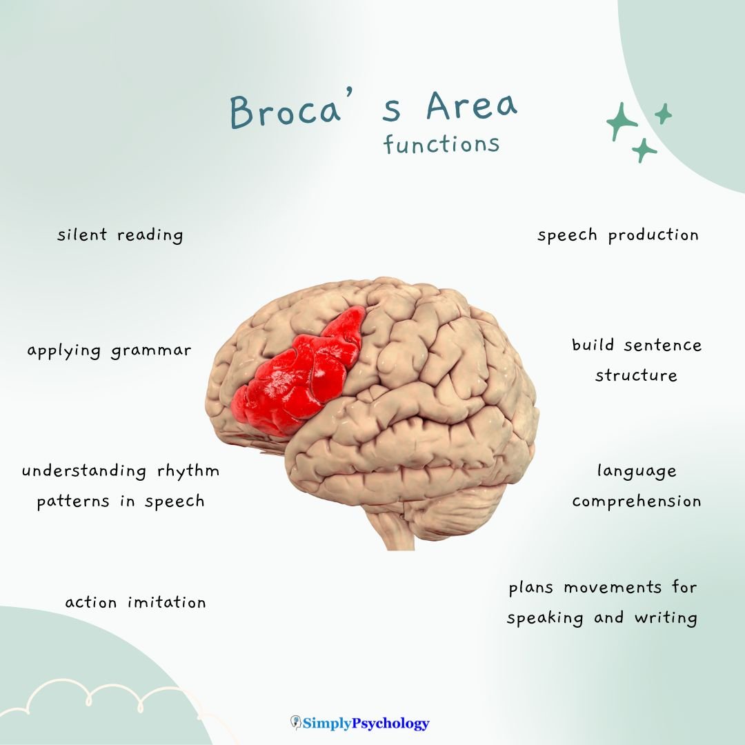
Where Is Broca’s Area Located?
Broca’s area sits in the posterior part of the inferior frontal gyrus (one of the long folds on the lower surface of the frontal lobe), typically in the left hemisphere.
This side of the brain controls language in about 97% of right-handed individuals and the majority of left-handed people as well.
The left hemisphere is known as the “dominant hemisphere” for language in most people.
Functions of Broca’s Area
Broca’s area plays a key role in:
- Speech and language production
- Understanding grammatically complex sentences, not just producing them
- Sentence structure and syntax
- Motor planning for spoken and written language
- Remembering verbal information
- Reading complex sentences
Research by Bookheimer et al. (1995) found that Broca’s area activates even during silent reading, not just aloud speech.
It plans speech before we speak. It assembles the sounds and order a word needs, then hands that plan to the neighboring motor cortex to execute.
Damage here does not paralyze the mouth. It disorganizes the plan behind fluent words, so speech turns effortful and halting instead.
Long sentences need memory too. Holding words in mind while parsing grammar draws on verbal working memory, which the left inferior frontal cortex, including Broca’s area, helps support (Gabrieli et al., 1998).
Syntax leans on it too. Imaging shows Broca’s area working hardest when sentences turn grammatically complex, tracking parsing demands more than speaking effort (Cooke et al., 2002).
Speech is not the only thing it does. Broca’s area, especially BA44, is part of the brain’s mirror system: active when we plan an action, and when we watch someone do it (Nishitani & Hari, 2002).
This overlap may explain why speech evolved from gesture. The idea fits how much the frontal cortex has grown across primate evolution (Chaplin et al., 2020).
Connections to Other Language Regions
Broca’s area is one part of a broader language network. It works closely with Wernicke’s area, which is responsible for understanding language.
These two areas are connected by the arcuate fasciculus, a bundle of nerve fibers that carries information between them.
This connection helps translate thoughts into coherent speech: Wernicke’s area finds the right words, and Broca’s area helps produce them aloud.
What Happens When Broca’s Area Is Damaged?
Damage to this region can result in Broca’s aphasia, a condition marked by:
- Difficulty speaking fluently
- Struggling to form grammatically correct sentences
- Repetitive or broken speech
- Understanding language better than producing it
- Abnormal tone or rhythm
For example, someone with Broca’s aphasia might want to say “I am going to the store” but instead say “go… store” with effort and frustration. Speech may be slow, halting, and limited to key words.
History of Broca’s Area
In 1861, French physician Paul Broca studied a patient named Louis Victor Leborgne, who could only say one word: “Tan.” Despite understanding others, he struggled to speak.
After Leborgne’s death, Broca examined his brain and found damage in the left frontal lobe. This was the first clear evidence that specific brain regions control specific functions—in this case, language production.
Broca studied more patients and found the same pattern: damage in the left frontal region consistently resulted in speech impairments.
Broca’s work launched two enduring ideas. First, mental functions are localized to specific brain regions rather than spread evenly across the whole brain. Second, language itself is lateralized: it relies more on one hemisphere, usually the left, than on the other.
Critical Evaluation of Broca’s Area
Few brain structures inspire as much textbook confidence as Broca’s area. Yet several serious problems complicate this classic story.
- Aphasia Doesn’t Match the Area: Broca’s aphasia usually needs damage well beyond Broca’s area itself, and reimaging of Broca’s own historic patients found lesions reaching far past the region he described.
- Production Isn’t Fully Separate From Comprehension: Patients with Broca’s aphasia often struggle to understand grammatically complex sentences, not just to produce fluent speech.
- Language Is a Network, Not Two Centers: Careful brain imaging finds no single region, Broca’s area included, uniquely dedicated to grammar; syntax and meaning are processed together across a wider network.
- Individual Variation and Terminology: The exact size, location, and even the hemisphere of language regions vary between people, and researchers do not all draw Broca’s area’s borders the same way.
Aphasia Doesn’t Match the Area
The most damaging criticism targets the syndrome itself. Persistent, severe Broca’s aphasia rarely follows damage confined to Broca’s area alone; it generally needs a much larger lesion, reaching into the surrounding cortex, the white matter, and the insula.
Damage confined to Broca’s area proper is often just mild. It typically recovers within weeks, unlike the severe, lasting syndrome the textbooks describe.
This gap became clear when researchers re-examined the preserved brains of Broca’s own patients, Leborgne and Lelong. The MRI was modern; the brains were not.
Dronkers and colleagues (2007) found that both lesions reached well beyond the surface area Broca had described, extending into deeper structures he could never have seen. The region he originally identified does not even coincide exactly with what neuroscientists now call “Broca’s area” today.
The founding cases don’t cleanly fit the theory.
Production Isn’t Fully Separate From Comprehension
The double dissociation between Broca’s and Wernicke’s aphasia is the strongest evidence for a clean production/comprehension split. But that split doesn’t fully hold up.
The classic model is neat: Broca’s area handles production, Wernicke’s area handles comprehension. Real patients are messier than that.
When understanding a sentence depends on grammar rather than word meaning, Broca’s aphasics often struggle. Take “the boy that the girl pushed was tall”: working out who did what to whom takes real syntactic parsing, not just knowing the words.
This shows Broca’s area supports comprehension too. So the two classic syndromes were never quite the mirror images the textbook model implies. The exception matters because it complicates how much weight the classic double dissociation can bear.
Language Is a Network, Not Two Centers
Modern imaging increasingly locates language not in two separate centers but in a network spanning the frontal and temporal lobes. Broca’s area is only one node in it.
When researchers map the language system in each person’s own brain, no single region stands out as a dedicated “syntax module,” separate from word meaning (Fedorenko et al., 2020). Grammar and meaning turn out to be tangled together across the whole network.
Treating Broca’s area as a self-contained “grammar box” makes a common mistake. It mistakes one necessary part for the whole mechanism.
Even the classic Broca-Wernicke model depended on a connecting pathway, the arcuate fasciculus. Cutting it produces its own disorder, conduction aphasia: fluent speech and normal understanding, but an inability to repeat back what was just heard.
The tracts matter as much as the regions they connect.
Individual Variation and Terminology
No two brains are quite alike. The exact location, size, and even the hemisphere of the language regions vary substantially from person to person.
Language sits on the right side, or on both sides, in a meaningful minority of people. And the brain can reorganize after damage, especially in children, so a function once served by an injured left-frontal region may shift elsewhere. That flexibility is real.
A single, fixed map understates this scatter and this capacity for change. The label itself adds to the confusion.
Different studies draw the borders of “Broca’s area” differently: some mean Brodmann area 44 alone, others mean areas 44 and 45 together. Because Broca’s own identified region does not map exactly onto the label that bears his name, two papers naming “Broca’s area” may not mean the same tissue.
Contemporary Research
The most important modern re-examination of Broca’s area recorded brain activity directly from the cortical surface as people spoke. The timing changed everything.
- Aim: To determine when, within a fraction of a second, Broca’s area is actually recruited during speech, and how it interacts with the neighboring sound and motor regions.
- Method: Flinker et al. (2015) used electrocorticography, electrodes placed directly on the cortex of neurosurgical patients, to record brain activity as participants repeated real words and produced new sounds, tracking the exact order in which each region activated.
- Results: Word production unfolded as a cascade: activity flowed from sound representations in the temporal cortex, through Broca’s area, to the motor cortex that drives articulation, and Broca’s area fell silent before speaking actually began.
- Conclusion: Broca’s area does not drive articulation itself; it coordinates the plan that motor cortex then executes, recasting a century-old “motor speech center” as a planning and coordinating hub (Flinker et al., 2015).
This does not overturn Broca’s basic insight. It refines it, keeping the region indispensable to fluent speech while rewriting the job it does.
Why Broca’s Area Matters
Broca’s area is central to how we express our thoughts in words. It coordinates mental ideas with physical actions, enabling fluent, structured speech.
Understanding this region not only helps clinicians treat language disorders but also gives us insight into how the brain turns thoughts into language.
Key Takeaways
- Location: Broca’s area sits in the posterior part of the inferior frontal gyrus (Brodmann areas 44 and 45), almost always in the left hemisphere.
- Function: It assembles the motor plan for speech, sequencing sounds and grammar into a fluent, articulate sentence.
- Discovery: Paul Broca linked damage here to a specific loss of speech in 1861, launching the study of brain localization and language lateralization.
- Damage: Injury to this area causes Broca’s aphasia: halting, effortful, grammar-stripped speech with fairly preserved understanding.
- Network: The arcuate fasciculus connects Broca’s area to Wernicke’s comprehension region, forming a language network rather than one stand-alone speech center.
- Modern View: Current research treats Broca’s area as a planning hub within a distributed language network, not a self-contained speech center.
References
Bookheimer, S. (2002). Functional MRI of language: new approaches to understanding the cortical organization of semantic processing. Annual review of neuroscience, 25 (1), 151-188.
Bookheimer, S. Y., Zeffiro, T. A., Blaxton, T., Gaillard, W., & Theodore, W. (1995). Regional cerebral blood flow during object naming and word reading. Human Brain Mapping, 3(2), 93-106.
Chaplin, T. A., Rosa, M. G. P., & Yu, H. H. (2020). Scaling up the simian primate cortex: A conserved pattern of expansion across brain sizes. In Evolutionary Neuroscience (pp. 533-545). Academic Press.
Cooke, A., Zurif, E. B., DeVita, C., Alsop, D., Koenig, P., Detre, J., Gee, J., Pinãngo, M., Balogh, J. & Grossman, M. (2002). Neural basis for sentence comprehension: Grammatical and short‐term memory components. Human brain mapping, 15 (2), 80-94.
Dronkers, N. F., Plaisant, O., Iba-Zizen, M. T., & Cabanis, E. A. (2007). Paul Broca’s historic cases: High resolution MR imaging of the brains of Leborgne and Lelong. Brain, 130(5), 1432-1441. https://doi.org/10.1093/brain/awm042
Fedorenko, E., Blank, I. A., Siegelman, M., & Mineroff, Z. (2020). Lack of selectivity for syntax relative to word meanings throughout the language network. Cognition, 203, 104348. https://doi.org/10.1016/j.cognition.2020.104348
Flinker, A., Korzeniewska, A., Shestyuk, A. Y., Franaszczuk, P. J., Dronkers, N. F., Knight, R. T., & Crone, N. E. (2015). Redefining the role of Broca’s area in speech. Proceedings of the National Academy of Sciences, 112(9), 2871-2875. https://doi.org/10.1073/pnas.1414491112
Gabrieli, J. D., Poldrack, R. A., & Desmond, J. E. (1998). The role of left prefrontal cortex in language and memory. Proceedings of the national Academy of Sciences, 95 (3), 906-913.
Guy-Evans, O. (2021, May 18). Lateralization of brain function. Simply Psychology.
Nishitani, N., & Hari, R. (2002). Viewing lip forms: cortical dynamics. Neuron, 36 (6), 1211-1220.
Nishitani, N., Schurmann, M., Amunts, K., & Hari, R. (2005). Broca’s region: from action to language. Physiology, 20 (1), 60-69.
FAQs
Broca’s area is located in which lobe of the brain?
Broca’s area is located in the frontal lobe of the brain, specifically in the left hemisphere for most right-handed individuals and a significant portion of left-handed individuals. This region is essential for language production and speech control.
What is the function of Broca’s area in the brain?
Broca’s area plays a key role in language development by supporting speech motor planning and articulation.
As children acquire language, Broca’s area develops to coordinate the complex mouth and vocal tract movements needed for fluent speech production.
Does Broca’s area only handle speech?
No. While it’s best known for producing speech, it’s also involved in processing grammar, verbal memory, and even some aspects of movement and gesture.


