Lateralization of brain function is the view that distinct brain regions perform certain functions.
Different brain areas control specific functions, such as language, memory, and movement.
Damage to one of these areas affects its associated function.
Understanding brain lateralization helps explain everything from how we speak to how we process emotions.
Key Takeaways
- Lateralization: Each hemisphere specializes in certain tasks rather than sharing every function equally.
- Language: Typically left-lateralized, while spatial and emotional functions often rely more on the right hemisphere.
- Key Studies: Broca’s and Wernicke’s areas, split-brain research, and case studies like Phineas Gage revealed these patterns.
- Development: Lateralization strengthens with age and varies by handedness, sex, and developmental stage.
- Cooperation: The hemispheres always work together, even when they specialize.
What Is Brain Lateralization?
Brain lateralization refers to how certain mental processes are more dominant in one hemisphere of the brain than the other.
While both hemispheres communicate constantly through a bundle of nerve fibers called the corpus callosum, they often specialize in different tasks.
This view contrasts with the holistic theory that every brain function is distributed evenly across both hemispheres.
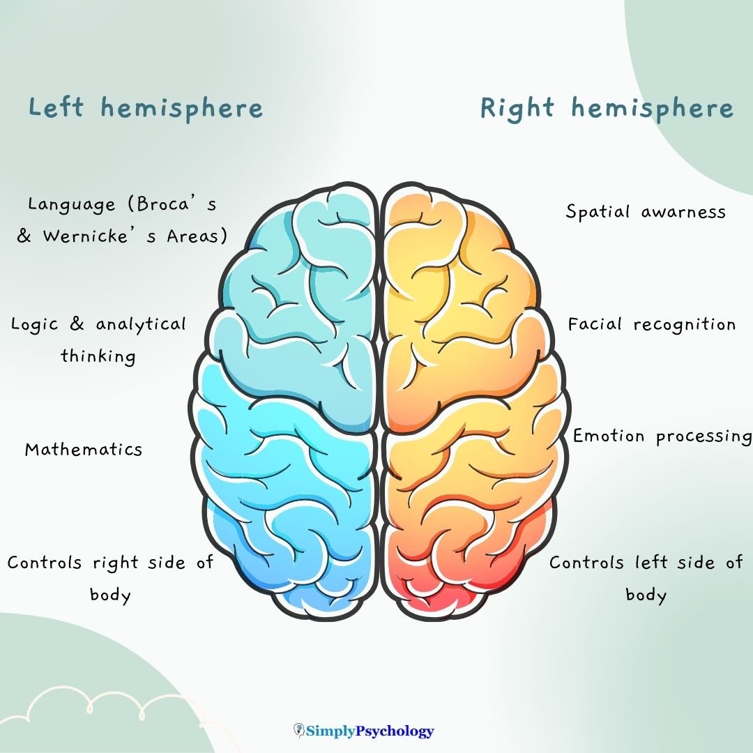
For example, the left hemisphere typically handles tasks related to language, logic, and analysis, while the right hemisphere tends to support spatial reasoning, creativity, and emotional recognition.
Left Brain vs. Right Brain
The idea of a left-brained or right-brained personality is a myth, but hemispheric specialization is real.
Here’s what science tells us:
- Left Hemisphere: Processes language (including grammar and vocabulary), handles math and logic, and controls the right side of the body. It includes Broca’s area, which manages speech production, and Wernicke’s area, which supports language comprehension.
- Right Hemisphere: Specializes in spatial awareness, facial recognition, emotional processing, and controls the left side of the body. It is also more active in interpreting negative emotions and visualizing complex patterns.
Although these functions are typically lateralized, most tasks involve cooperation between both hemispheres.
For example, while the left hemisphere may handle grammar, the right helps interpret tone and emotional context.

How Do We Know This? Landmark Research and Case Studies
Much of what we know about lateralization comes from early case studies and neurological research:
- Broca’s area: In 1861, French physician Paul Broca studied a patient known as “Tan,” who could only utter that single word. Upon his death, Broca discovered damage to the left frontal cortex. Broca found that other patients with similar symptoms had lesions in the same area. He concluded this region, now called Broca’s area, was essential for speech production and language comprehension.
- Wernicke’s area: In 1876, German neurologist Carl Wernicke studied patients who could speak fluently but could not understand language. He found that these individuals had damage to a region in the upper left temporal lobe. This area, now called Wernicke’s area, enables us to understand spoken and written language and select appropriate words for speech.
- Split-brain studies: Roger Sperry and Michael Gazzaniga conducted experiments in the 1960s with patients who had undergone a severing of the corpus callosum to treat epilepsy. They found that the left hemisphere could name objects shown in the right visual field, but not those shown in the left. However, patients could draw or pick up objects seen in the left field using the left hand, revealing the right hemisphere’s strengths in spatial and motor tasks.
- Gazzaniga’s facial recognition study: In 1983, Gazzaniga found that faces shown to the left visual field, processed by the right hemisphere, were recognized more easily. This supported the idea that the right hemisphere excels at facial processing.
- Phineas Gage: After an 1848 accident destroyed part of his left frontal lobe, American railway worker Phineas Gage survived, but his personality changed dramatically. He became impulsive and unreliable, providing strong support for localization of function in the frontal cortex.
Sperry and Gazzaniga’s Split-Brain Study
The split-brain research named above is the article’s key evidence for lateralization, so it is worth examining in more detail (Gazzaniga, 1967).
- Aim: Sperry and Gazzaniga wanted to find out whether the two hemispheres have different, specialized functions once surgically disconnected from each other.
- Method: Split-brain patients fixated on a central dot while pictures were flashed briefly to their left or right visual field, reaching only the opposite hemisphere. They also felt hidden objects by hand, since each hand is controlled by the opposite hemisphere too.
- Results: Patients could not name a spoon shown only to their right hemisphere, but picked it out correctly by touch with their left hand. Their left-hand drawings were also far more accurate than their right-hand ones, showing a right-hemisphere strength for visuospatial tasks.
- Conclusion: The left hemisphere is dominant for language production, but this dominance is not absolute. The right hemisphere showed some ability to understand words, even though it could not produce speech itself.
Because split-brain patients are a small, unusual clinical group, it’s important not to over-generalize these findings to typical brains.

Language and Emotion Lateralization
Lateralization is especially clear in language processing. For most people (especially right-handers), language is controlled by the left hemisphere. This includes both understanding and producing speech.
However, language lateralization develops over time. A 2020 fMRI study by Olulade et al. found that children aged 4–6 showed language activation in both hemispheres.
As children aged, right hemisphere involvement decreased, and left hemisphere dominance became more apparent, suggesting that lateralization strengthens with development.
Emotion is also lateralized (Silberman & Weingartner, 1986):
- The right hemisphere plays a larger role in recognizing facial expressions and managing negative emotions.
- The left hemisphere appears more active during experiences of happiness and optimism.
Patients with damage to the left frontal lobe were more likely to experience depression (Paradiso et al., 1999).
Those with right frontal lobe damage, by contrast, often showed signs of mania or inappropriate cheerfulness (Starkstein et al., 1989).
Does Lateralization Differ Across People?
Yes, lateralization varies by handedness, sex, and age:
- Handedness: A 2002 fMRI study by Szaflarski et al. found that left-handed individuals often show more bilateral activation during language tasks, meaning they may use both hemispheres for language.
- Sex differences: A study by Tomasi and Volkow (2012) found that males had increased right-lateralized connectivity in temporal, frontal, and occipital cortices, while females showed more left-lateralized connectivity in the frontal cortex.
- Developmental differences: As shown in Olulade et al.’s (2020) study, children show more balanced hemispheric activation that gradually becomes more lateralized with age.
Additionally, a review by Reber and Tranel (2017) found that men and women differ in how they lateralize emotional and decision-making functions in the ventromedial prefrontal cortex (vmPFC).
Male patients with right vmPFC damage, for instance, showed more behavioral deficits than those with left-side damage.
This reverse pattern was not observed in female patients (Tranel et al., 2002).
Why Brain Lateralization Matters
Understanding lateralization helps clinicians diagnose and treat brain injuries, strokes, aphasia, and emotional disorders.
It also deepens our insight into how brain structure shapes cognition and behavior.
Although lateralization provides important insights, it’s essential to avoid oversimplifications.
We are not simply “left-brained” or “right-brained”.
We are whole-brained humans whose hemispheres work in constant collaboration.
Critical Evaluation
Split-brain research has shaped how psychologists think about hemispheric specialization, but the method carries specific strengths and limitations worth weighing.
- Rare Sample: The classic split-brain studies used a small, unique clinical sample of epilepsy patients rather than typical participants, so it’s unclear how far the findings generalize to typical brains.
- Reveals Hidden Specialization: Cutting the corpus callosum lets each hemisphere’s abilities be tested separately, revealing specialization that is normally invisible because the intact brain shares information between hemispheres almost instantly.
- History Confound: Split-brain patients had often endured years of severe, treatment-resistant epilepsy and possible brain reorganization before surgery, so their unusual medical history may affect the results.
- Individual Variation: Right-hemisphere language ability differed noticeably between patients: some could spell or name simple words with the right hemisphere, others could not. This means lateralization is not a strict, all-or-nothing rule.
- Risk of Over-Interpretation: The clean-looking dissociations from split-brain testing can make the hemispheres seem like two entirely separate minds, but everyday behavior like conversation still depends on both hemispheres cooperating closely.
- Relative, Not Absolute: The findings fit neither pure holism nor strict localization: some functions, like language production, are strongly lateralized, while others, like emotion, appear to be shared by both hemispheres.
Contemporary Research
A 2019 whole-brain mapping study puts these classic split-brain findings into a broader modern context (Karolis et al., 2019).
- Aim: Karolis, Corbetta and Thiebaut de Schotten (2019) aimed to build the first whole-brain map of functional lateralization and relate it to the strength of the callosal connections between hemispheres.
- Method: The researchers combined a meta-analysis of thousands of brain-activation studies with structural and functional connectivity data from healthy adults. They then tested whether more strongly lateralized brain regions had weaker direct connections to their mirror-image region on the other side.
- Findings: Functional lateralization across the brain was not a simple left-versus-right split, but followed four largely independent patterns covering language, perception and action, emotion, and decision-making. Regions with stronger lateralization also tended to have weaker direct connections to their opposite-side counterpart.
- Conclusion: Lateralization has a multi-dimensional structure rather than one simple left/right split. This fits an evolutionary account in which, as brains grew larger, some functions became more strongly lateralized to cut the cost of constantly relaying information across the hemispheres.
This modern mapping helps explain why Gazzaniga’s split-brain patients could independently generate emotional reactions in both hemispheres, even though language stayed firmly left-lateralized. Emotion and language, in other words, sit on different lateralization patterns with different underlying connectivity.
References
Clements, A. M., Rimrodt, S. L., Abel, J. R., Blankner, J. G., Mostofsky, S. H., Pekar, J. J., Denckla, M. B. & Cutting, L. E. (2006). Sex differences in cerebral laterality of language and visuospatial processing. Brain and Language, 98 (2), 150-158.
Gazzaniga, M. S. (1967). The split brain in man. Scientific American, 217(2), 24–29. https://doi.org/10.1038/scientificamerican0867-24
Gazzaniga, M. S., & Smylie, C. S. (1983). Facial recognition and brain asymmetries: Clues to underlying mechanisms. Annals of Neurology: Official Journal of the American Neurological Association and the Child Neurology Society, 13 (5), 536-540.
Karolis, V. R., Corbetta, M., & Thiebaut de Schotten, M. (2019). The architecture of functional lateralisation and its relationship to callosal connectivity in the human brain. Nature Communications, 10, 1417. https://doi.org/10.1038/s41467-019-09344-1
Olulade, O. A., Seydell-Greenwald, A., Chambers, C. E., Turkeltaub, P. E., Dromerick, A. W., Berl, M. M., Gaillard, W. D. & Newport, E. L. (2020). The neural basis of language development: Changes in lateralization over age. Proceedings of the National Academy of Sciences, 117 (38), 23477-23483.
Paradiso, S., Johnson, D. L., Andreasen, N. C., O’Leary, D. S., Watkins, G. L., Boles Ponto, L. L., & Hichwa, R. D. (1999). Cerebral blood flow changes associated with attribution of emotional valence to pleasant, unpleasant, and neutral visual stimuli in a PET study of normal subjects. American Journal of Psychiatry, 156 (10), 1618-1629.
Reber, J., & Tranel, D. (2017). Sex differences in the functional lateralization of emotion and decision making in the human brain. Journal of Neuroscience Research, 95 (1-2), 270-278.
Silberman, E. K., & Weingartner, H. (1986). Hemispheric lateralization of functions related to emotion. Brain and Cognition, 5 (3), 322-353.
Sperry, R. W. (1967). Split-brain approach to learning problems. The neu.
Starkstein, S. E., Robinson, R. G., Honig, M. A., Parikh, R. M., Joselyn, J., & Price, T. R. (1989). Mood changes after right-hemisphere lesions. The British Journal of Psychiatry, 155 (1), 79-85.
Szaflarski, J. P., Binder, J. R., Possing, E. T., McKiernan, K. A., Ward, B. D., & Hammeke, T. A. (2002). Language lateralization in left-handed and ambidextrous people: fMRI data. Neurology, 59 (2), 238-244.
Tomasi, D., & Volkow, N. D. (2012). Laterality patterns of brain functional connectivity: gender effects. Cerebral Cortex, 22 (6), 1455-1462.
Tranel, D., Bechara, A., & Denburg, N. L. (2002). Asymmetric functional roles of right and left ventromedial prefrontal cortices in social conduct, decision-making, and emotional processing. Cortex, 38 (4), 589-612.
Further Reading
- Gainotti, G. (2014). Why are the right and left hemisphere conceptual representations different?. Behavioral neurology, 2014.
- Macdonald, K., Germine, L., Anderson, A., Christodoulou, J., & McGrath, L. M. (2017). Dispelling the myth: Training in education or neuroscience decreases but does not eliminate beliefs in neuromyths. Frontiers in psychology, 8, 1314.
- Corballis, M. C. (2014). Left brain, right brain: facts and fantasies. PLoS Biol, 12(1), e1001767.
- Nielsen, J. A., Zielinski, B. A., Ferguson, M. A., Lainhart, J. E., & Anderson, J. S. (2013). An evaluation of the left-brain vs. right-brain hypothesis with resting state functional connectivity magnetic resonance imaging. PloS one, 8(8), e71275.

