The brainstem is the stalk-like lower part of the brain that connects the main brain (cerebrum) to the spinal cord. It sits at the base of the skull, just above the spinal cord, and is considered part of the central nervous system (CNS).
In plain language, the brainstem is like the body’s life support system. It controls basic functions that keep you alive.
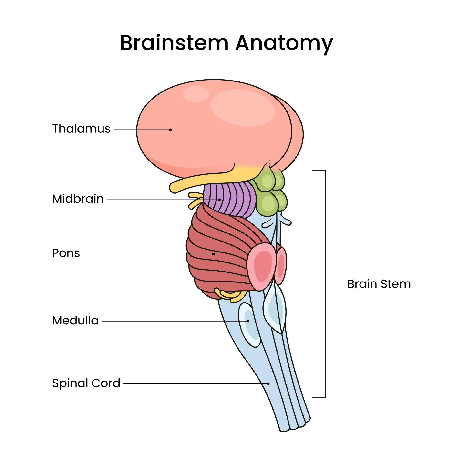
The brainstem has three major parts, stacked from top to bottom. The midbrain (mesencephalon) is the top tier, the pons (Latin for “bridge”) is the middle tier, and the medulla oblongata is the lowest, continuing into the spinal cord.
Each part plays a different role, as explained below. Together, they manage functions you don’t have to think about, such as breathing, heart rate, and consciousness.
Its nerve pathways also link the brain to the body, carrying messages to and from the spinal cord.
Key Takeaways
- The brainstem connects the brain to the spinal cord and regulates automatic, life-sustaining functions.
- It controls breathing, heart rate, blood pressure, consciousness, and reflexes like swallowing.
- It acts as a communication bridge between the brain and body.
- Most cranial nerves emerge from the brainstem, supporting facial movement, hearing, and swallowing.
- In psychology, the brainstem is essential for consciousness, sleep, and instinctive survival behaviors.
- Damage can result in coma, paralysis, or death, showing how vital the brainstem is to everyday functioning.
Main Functions of the Brainstem
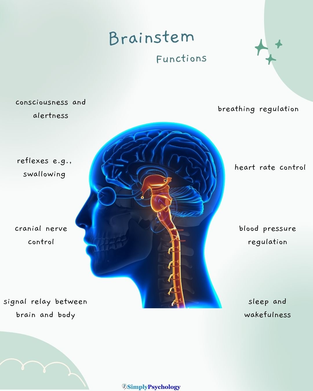
The brainstem controls many automatic, life-sustaining functions that keep us alive without conscious effort.
It works as the body’s autopilot for critical processes. Key functions of the brainstem include:
- Breathing: Regulates the rate and depth of breathing. Neurons in the medulla (with help from the pons) automatically control our breathing rhythm.
- Heart rate & blood pressure: Adjusts heartbeats and blood vessel diameter to maintain blood pressure. The medulla contains cardiovascular centers that manage heart rate and blood pressure reflexively.
- Sleep and arousal: Maintains consciousness, alertness, and sleep cycles. The brainstem’s reticular activating system (RAS) stimulates wakefulness and helps regulate the sleep-wake cycle.
- Reflexes: Coordinates involuntary reflex actions like swallowing, coughing, gagging, and blinking. For example, the brainstem triggers the gag and swallowing reflex to protect your airway, and adjusts pupil size in response to light.
- Relay of signals: Serves as a communication bridge between brain and body. Sensory signals such as touch and pain travel up, and motor commands travel down, all through the brainstem, a two-way highway.
These functions happen automatically.
You do not have to “think” to breathe or keep your heart beating. Your brainstem handles it in the background. That is why it is crucial for survival.
Parts of the Brainstem
The brainstem is made up of three main parts. Each part has specific structures and roles.
Location and Layout
The brainstem lies deep in the head, on the midline. It sits beneath the cerebral hemispheres and in front of the cerebellum, joined to it by three pairs of thick fiber bundles called cerebellar peduncles.
Above, it continues into the diencephalon, which holds the thalamus and hypothalamus. Below, it passes into the spinal cord at the foramen magnum, the large opening at the base of the skull.
A narrow channel, the cerebral aqueduct, runs through the midbrain. It links the third ventricle above to the fourth ventricle, a fluid-filled space behind the pons and medulla.
Anatomists disagree slightly about what counts as the brainstem. Some also include the thalamus and hypothalamus. This article uses the stricter, more common definition: the midbrain, pons and medulla only.
In the developmental scheme of the forebrain, midbrain and hindbrain, the midbrain forms the mesencephalon on its own. The pons and medulla belong to the hindbrain.
Each tier has the same front-to-back layering:
- Front: the great descending motor tracts.
- Middle (tegmentum): cranial-nerve nuclei, the reticular formation, and ascending and descending pathways.
- Back: sensory and reflex structures, such as the tectum in the midbrain.
This layering helps a neurologist work out where a lesion has landed.
Midbrain (Mesencephalon)
This is the upper part of the brainstem.
It controls eye movements and certain reflexes to visual and auditory stimuli.
The roof of the midbrain, the tectum, holds two pairs of bumps called colliculi. The superior colliculi take direct input from the eyes and help orient the eyes and head toward a sudden movement.
The inferior colliculi relay sound to the thalamus. They also trigger reflexes, such as turning your head toward a bang.
The midbrain also houses dopamine-producing neurons in two key places. The substantia nigra (“black substance,” a darkly pigmented nucleus) sends dopamine to the basal ganglia to regulate voluntary movement. Its gradual cell death causes Parkinson’s disease.
The ventral tegmental area sends dopamine to the limbic system and prefrontal cortex. This forms the core of the brain’s reward and motivation circuitry.
The larger, ventral region is the tegmentum. It holds the red nucleus, which helps coordinate limb movement. It also holds the nuclei of the nerves that move the eyes.
Midbrain damage therefore often disturbs eye position and the pupil’s response to light. Descending motor fibers also run through it, so a one-sided lesion can weaken the opposite side of the body.
Pons
The pons is the middle section of the brainstem (its name means “bridge”).
It helps regulate breathing by working with the medulla to maintain a smooth respiratory rhythm.
It serves as a bridge linking the cerebellum with the rest of the brain, and relays signals for facial movement and sensation, as well as for hearing and balance.
The pons earns its name from a massive relay. Motor plans from the cortex descend into the pons, where most fibers synapse onto neurons that cross to the opposite half of the cerebellum.
This cortico-ponto-cerebellar relay carries tens of millions of axons. It gives the cerebellum a running copy of the movements the cortex intends, so it can smooth and correct them.
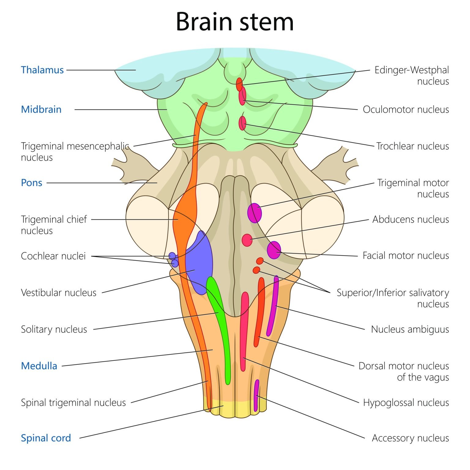
The pons also houses four sets of cranial-nerve nuclei:
- Trigeminal: facial sensation and chewing.
- Abducens: sideways eye movement.
- Facial: the muscles of facial expression.
- Vestibulocochlear: hearing and balance.
The pons is also central to sleep. Cell groups in its tegmentum switch rapid-eye-movement (REM) sleep on and off. They drive the vivid dreaming, muscle paralysis and rapid eye movements of that state.
Medulla Oblongata
The medulla is the lowest part of the brainstem, directly continuous with the spinal cord.
It controls critical automatic functions: it regulates heart rate, blood pressure, and breathing.
It also manages reflexes like swallowing, coughing, sneezing, and vomiting. Because the medulla governs such vital processes, damage to it can be fatal.
Two vital centres work here around the clock. A cardiovascular centre adjusts heart rate and the force of each heartbeat, and controls blood pressure by changing the width of blood vessels. Sensors reporting blood pressure and blood chemistry keep it updated.
A respiratory centre sets the basic rhythm and depth of breathing. Smith et al. (1991) traced the source of that rhythm by progressively slicing the brainstem of newborn rats. They found a small region of the ventral medulla, the pre-Bötzinger complex.
Removing it abolished rhythmic breathing. A thin slice containing it kept producing a breathing-like rhythm on its own, which suggests it holds pacemaker neurons that drive each breath.
Motor fibers descending from the cortex cross the midline near the base of the medulla, at the pyramidal decussation. This crossing is why each half of the cerebral cortex controls the muscles on the opposite side of the body.

Cranial Nerves and the Brainstem
Twelve pairs of cranial nerves arise from the brain to control the head and internal organs.
Importantly, 10 of the 12 cranial nerves (III through XII) connect to the brainstem. Only the first two, for smell and vision, bypass it.
Most nerves that let you blink, smile, cry, hear, taste, speak, and swallow trace back to the brainstem. Their control centers, called nuclei, sit inside it.
The brainstem acts as a command center for these cranial nerves, sending signals between the brain and the face, throat, and sensory organs.
This is why damage to the brainstem can cause widespread problems. It can affect hearing, balance, facial movements, and even basic abilities like swallowing or speaking.
The nuclei sit in an orderly sequence down the brainstem:
- Midbrain: eye movement.
- Pons: facial sensation, chewing, facial expression, hearing and balance.
- Medulla: swallowing, speech, taste and tongue movement.
Because the order is known, failing nerves reveal where the brainstem is damaged. A drooping eyelid, a deviating eye, a paralyzed half of the face or a lost gag reflex each points to a different level.
Why the Brainstem Matters in Psychology
In psychology and neuroscience, the brainstem is seen as the foundation for consciousness and basic survival behavior.
The reticular formation, particularly the reticular activating system (RAS), plays a key role in alertness, attention, and wakefulness.
Damage to this area can lead to coma, as the brain’s internal “alarm system” shuts down.
The brainstem is sometimes called the “reptilian brain.” The label reflects its ancient lineage and its control of basic survival functions. It comes from an older, three-part model of brain evolution that neuroscientists now regard as an oversimplification.
The brainstem is not a separate primitive layer. It is continuously and reciprocally interwoven with the rest of the brain.
The brainstem works closely with the autonomic nervous system to produce physical reactions to emotions.
For instance, during fear, the limbic system signals the brainstem to trigger a fight-or-flight response. Heart rate rises and breathing quickens.
In short, the brainstem supports automatic responses that form the basis of more complex behaviors.
Without it, higher thinking cannot occur. That makes the brainstem essential for both survival and psychological functioning.
The Reticular Activating System
The reticular formation is a net-like web of neurons running through the core of the whole brainstem, from the medulla to the midbrain. Its name comes from the Latin reticulum, “little net.”
The part that sustains alertness is the ascending reticular activating system (RAS). Its neurons receive input from nearly every sensory pathway passing through the brainstem.
They then project upward, via the thalamus and directly to the cortex. This broad activation carries no particular sensation. It sets the overall level of arousal on which conscious perception depends.
When ascending activity is high, the cortex is awake and alert. When it falls, the brain drifts into drowsiness and sleep. If the system is damaged, coma can follow.
Moruzzi and Magoun (1949) provided the founding evidence.
- Aim: To test whether a diffuse brainstem system, rather than specific sensory pathways, keeps the cortex awake.
- Method: In anesthetized animals, the researchers recorded the cortex’s electroencephalogram (EEG). They then electrically stimulated the brainstem reticular formation.
- Results: Stimulation immediately turned slow, synchronized cortical waves into the fast, low-voltage pattern of an alert brain. The effect was widespread and did not depend on the classic sensory pathways.
- Conclusion: A diffuse activating system in the brainstem reticular formation drives arousal of the whole cortex. Damage to it explains why small brainstem lesions can cause coma.
Later work added detail. Arousal also depends on chemically distinct cell groups, such as noradrenergic neurons of the locus coeruleus and serotonergic neurons of the raphe nuclei. These reach beyond the brainstem into the hypothalamus and basal forebrain.
Disorders and Damage to the Brainstem
The brainstem controls vital functions. Injuries here can be life-threatening.
A brainstem stroke may cause locked-in syndrome, where a person is fully conscious but paralyzed except for eye movements.
The damage typically sits in the ventral pons, the front part of the pons where descending motor tracts run. It cuts almost all voluntary movement but spares the tegmentum behind it, and with it the arousal system.
Awareness survives, but action does not. The person usually keeps only vertical eye movements and blinking.
Severe damage, whether from trauma, swelling, or disease, can result in coma or brain death.
Coma follows when a lesion interrupts the arousal system in the brainstem tegmentum. Even a small lesion can do it.
Parvizi and Damasio (2003) tested where in the human brainstem this happens.
- Aim: To find which human brainstem regions cause coma when damaged.
- Method: In 47 patients with brainstem stroke, researchers retrospectively mapped each MRI-defined lesion onto a common reference brainstem. They then compared patients who fell into coma with those who did not.
- Results: Comatose patients mostly had bilateral lesions centered on the tegmentum of the upper pons. Patients who stayed conscious had spared the tegmentum or damaged it only slightly and on one side.
- Conclusion: Human consciousness depends on a specific set of nuclei in the tegmentum of the upper pons and midbrain, not on an undifferentiated reticular mass.
The study turned the animal-based activating-system idea into precise human anatomy.
Since the brainstem controls breathing and consciousness, damage may cause loss of awareness and inability to breathe independently.
In cases of brainstem death, recovery is not possible.
Doctors diagnose brain death by testing brainstem reflexes at the bedside:
- the pupil’s response to light
- the corneal (blink) reflex
- two eye-movement responses, called oculocephalic and oculovestibular
- the gag and cough reflexes
An apnea test then confirms there is no spontaneous drive to breathe (Wijdicks, 2001). Together, absent reflexes and no breathing drive mark irreversible brainstem failure.
Other symptoms of brainstem damage include:
- Difficulty swallowing
- Impaired balance
- Slurred speech
- Paralysis
Recovery is often limited, and many brainstem injuries require urgent life support. Even survivors may face lasting disabilities.


Critical Evaluation of Brainstem Research
Few brain structures are as securely mapped as the brainstem. Even so, the evidence has limits, and recent work is redrawing the textbook picture.
Contemporary Research
Recent studies have moved from asking whether the brainstem sustains consciousness to mapping the networks through which it does so. The unifying theme is that arousal comes not from one diffuse core but from a defined network of brainstem nuclei wired to specific cortical and subcortical partners.
Lesion-Network Mapping of Coma
- Aim: To find the brainstem site whose damage causes coma, and map the brain network connected to it.
- Method: Fischer et al. (2016) compared 12 coma-causing brainstem lesions with 24 that did not. They then used resting-state connectivity data from healthy brains to map the network linked to the coma site.
- Results: A small site in the dorsolateral pontine tegmentum was tied to coma. It connected to two cortical hubs, the ventral anterior insula and the pregenual anterior cingulate cortex, whose link was disrupted in disorders of consciousness.
- Conclusion: Consciousness appears to rely on a specific brainstem-to-cortex network, not a vague reticular core. Arousal becomes a connectivity problem.
A Default Ascending Arousal Network
Edlow et al. (2024) added fine anatomical detail. They combined diffusion MRI of three post-mortem human brains with immunostaining of the tissue.
This revealed a “default ascending arousal network.” Its connected nodes sit in the brainstem, hypothalamus, thalamus and basal forebrain. Tractography showed pathways linking them to one another and to the default mode network, which supports self-awareness.
Resting-state fMRI from living people then singled out one hub. It was the dopaminergic ventral tegmental area of the midbrain. It connects widely to both arousal and awareness networks.
Together, these studies point one way. The old image of a diffuse brainstem “switch” gives way to a mapped network of nodes and pathways.
That network still rests largely on lesion and post-mortem evidence, not on direct causal tests in healthy people.
Distributed Networks, Not Single Centers
Textbooks often assign “the breathing centre” or “the arousal centre” to a neat spot in the brainstem. That is a useful shortcut, but it can mislead.
These functions are carried by distributed networks of interacting nuclei. Respiratory rhythm emerges from interacting cell groups, of which the pre-Bötzinger complex is one node (Smith et al., 1991).
Arousal depends on several chemically distinct populations. They reach from the brainstem into the hypothalamus and basal forebrain (Edlow et al., 2024; Fischer et al., 2016).
Placing a function at a single point is a reductionist error. It ignores the network. A lesion’s effect depends on which parts of that network it interrupts.
A network-level reading, with brainstem nuclei as nodes in circuits that also involve the diencephalon and cortex, fits the modern evidence far better.
Reliance on Lesion and Animal Evidence
Much of what we know comes from animals. The founding arousal experiment used anesthetized animals (Moruzzi & Magoun, 1949). The breathing pacemaker was isolated in neonatal rat tissue (Smith et al., 1991).
Human data have limits too. Mapping rests on retrospective series of stroke patients (Fischer et al., 2016; Parvizi & Damasio, 2003). Lesions show what a region is necessary for, not everything it normally does.
Animal circuits may not map cleanly onto human experience. Retrospective lesion samples are also shaped by which strokes happen to occur and survive.
The brainstem is small and deeply buried. It is also too vital to manipulate in healthy people. So the strongest recent human evidence comes from donated post-mortem brains (Edlow et al., 2024).
Convergence across methods, not any single study, makes the account credible.
Brain Death Criteria Differ Worldwide
Using irreversible loss of brainstem function to define death is medically entrenched. Yet countries do not agree on the rules.
A survey of international guidelines found wide variation (Wijdicks, 2002). Some jurisdictions, including the United Kingdom, rest the diagnosis on brainstem function. Others, including the United States, require the whole brain to have failed.
The details differ too. Countries vary on observation periods, the number of examiners and ancillary tests such as EEG.
Both traditions use the same bedside reflex and apnea tests (Wijdicks, 2001). Their reasoning differs.
| Brainstem-only criteria | Whole-brain criteria | |
|---|---|---|
| Example | United Kingdom | United States |
| Diagnosis rests on | Irreversible loss of brainstem function and reflexes | Loss of function of the whole brain |
| Reasoning | Brainstem loss alone removes both consciousness and the drive to breathe | The brainstem is necessary, but independent evidence must show the cerebral hemispheres have also ceased to function |
Neither position is universally adopted, so the definition of death drawn from this one structure remains legally and ethically unsettled.
References
Edlow, B. L., Olchanyi, M., Freeman, H. J., Li, J., Maffei, C., Snider, S. B., Zöllei, L., Iglesias, J. E., Augustinack, J., Bodien, Y. G., Haynes, R. L., Greve, D. N., Diamond, B. R., Stevens, A., Giacino, J. T., Destrieux, C., van der Kouwe, A., Brown, E. N., Folkerth, R. D., . . . Kinney, H. C. (2024). Multimodal MRI reveals brainstem connections that sustain wakefulness in human consciousness. Science Translational Medicine, 16(745), Article eadj4303. https://doi.org/10.1126/scitranslmed.adj4303
Fischer, D. B., Boes, A. D., Demertzi, A., Evrard, H. C., Laureys, S., Edlow, B. L., Liu, H., Saper, C. B., Pascual-Leone, A., Fox, M. D., & Geerling, J. C. (2016). A human brain network derived from coma-causing brainstem lesions. Neurology, 87(23), 2427–2434. https://doi.org/10.1212/WNL.0000000000003404
Moruzzi, G., & Magoun, H. W. (1949). Brain stem reticular formation and activation of the EEG. Electroencephalography and Clinical Neurophysiology, 1(4), 455–473. https://doi.org/10.1016/0013-4694(49)90219-9
Parvizi, J., & Damasio, A. R. (2003). Neuroanatomical correlates of brainstem coma. Brain, 126(7), 1524–1536. https://doi.org/10.1093/brain/awg166
Smith, J. C., Ellenberger, H. H., Ballanyi, K., Richter, D. W., & Feldman, J. L. (1991). Pre-Bötzinger complex: A brainstem region that may generate respiratory rhythm in mammals. Science, 254(5032), 726–729. https://doi.org/10.1126/science.1683005
Wijdicks, E. F. M. (2001). The diagnosis of brain death. New England Journal of Medicine, 344(16), 1215–1221. https://doi.org/10.1056/NEJM200104193441606
Wijdicks, E. F. M. (2002). Brain death worldwide: Accepted fact but no global consensus in diagnostic criteria. Neurology, 58(1), 20–25. https://doi.org/10.1212/WNL.58.1.20

