Intelligence isn’t controlled by one single “intelligence center” in the brain. Instead, it emerges from a network of interconnected brain areas working together.
Psychologists define intelligence as the mental capacity to learn from experience, adapt to new situations, and understand abstract concepts.
This has practical importance, from interpreting brain scans to knowing how injuries or development might affect intelligence.
Key Takeaways
- Network-Based: Intelligence depends on coordinated activity across many brain regions, not one “IQ spot.”
- Multiple Areas: Frontal, parietal, and temporal lobes, plus deeper structures, all play roles in thinking and problem-solving.
- Connections: Fast, efficient white matter links are associated with quicker processing and better cognitive performance.
- Brain Size: Organization and wiring quality often outweigh overall volume, which explains only about 6% of IQ differences.
- IQ Limits: Tests miss creativity, practical skills, and emotional intelligence.
- Correlational Evidence: Most brain-imaging findings show association, not cause.
Intelligence as a Brain Network, Not a Single Spot
It’s a common myth that there’s a single “intelligence lobe” or brain region responsible for IQ. Modern research strongly refutes this.
One leading model, the Parieto-Frontal Integration Theory (P-FIT), proposes that intelligence depends on how well different brain regions integrate their activity (Jung & Haier, 2007). It rests on converging structural and functional imaging evidence.
In this view, large-scale networks form the biological basis of human intelligence. They link the frontal lobes and parietal lobes, along with parts of the temporal lobes and cingulate cortex. White-matter tracts connect these regions.
One such network is the brain’s multiple-demand (MD) network, a set of frontal and parietal brain regions that become active during many challenging tasks.
Studies show that this fronto-parietal MD network (including lateral frontal cortex and areas along the intraparietal sulcus) works as an integrated system. Its parts work in sync during complex problem-solving.
In a large fMRI study (n = 216), stronger MD activity during a spatial working memory task was linked to faster, more accurate responses (Assem et al., 2020). It was also linked to higher fluid intelligence, measured separately in 114 participants.
Damage to fronto-parietal areas is selectively linked to drops in problem-solving ability.
This evidence busts the myth of a single “intelligence center.” Instead, intelligence appears to emerge from coordinated activity across a network of regions, especially in the frontal and parietal areas.
Key Brain Regions Involved in Intelligence
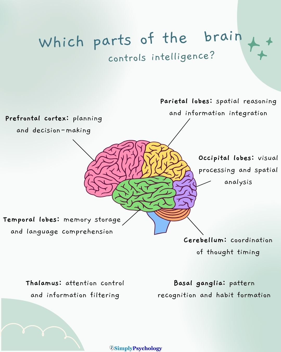
While no single region alone “controls” intelligence, certain brain areas play especially important roles within the broader network. Key regions include:
- Prefrontal Cortex: Plans, makes decisions and holds information in working memory, which makes it central to fluid intelligence.
- Parietal Lobes: Integrate sensory information, abstract it into patterns, and support spatial reasoning and attention.
- Temporal Lobes: Store memories and support language comprehension, drawing on learned facts and vocabulary.
- Occipital Lobes: Process the visual information that many reasoning tasks depend on.
Prefrontal Cortex (Frontal Lobes)
Often called the brain’s “executive,” the prefrontal cortex is critical for planning, decision-making, and working memory.
It helps you hold and manipulate information in mind. For example, it lets you consider different steps in a puzzle or reason through a problem.
Strong reasoning and problem-solving skills are linked to efficient functioning of the dorsolateral prefrontal cortex, which helps coordinate other brain areas and focus attention on goals.
This region is heavily engaged when solving novel problems or inhibiting incorrect responses, making it central to fluid intelligence.
Parietal Lobes
The parietal regions specialize in processing and integrating information.
They handle sensory input (like visual-spatial information) and help in abstracting patterns.
For instance, the parietal cortex helps combine visual data and prior knowledge to understand relationships, which is useful in geometry or assembling a puzzle.
Parietal areas also contribute to spatial reasoning and attention. Both support many intellectual tasks.
Temporal Lobes
The temporal lobes are key to memory storage and language comprehension.
They contribute more to understanding vocabulary, recalling factual information, or comprehending language.
Temporal regions also relate to crystallized intelligence. This is the knowledge and vocabulary people accumulate. Among 76 healthy adults aged 65 to 75, higher blood levels of the nutrient lutein went with higher crystallized intelligence (Zamroziewicz et al., 2016).
Gray matter in the right parahippocampal cortex, a temporal region, partly accounted for that link. The design was correlational.
In essence, the temporal lobes act as a vast library of knowledge and meaning. The more facts and language we learn, the more we draw on these regions.
Occipital Lobes
The occipital lobes process visual information.
While they aren’t “thinking centers” per se, they provide the visual groundwork for many cognitive tasks.
Solving a complex diagram, recognizing patterns, or mentally rotating an object engages the occipital visual cortex.
In fact, brain imaging has found that even visual areas activate during reasoning tasks, especially tasks that involve pattern analysis or spatial puzzles.
Beyond the Cortex: Other Contributors
Intelligence involves more than just the cerebral cortex (the brain’s outer layer). Other brain structures play supporting roles in cognitive performance, often behind the scenes:
- Cerebellum: Times and sequences thoughts, much as it coordinates movement.
- Thalamus: Relays signals between the senses and the cortex, and helps regulate attention and alertness.
- Basal Ganglia: Support feedback-based learning, pattern recognition and habit formation alongside the frontal cortex.
Cerebellum
The cerebellum is best known for coordinating movement and balance.
However, it also contributes to cognition, particularly the timing and sequencing of thoughts. Think of it as “smoothing out” mental operations. It fine-tunes thinking much as it fine-tunes physical movements.
Studies of stroke patients have shown that cerebellar damage can lead to a range of cognitive deficits, including problems with attention, executive function, spatial reasoning, memory, and even language.
In short, the cerebellum supports intelligence by keeping our mental processes well-timed and coordinated.
Thalamus
The thalamus is a deep-brain structure that acts like a central relay station.
It receives signals from various senses and from different brain regions, then sends them to where they need to go.
In doing so, the thalamus helps regulate attention and alertness. It essentially decides what information is important enough to pass along for higher processing.
By filtering and amplifying signals, the thalamus ensures that critical data (a math problem in front of you, for instance) gets priority in the cortex.
Basal ganglia
The basal ganglia are traditionally known for their role in movement (as affected in Parkinson’s disease).
But they also contribute significantly to cognitive functions like learning, pattern recognition, and habit formation.
They work with the frontal cortex in feedback-based learning. Examples include learning from trial and error or recognizing a sequence or category through repetition.
Moreover, they form circuits with the cortex that influence higher-order thinking; studies have found basal ganglia involvement in attention, language, and even planning and logical reasoning.
In essence, these subcortical structures provide the substructure for intelligence. They route information and fine-tune the learning of rules and patterns that underlie smart behavior.
How Brain Connectivity Supports Intelligence
Intelligence depends on networks of brain regions, making the connections between them crucial. These connections run through white matter tracts, which are bundles of nerve fibers that act like the brain’s wiring.
White matter wrapped in a fatty insulating layer called myelin carries signals quickly and reliably, much like a high-speed internet network linking computers.
Reviews of the neuroscience of intelligence identify white matter integrity as a candidate mechanism (Deary et al., 2010). Intact tracts allow efficient communication between regions.
People with more coherent white matter often process information faster and score higher on certain cognitive tests. One interpretation is that their networks exchange information without bottlenecks. The evidence is correlational. It cannot show that better wiring causes higher scores.
Common Misconceptions About Intelligence and the Brain
“Bigger brains mean smarter people.”
Brain size has only a modest link with IQ. A meta-analysis of 88 studies covering over 8,000 people found a correlation of about r = .24 (Pietschnig et al., 2015).
That is roughly 6% of the differences in IQ. Published estimates were also inflated, because strong positive results were reported more often than small or null ones.
Some people with large brains have average intelligence, and vice versa.
Organization and efficiency matter more than sheer volume. Think of a smaller, well-designed processor outperforming a bulkier one.
The quality of connections, balance between regions, and flexible recruitment of different areas are key.
Einstein’s brain wasn’t much larger than average; its unique connectivity and structure likely mattered more. Size is just one piece of the puzzle.
“IQ tests measure all aspects of intelligence.”
IQ tests mainly assess analytical and problem-solving skills: logic, math, verbal reasoning, spatial ability, and memory.
They don’t fully capture creativity, practical intelligence (handling real-world problems), or emotional intelligence (managing emotions and relationships).
Theories like Howard Gardner’s multiple intelligences highlight talents such as musical or kinesthetic skills that standard tests overlook.
Not all of Gardner’s categories are universally accepted.
Visser, Ashton and Vernon (2006) gave 200 adults two tests for each of Gardner’s eight domains. A large general factor emerged.
It loaded strongly on the cognitive domains, such as linguistic, logical–mathematical and spatial ability, and weakly on bodily–kinaesthetic ability. The intelligences were therefore not independent.
Gardner and Moran (2006) reply that paper-and-pencil tests cannot fairly test a theory that rejects the psychometric tradition’s measurement assumptions.
Intelligence is still multi-faceted. A high IQ doesn’t mean someone has no weaknesses, and a low IQ doesn’t mean they lack valuable skills.
Critical Evaluation of the Evidence
Most of the evidence is correlational. Brain differences accompany differences in intelligence, but the findings cannot show what causes them.
Contemporary Research
Genetics now offers the strongest evidence that intelligence reflects something biologically coherent. Genome-wide studies scan every chromosome for variants linked to a trait. Savage et al. (2018) is the largest so far.
- Aim: To find genetic variants linked to intelligence in a sample large enough to detect very small effects, and to see whether the genes involved point to specific brain biology.
- Method: Savage et al. (2018) pooled 14 cohorts (n = 269,867) that each measured general cognitive function. They then mapped associated variants to genes and tested which tissues and cell types expressed them.
- Results: They found 205 associated regions of the genome (190 previously unreported) and 1,016 genes (939 new). The genes were strongly expressed in brain tissue, especially striatal medium spiny neurons and hippocampal pyramidal neurons.
- Conclusion: Intelligence is highly polygenic, meaning many genes each contribute a tiny effect. Those genes converge on brain development and on synapses, the connections between neurons.
The study has limits. Samples were overwhelmingly of European ancestry, so polygenic scores built from them predict poorly in other populations.
Individual variant effects are minute, and the variance explained remains far below twin-study heritability (Plomin & von Stumm, 2018). Association is also not mechanism. Genes influence the environments people encounter and select.
Even so, the findings show that differences in intelligence are not just an artefact of how tests are built.
Why Cognitive Abilities Correlate
People who do well on one cognitive test tend to do well on others. This pattern is called the positive manifold, and it is why intelligence is often summarised as one general factor, or g.
Three accounts explain the pattern without assuming a single “intelligence center”:
- Sampling: Each test draws on a different subset of a huge pool of neural elements, and overlap between subsets produces positive correlations (Bartholomew et al., 2009).
- Mutualism: Abilities that start out uncorrelated help each other grow, so correlations emerge over development (van der Maas et al., 2006).
- Process overlap: Many but not all tests tap the same domain-general executive processes, producing a general factor that emerges from the overlap (Kovacs & Conway, 2016).
All three reproduce the observed data, and none has been decisively established. Neuroimaging findings do not currently discriminate between them.
Stability and Change in Intelligence
Rank order in intelligence is strikingly stable. The Lothian Birth Cohort of 1921 took the same test at ages 11 and 90 (n = 106). Scores correlated .54 across 79 years, or .67 after statistical correction for a narrower range of scores (Deary et al., 2013).
Stable rank order is not the same as fixed ability.
The childhood score leaves roughly half to two-thirds of the variance at age 90 unexplained, and the survivors were plausibly healthier than their birth cohort.
Education appears to raise measured intelligence. A meta-analysis pooled 142 effect sizes from 42 datasets and over 600,000 participants. Each extra year of education was linked to a gain of roughly 1 to 5 IQ points (Ritchie & Tucker-Drob, 2018).
Working memory training does not appear to help. Working memory is a prefrontal function, but a meta-analysis of 145 comparisons found reliable gains only on working memory tests (Melby-Lervåg et al., 2016).
Gains did not carry over to non-verbal ability, verbal ability or reading comprehension when compared with control groups that did other activities.
References
Assem, M., Blank, I. A., Mineroff, Z., Ademoğlu, A., & Fedorenko, E. (2020). Activity in the fronto-parietal multiple-demand network is robustly associated with individual differences in working memory and fluid intelligence. Cortex, 131, 1–16. https://doi.org/10.1016/j.cortex.2020.06.013
Bartholomew, D. J., Deary, I. J., & Lawn, M. (2009). A new lease of life for Thomson’s bonds model of intelligence. Psychological Review, 116(3), 567–579. https://doi.org/10.1037/a0016262
Deary, I. J., Pattie, A., & Starr, J. M. (2013). The stability of intelligence from age 11 to age 90 years: The Lothian Birth Cohort of 1921. Psychological Science, 24(12), 2361–2368. https://doi.org/10.1177/0956797613486487
Deary, I. J., Penke, L., & Johnson, W. (2010). The neuroscience of human intelligence differences. Nature Reviews Neuroscience, 11(3), 201–211. https://doi.org/10.1038/nrn2793
Gardner, H., & Moran, S. (2006). The science of multiple intelligences theory: A response to Lynn Waterhouse. Educational Psychologist, 41(4), 227–232. https://doi.org/10.1207/s15326985ep4104_2
Jung, R. E., & Haier, R. J. (2007). The parieto-frontal integration theory (P-FIT) of intelligence: Converging neuroimaging evidence. Behavioral and Brain Sciences, 30(2), 135–154. https://doi.org/10.1017/S0140525X07001185
Kovacs, K., & Conway, A. R. A. (2016). Process overlap theory: A unified account of the general factor of intelligence. Psychological Inquiry, 27(3), 151–177. https://doi.org/10.1080/1047840X.2016.1153946
Melby-Lervåg, M., Redick, T. S., & Hulme, C. (2016). Working memory training does not improve performance on measures of intelligence or other measures of “far transfer”: Evidence from a meta-analytic review. Perspectives on Psychological Science, 11(4), 512–534. https://doi.org/10.1177/1745691616635612
Moretti, R., Caruso, P., Crisman, E., & Gazzin, S. (2017). Basal ganglia: Their role in complex cognitive procedures in experimental models and in clinical practice. Neurology India, 65(4), 814–825. https://doi.org/10.4103/neuroindia.ni_850_16
Pietschnig, J., Penke, L., Wicherts, J. M., Zeiler, M., & Voracek, M. (2015). Meta-analysis of associations between human brain volume and intelligence differences: How strong are they and what do they mean? Neuroscience & Biobehavioral Reviews, 57, 411–432. https://doi.org/10.1016/j.neubiorev.2015.09.017
Plomin, R., & von Stumm, S. (2018). The new genetics of intelligence. Nature Reviews Genetics, 19(3), 148–159. https://doi.org/10.1038/nrg.2017.104
Ritchie, S. J., & Tucker-Drob, E. M. (2018). How much does education improve intelligence? A meta-analysis. Psychological Science, 29(8), 1358–1369. https://doi.org/10.1177/0956797618774253
Savage, J. E., Jansen, P. R., Stringer, S., Watanabe, K., Bryois, J., de Leeuw, C. A., Nagel, M., Awasthi, S., Barr, P. B., Coleman, J. R. I., Grasby, K. L., Hammerschlag, A. R., Kaminski, J. A., Karlsson, R., Krapohl, E., Lam, M., Nygaard, M., Reynolds, C. A., Trampush, J. W., . . . Posthuma, D. (2018). Genome-wide association meta-analysis in 269,867 individuals identifies new genetic and functional links to intelligence. Nature Genetics, 50(7), 912–919. https://doi.org/10.1038/s41588-018-0152-6
van der Maas, H. L. J., Dolan, C. V., Grasman, R. P. P. P., Wicherts, J. M., Huizenga, H. M., & Raijmakers, M. E. J. (2006). A dynamical model of general intelligence: The positive manifold of intelligence by mutualism. Psychological Review, 113(4), 842–861. https://doi.org/10.1037/0033-295X.113.4.842
Visser, B. A., Ashton, M. C., & Vernon, P. A. (2006). Beyond g: Putting multiple intelligences theory to the test. Intelligence, 34(5), 487–502. https://doi.org/10.1016/j.intell.2006.02.004
Zamroziewicz, M. K., Paul, E. J., Zwilling, C. E., Johnson, E. J., Kuchan, M. J., Cohen, N. J., & Barbey, A. K. (2016). Parahippocampal cortex mediates the relationship between lutein and crystallized intelligence in healthy, older adults. Frontiers in Aging Neuroscience, 8, Article 297. https://doi.org/10.3389/fnagi.2016.00297
Zhang, P., Duan, L., Ou, Y., Ling, Q., Cao, L., Qian, H., Zhang, J., Wang, J., & Yuan, X. (2023). The cerebellum and cognitive neural networks. Frontiers in Human Neuroscience, 17, 1197459. https://doi.org/10.3389/fnhum.2023.1197459