The brain is the coordinating center of the Central Nervous System (CNS), integrating sensory information to regulate both conscious thought and involuntary survival mechanisms.
It functions by processing inputs from sensory receptors and generating coordinated responses through effectors: muscles and glands.
Structurally, the brain is dominated by the cerebrum, which makes up roughly two-thirds of total brain mass.
This region is divided into specialized lobes. These lobes are responsible for “high-order” functions, including cognition, memory, and emotional response.
While the cerebrum handles conscious awareness, deeper structures like the cerebellum, hypothalamus, and medulla oblongata work in tandem to manage motor coordination and homeostasis.
Understanding this neuroanatomy is vital to seeing how localized regions contribute to the overall behavior and physiological stability of the organism.
Key Takeaways
- Three Main Parts: The brain is made up of the cerebrum, cerebellum, and brainstem, each handling different jobs, from higher thought to vital survival functions.
- Four Lobes: The cerebrum’s frontal, temporal, parietal, and occipital lobes each specialize in different functions, from movement and language to vision and touch.
- Subcortical Structures: Structures like the amygdala, hippocampus, thalamus, and hypothalamus sit beneath the cortex and handle emotion, memory, and basic drives.
- Ventricles & CSF: Fluid-filled ventricles produce cerebrospinal fluid, which cushions the brain and removes waste.
- Neurons: Neurons transmit information through electrochemical signals and communicate with each other across synapses.
- Cranial Nerves: Twelve pairs of cranial nerves connect directly to the brain, carrying sensory and motor signals to and from the face and head.
- Structure Predicts Function: Because each brain region has a distinctive role, damage to a specific area often produces a predictable pattern of symptoms.
Parts of the Brain
The brain is composed of the cerebrum, cerebellum, and brainstem (Fig. 1).
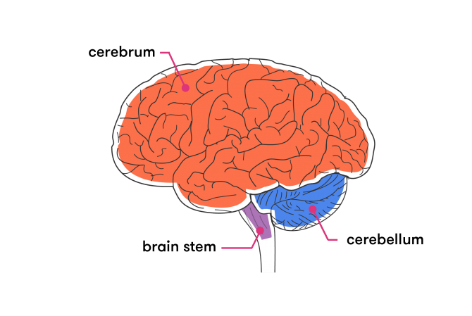
Cerebrum
The cerebrum is the largest and most recognizable part of the brain.
It consists of grey matter (the cerebral cortex ) and white matter at the center.
The cerebrum is divided into two hemispheres, the left and right, and contains the lobes of the brain (frontal, temporal, parietal, and occipital lobes).
The cerebrum produces higher functioning roles such as thinking, learning, memory, language, emotion, movement, and perception.
The Cerebellum
The cerebellum is located under the cerebrum and monitors and regulates motor behaviors, especially automatic movements.
The cerebellum helps regulate posture and balance, and has recently been suggested for being involved in learning and attention.
The cerebellum makes up about 11% of brain weight. Even so, it holds far more neurons than the rest of the brain combined.
A large-scale neuron count settled the question. It found the adult brain contains around 86 billion neurons in total. Roughly 69 billion of these, about four-fifths, sit in the cerebellum, compared with only around 16 billion in the cerebral cortex (Herculano-Houzel, 2009).
These figures overturned the long-repeated textbook estimate of 100 billion neurons spread fairly evenly across the brain.
Brainstem
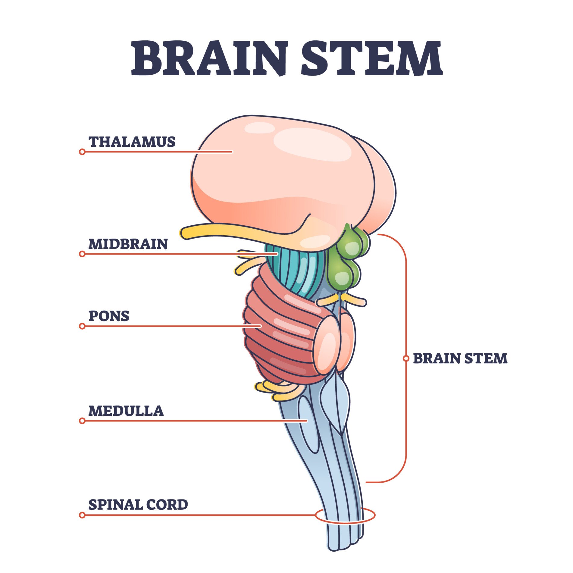
The brainstem is located at the base of the brain.
This area connects the cerebrum and the cerebellum to the spinal cord, acting as a relay station for these areas.
The brainstem regulates automatic functions such as sleep cycles, breathing, body temperature, digestion, coughing, and sneezing.
The Midbrain: Tectum and Tegmentum
Above the pons and below the cerebrum sits the midbrain, the smallest of the brain’s three main divisions. It relays visual, auditory, and motor signals and drives several automatic reflexes.
Two structures do most of the work here.
The midbrain’s roof is called the tectum. It has two pairs of rounded bumps, the colliculi. The superior colliculi trigger visual reflexes, turning the eyes toward a sudden sight. The inferior colliculi do the same for sound.
Below the tectum lies the tegmentum, the midbrain’s floor. It houses the substantia nigra, already introduced above, plus the red nucleus, which helps control limb movement, and the periaqueductal grey, a hub for the brain’s built-in pain-suppression system.
Together, the tectum and tegmentum make the midbrain a compact but vital hub.
The Reticular Formation: The Brain’s Arousal System
Running through the midbrain, pons, and medulla is a diffuse web of neurons called the reticular formation. Its ascending branch reaches up into the forebrain and earns it the nickname the brain’s “consciousness switch.”
That role has three parts.
It keeps us aroused and conscious, governing the sleep-wake cycle; severe damage causes coma. It filters out constant, unchanging background stimulation so the cortex notices only what changes. It also underlies habituation, the fading response to a stimulus that carries no new information.
- Aim: Moruzzi and Magoun (1949) wanted to find out what drives the brain’s shift from sleep to waking, and whether a distinct brainstem system was responsible.
- Method: Working with anaesthetised cats, they implanted electrodes in the brainstem reticular formation, recorded the cortex’s electrical activity, then stimulated the reticular formation directly.
- Results: Stimulating the reticular formation replaced the slow, synchronised EEG pattern of a resting brain with the fast, desynchronised pattern of full waking alertness.
- Conclusion: The brainstem reticular formation forms a distinct system that governs cortical arousal and the sleep-wake transition (Moruzzi & Magoun, 1949).
A 2024 high-resolution imaging study updated this classic finding. Edlow and colleagues mapped eighteen interconnected brainstem, thalamic, and basal-forebrain nodes that together sustain wakefulness (Edlow et al., 2024). The findings show arousal depends on a network, not one single switch.
That picture is still being refined today.
The Pons
The pons sits between the midbrain and the medulla, forming a rounded bulge of white matter on the front of the brainstem. It works as a switchboard. It links the two halves of the cerebellum and relays motor commands from the cortex down to them.
Several cranial nerves connect here, including the large trigeminal nerve, which carries facial sensation and controls chewing. The pons also helps regulate sleep, arousal, and the rhythm of breathing.
The pons keeps both sides of the brain in sync.
The Medulla Oblongata
The medulla oblongata is the lowest part of the brainstem, a direct continuation of the spinal cord. It is evolutionarily the oldest part of the brain.
It houses the vital reflex centers that keep us alive without conscious effort. These include the cardiovascular centre, which regulates heart rate and blood pressure, and the respiratory centre, which drives the rhythm of breathing.
Its job is simple: keep the vital signs running.
It also holds reflex centers for swallowing, vomiting, coughing, and sneezing. Because these functions are non-negotiable, damage to the medulla is often fatal. Opioid drugs can suppress its respiratory centre and stop breathing altogether.
🧠 Review: Major Brain Regions
- What are the three main parts of the brain, and what is one function of each?
- How does the cerebellum support movement and coordination?
- Which brain region connects to the spinal cord and regulates automatic functions like breathing?
Right Brain vs. Left Brain
The cerebrum is divided into two halves, the right and left hemispheres (Fig. 2).
The left hemisphere controls the right side of the body. The right hemisphere controls the left side.
The corpus callosum connects the two hemispheres. This thick band of neural fibers is made up of about 200 million axons.
The corpus callosum allows the two hemispheres to communicate and allows information being processed on one side of the brain to be shared with the other.
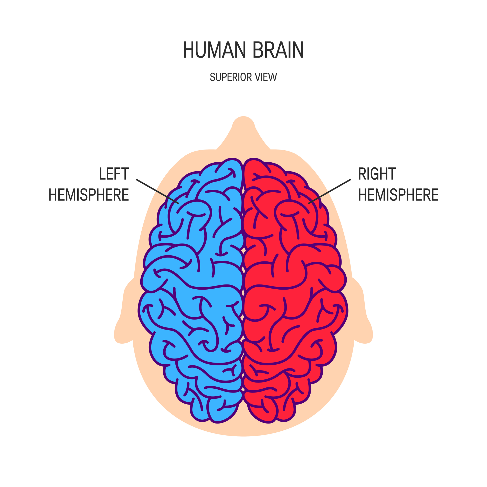
Hemispheric Lateralization
Hemispheric lateralization is the idea that each hemisphere is responsible for different functions.
Each of these functions is localized to either the right or left side.
The left hemisphere is associated with language functions, such as formulating grammar and vocabulary and containing different language centers (Broca’s and Wernicke’s area).
The right hemisphere is associated with more visuospatial functions such as visualization, depth perception, and spatial navigation.
These left and right functions are the case in most people, especially those who are right-handed.
Lobes of the Brain
Each cerebral hemisphere can be subdivided into four lobes, each associated with different functions.
The four lobes of the brain are the frontal, parietal, temporal, and occipital lobes (Figure 3).
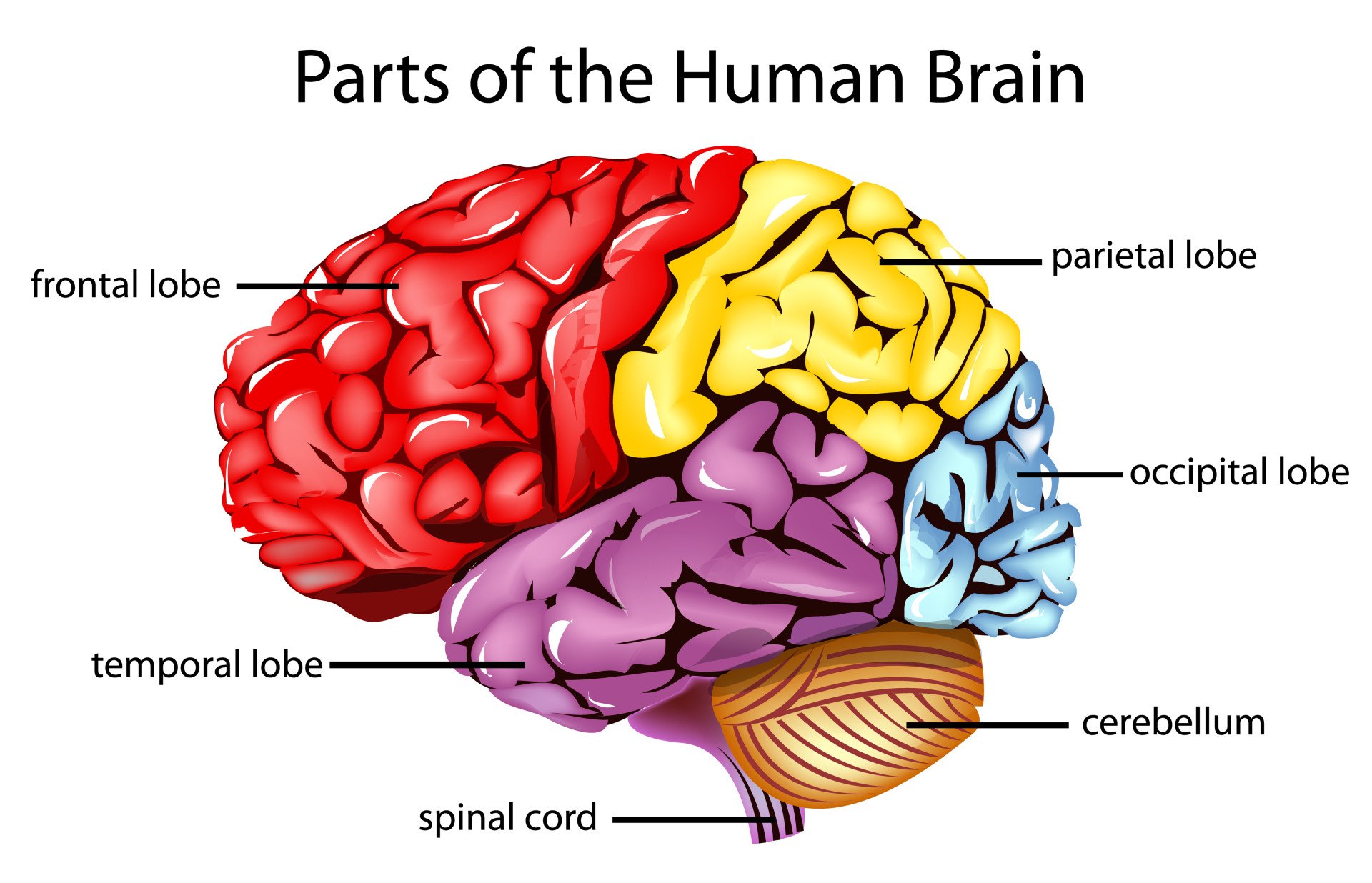
Frontal lobes
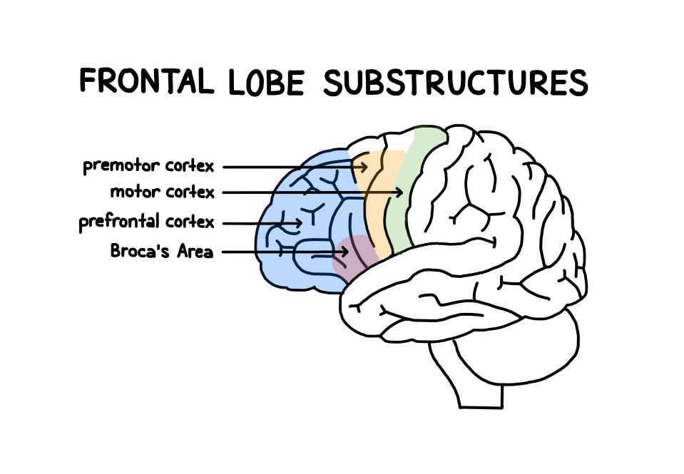
The frontal lobes sit at the front of the brain, behind the forehead (Figure 4).
They handle higher cognitive functions: problem-solving, decision-making, attention, intelligence, and voluntary behavior.
The frontal lobes contain the motor cortex responsible for planning and coordinating movements.
It also contains the prefrontal cortex, which is responsible for initiating higher-level cognitive functioning, and Broca’s Area, which is essential for language production.
Phineas Gage shows what frontal lobe damage can do. In 1848, an iron rod pierced his frontal lobes in a railway accident.
Gage survived. His personality and judgment changed permanently afterward (Harlow, 1868).
Damage to Broca’s area produces Broca’s aphasia: fluent, grammatical speech is lost, but comprehension stays intact. Broca (1861) first described this in a patient who could understand language but could barely speak.
Temporal lobes
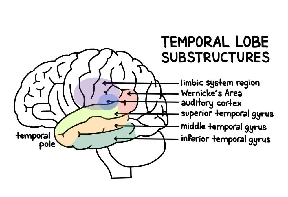
The temporal lobes (Figure 5) sit on both sides of the brain, near the temples. That is where their name comes from.
The main functions of these lobes include understanding, language, memory acquisition, face recognition, object recognition, perception, and auditory information processing.
There is a temporal lobe in both the left and right hemispheres.
The left temporal lobe, which is usually the most dominant in people, is associated with language, learning, memorizing, forming words, and remembering verbal information.
The left lobe also contains a vital language center known as Wernicke’s area, which is essential for language development.
The right temporal lobe is usually associated with learning and memorizing non-verbal information and determining facial expressions.
Parietal lobes
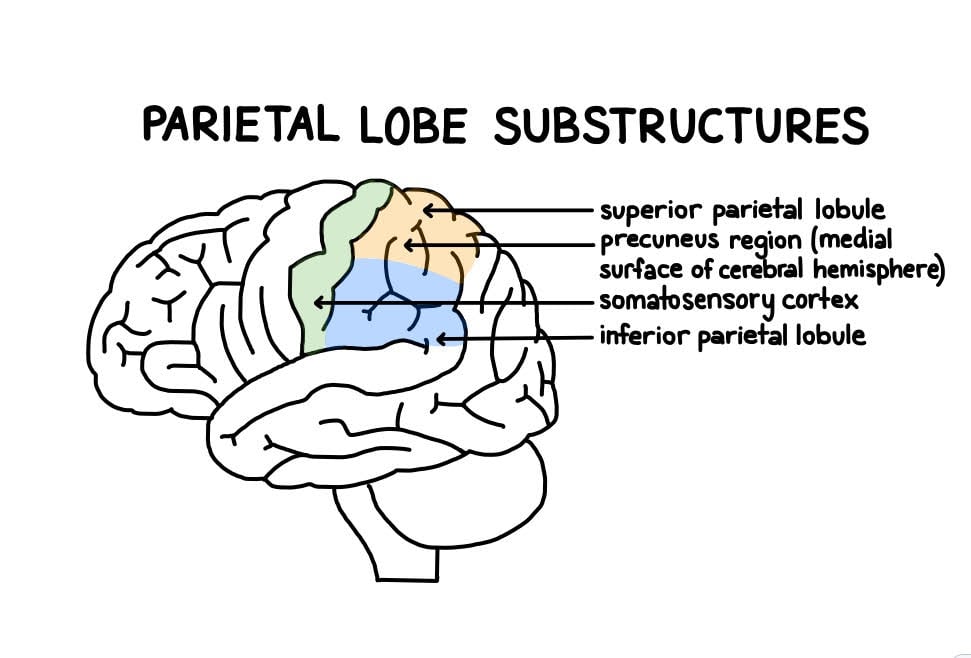
The parietal lobe sits at the top of the brain, between the frontal and occipital lobes, and above the temporal lobes (Figure 6).
It integrates information from the body’s senses. This builds a coherent picture of the world around us.
These lobes allow us to perceive our bodies through somatosensory information (e.g., through touch, pressure, and temperature). It can also help with visuospatial processing, reading, and number representations (mathematics).
The parietal lobes also contain the somatosensory cortex, which receives and processes sensory information, integrating this into a representational map of the body.
This means it can pinpoint the exact area of the body where a sensation is felt, as well as perceive the weight of objects, shape, and texture.
Occipital lobes
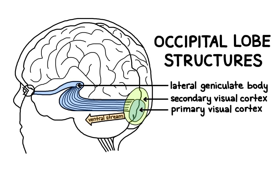
The occipital lobes sit at the back of the brain, behind the parietal and temporal lobes (Figure 7).
They receive sensory information from the eyes’ retinas. This input is then encoded into visual data such as form, color, and motion.
Some of the functions of the occipital lobes include being able to assess the size, depth, and distance, determine color information, object and facial recognition, and mapping the visual world.
The occipital lobes also contain the primary visual cortex. It receives sensory information from the retinas and encodes details like location, spatial data, motion, and color.
🧠 Review: Lobes of the Cerebral Cortex
- Which lobe is responsible for decision-making and voluntary movement?
- What is the primary function of the occipital lobe?
- How does the parietal lobe help us perceive touch and spatial awareness?
Cerebral Cortex
Gyri and Sulci
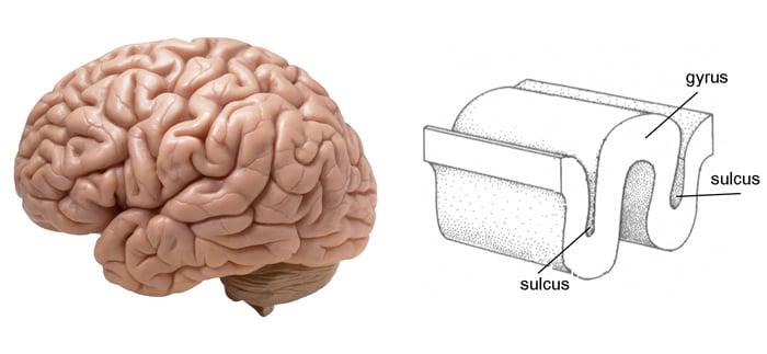
The surface of the cerebrum (Figure 8) is called the cerebral cortex. It has a wrinkled appearance, made up of bulges known as gyri and deep furrows known as sulci.
A gyrus (plural: gyri) is a raised fold. Gyri are the bumps and ridges on the cerebral cortex (the outermost layer of the brain).
A sulcus (plural: sulci) is another name for a groove in the cerebral cortex.
Grey Matter and Neuron Density
The cerebral cortex is primarily constructed of grey matter (neural tissue made up of neurons), with between 14 and 16 billion neurons found here.
The many folds and wrinkles increase surface area. This lets far more neurons fit inside the skull, so the brain can process much larger amounts of information.
Subcortical structures
Beneath the cerebral cortex lie several key subcortical brain regions that play essential roles in emotion, memory, sensory processing, and motor control.
These include the following:
Limbic System
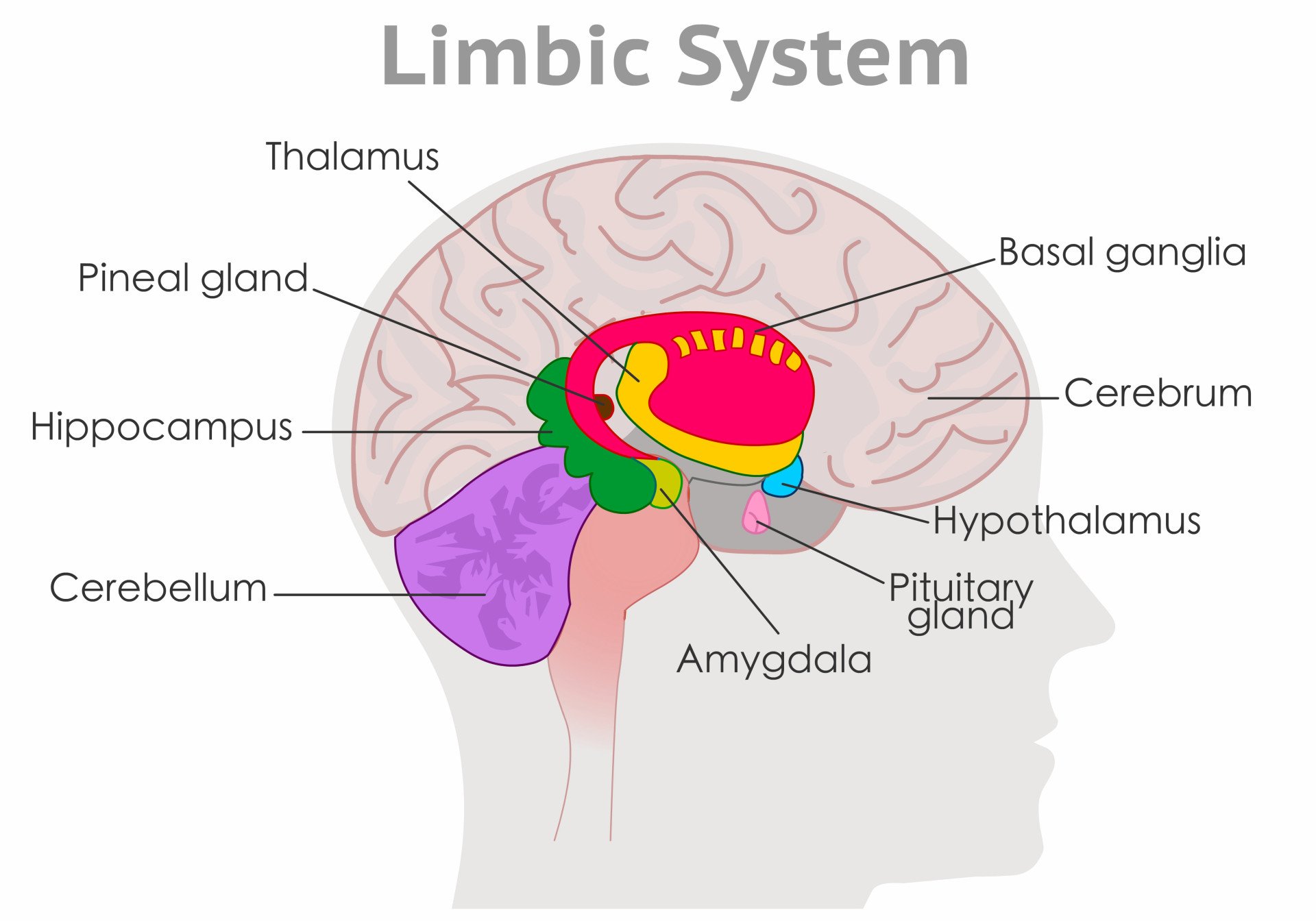
The limbic system is a network of interconnected structures deep within the brain that supports emotional regulation, memory, and motivation.
Key components include the amygdala, which processes emotional responses like fear; the hippocampus, involved in memory formation; and the hypothalamus, which helps regulate basic drives such as hunger and stress.
By linking emotional experience to memory and bodily responses, the limbic system plays a vital role in how we perceive and react to the world.
The limbic system is not one single structure. It is several closely interconnected forebrain regions that, seen from the side, seem to nest inside each other around the brainstem.
Because much of its evolution was tied to the sense of smell, some psychologists nickname it the “old mammalian brain.” It is closely involved in feeding, fighting, fleeing, and mating: the “four Fs” of motivated behavior.
The word “limbic” comes from the Latin for “border.”
In 1937, James Papez proposed that emotion runs through a closed loop connecting these structures (Papez, 1937). The circuit runs from the hippocampus to the fornix, on to the mammillary bodies and anterior thalamus, then to the cingulate gyrus and back again.
Amygdala
The amygdala is a brain region deep in the brain that is involved in the processing of emotions and fear learning.
The amygdala is part of the limbic system, a neural network that mediates emotion and memory (Figure 9).
It ties emotional meaning to memories. It also processes rewards and helps us make decisions.
This brain region has also been linked with the fight-or-flight response.
Damage here can cause Klüver-Bucy syndrome.
Affected individuals lose their normal fear response and become unusually calm, even around threats (Klüver & Bucy, 1939; Hayman et al., 1998).
Thalamus and Hypothalamus
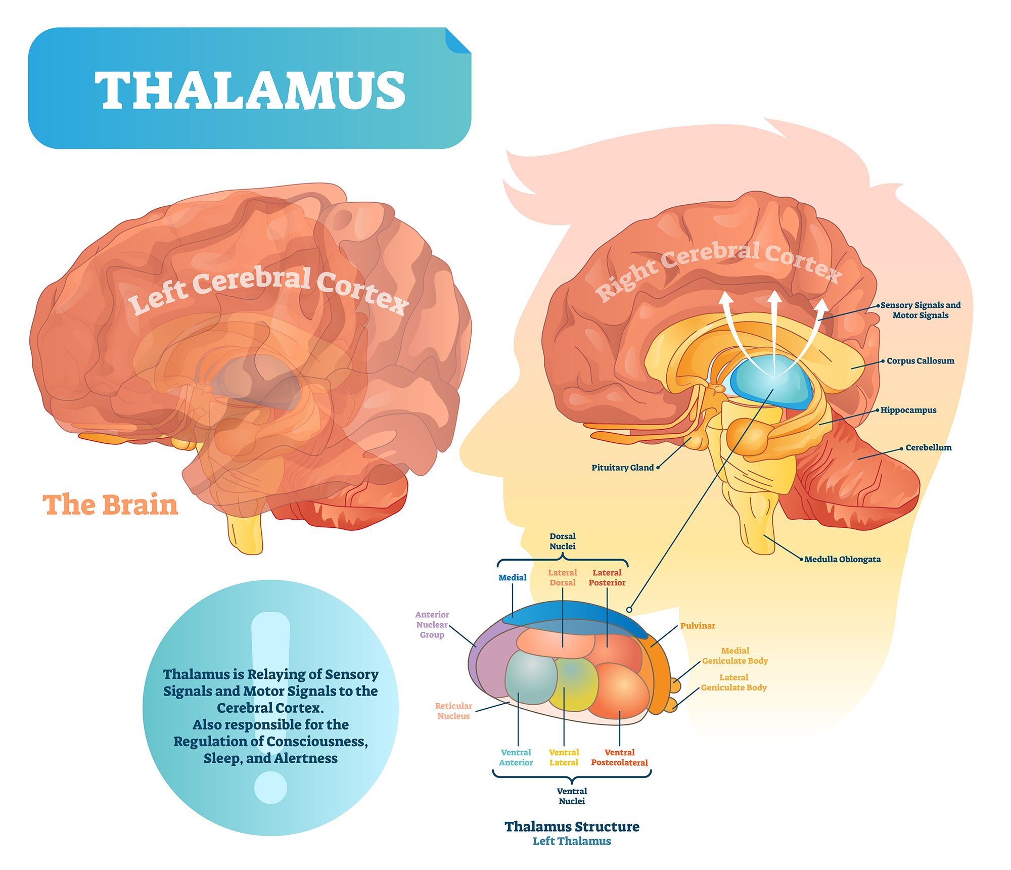
The thalamus is an egg-shaped mass of grey matter that relays information between the cerebral cortex, brain stem, and other brain regions (Figure 10).
Because of its interactive role in relaying sensory and motor information, the thalamus contributes to many processes, including attention, perception, timing, and movement.
The thalamus is not just a passive relay. It actively gates and coordinates the ongoing activity between cortical areas, making it a hub within brain-wide networks rather than a simple switchboard (Sherman, 2016).
The hypothalamus modulates a range of behavioral and physiological functions.
It controls autonomic functions such as hunger, thirst, body temperature, and sexual activity.
To do this, the hypothalamus integrates information from different brain parts and responds to various stimuli such as light, odor, and stress.
The tiny pineal gland sits nearby, on the midline. It lies behind the third ventricle and secretes the hormone melatonin in response to darkness, timing the sleep-wake cycle and syncing the body’s internal clock to the day-night cycle.
Hippocampus
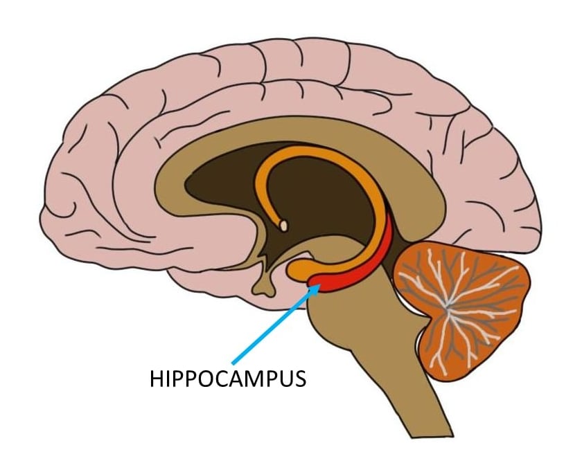
The hippocampus is a curved-shaped structure in the limbic system associated with learning and memory (Figure 11).
The hippocampus plays a key role in forming memories.
It serves as an early storage system for long-term memory and helps convert them into permanent memory.
The clearest evidence for this comes from patient H.M.
After surgeons removed both his hippocampi to treat severe epilepsy, he lost the ability to form new long-term memories (Scoville & Milner, 1957). His memories from before the surgery stayed intact.
Basal Ganglia
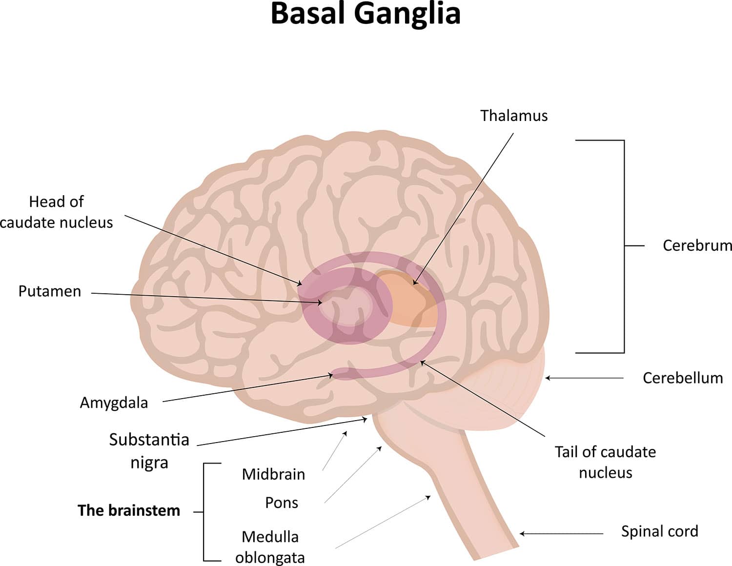
The basal ganglia are a group of structures that regulate the coordination of fine motor movements, balance, and posture alongside the cerebellum.
The basal ganglia are connected to other motor areas and link the thalamus with the motor cortex.
The basal ganglia are also involved in cognitive and emotional behaviors, as well as playing a role in reward and addiction.
Damage to the basal ganglia produces two well-known movement disorders.
In Parkinson’s disease, neurons in the substantia nigra (a small, dopamine-producing structure in the midbrain) degenerate. This loss of dopamine causes the tremor, rigidity, and slowness (bradykinesia) that define the condition (Ehringer & Hornykiewicz, 1960).
In Huntington’s disease, degeneration of the caudate nucleus and putamen instead produces involuntary, jerky movements.
🧠 Review: Subcortical Structures
- What role does the hippocampus play in memory?
- How does the hypothalamus help regulate body functions?
- Which structure helps process fear and emotional responses?
Ventricles and Cerebrospinal Fluid
Within the brain, there are fluid-filled interconnected cavities called ventricles, which are extensions of the spinal cord.
The ventricles produce cerebrospinal fluid, and also transport and remove it. They do not have one unique function of their own.
Instead, they cushion the brain and help mark the location of other brain regions.
Cerebrospinal fluid is a clear, colorless liquid. It circulates through the brain and spinal cord and cushions the brain within the skull.
If the skull is damaged, this fluid acts as a shock absorber, helping to protect the brain from injury.
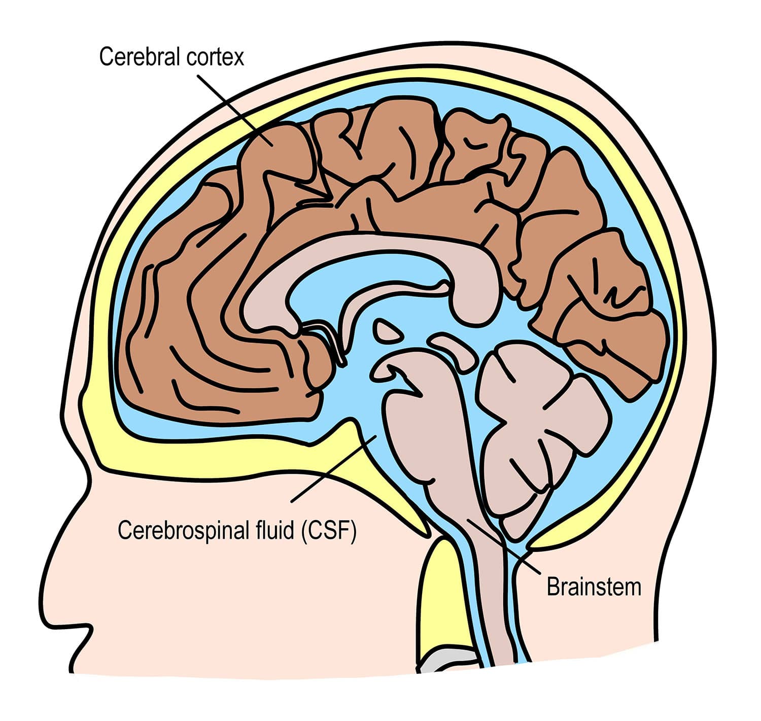
Beyond cushioning, cerebrospinal fluid circulates nutrients and chemicals filtered from the blood, and removes waste products from the brain. It is constantly absorbed and replenished by the ventricles.
A disruption or blockage in this cycle can cause cerebrospinal fluid to build up, enlarging the ventricles.
Neurons
Neurons are the nerve cells of the central nervous system that transmit information through electrochemical signals throughout the body.
Neuron Structure and Types
Neurons contain a soma. This cell body is where the axon extends from, the longest part of the neuron, conducting electrical impulses away from the soma.

Dendrites sit at the other end of the neuron. These branch-like structures send and receive information from other neurons.
A myelin sheath, a fatty insulating layer, forms around the axon. It lets nerve impulses travel down the axon quickly.
There are different types of neurons. Sensory neurons transmit sensory information, motor neurons transmit motor information, and relay neurons allow sensory and motor neurons to communicate.
Synaptic Transmission
The communication between neurons is called a synapse. Neurons communicate via synaptic clefts, small gaps between the endings of neurons.
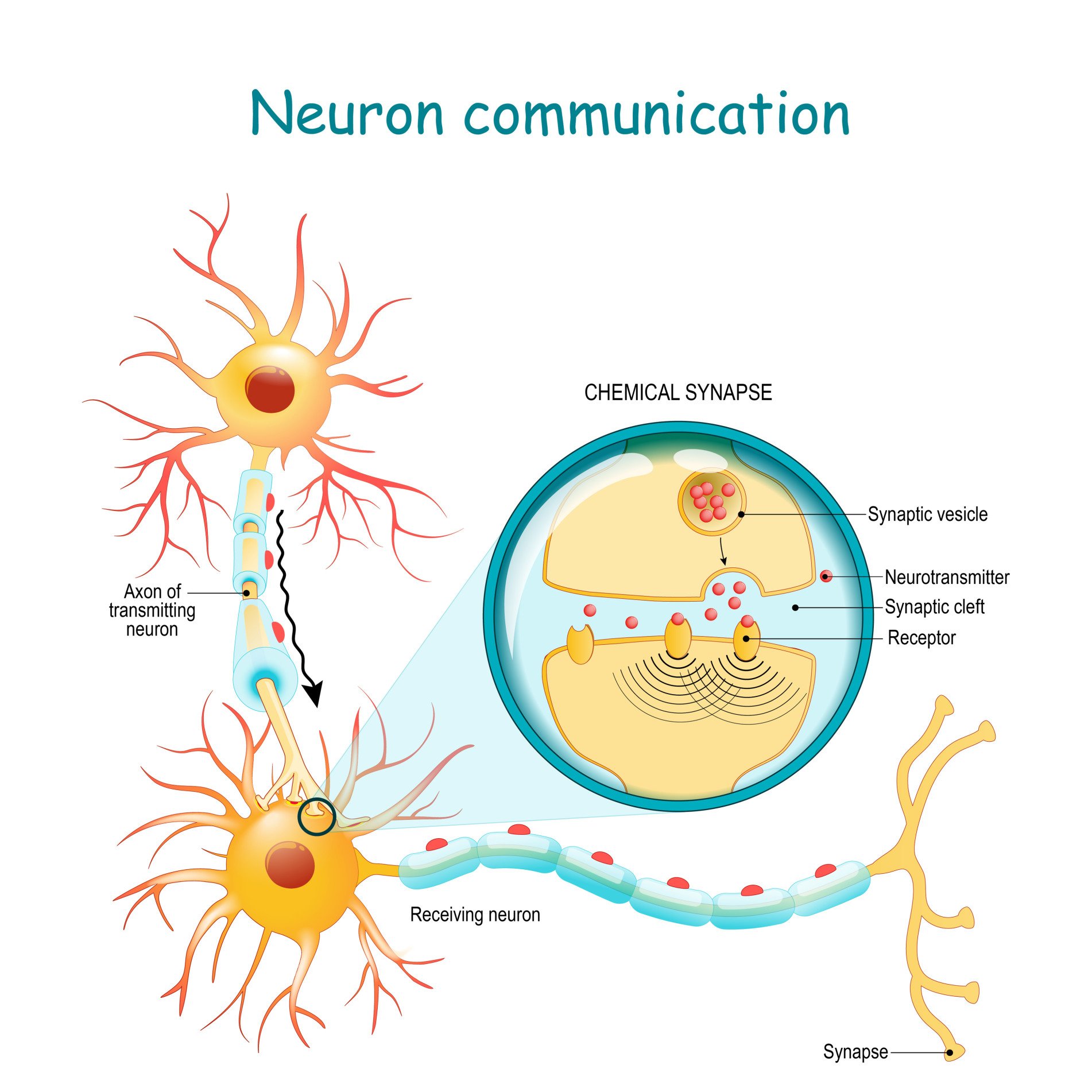
During synaptic transmission, chemicals called neurotransmitters are released from the presynaptic neuron. These chemicals cross the synaptic cleft to reach receptors on the next, postsynaptic neuron.
The message travels on. It influences functions such as behavior and movement, and depending on the neurotransmitter and receptor involved, can excite or inhibit the receiving neuron.
Glial Cells
Glial cells are non-neuronal cells in the central nervous system which work to provide the neurons with nourishment, support, and protection.
Astrocytes
These are star-shaped cells that function to maintain the environment for neuronal signaling by controlling the levels of neurotransmitters surrounding the synapses.
They also work to clean up what is left behind after synaptic transmission, either recycling any leftover neurotransmitters or cleaning up when a neuron dies.
Oligodendrocytes
These types of glial have the appearance of balls with spikes all around them.
They function by wrapping around the axons of neurons to form a protective layer called the myelin sheath.
This is a substance that is rich in fat and provides insulation to the neurons to aid neuronal signaling.
Microglial
Microglial cells have oval bodies and many branches projecting out of them.
The primary function of these cells is to respond to injuries or diseases in the central nervous system.
They respond by clearing away any dead cells or removing any harmful toxins or pathogens that may be present, so they are, therefore, important to the brain’s health.
Ependymal cells
These cells are column-shaped and usually line up together to form a membrane called the ependyma.
The ependyma is a thin membrane lining the spinal cord and ventricles of the brain.
In the ventricles, these cells have small hairlike structures called cilia, which help encourage the flow of cerebrospinal fluid.
🧠Review: Neurons and Glial Cells
- What are the three main parts of a neuron, and what does each do?
- What is the function of the myelin sheath?
- Name two types of glial cells and explain their roles.
Cranial Nerves
There are 12 types of cranial nerves which are linked directly to the brain without having to pass through the spinal cord. These allow sensory information to pass from the organs of the face to the brain:
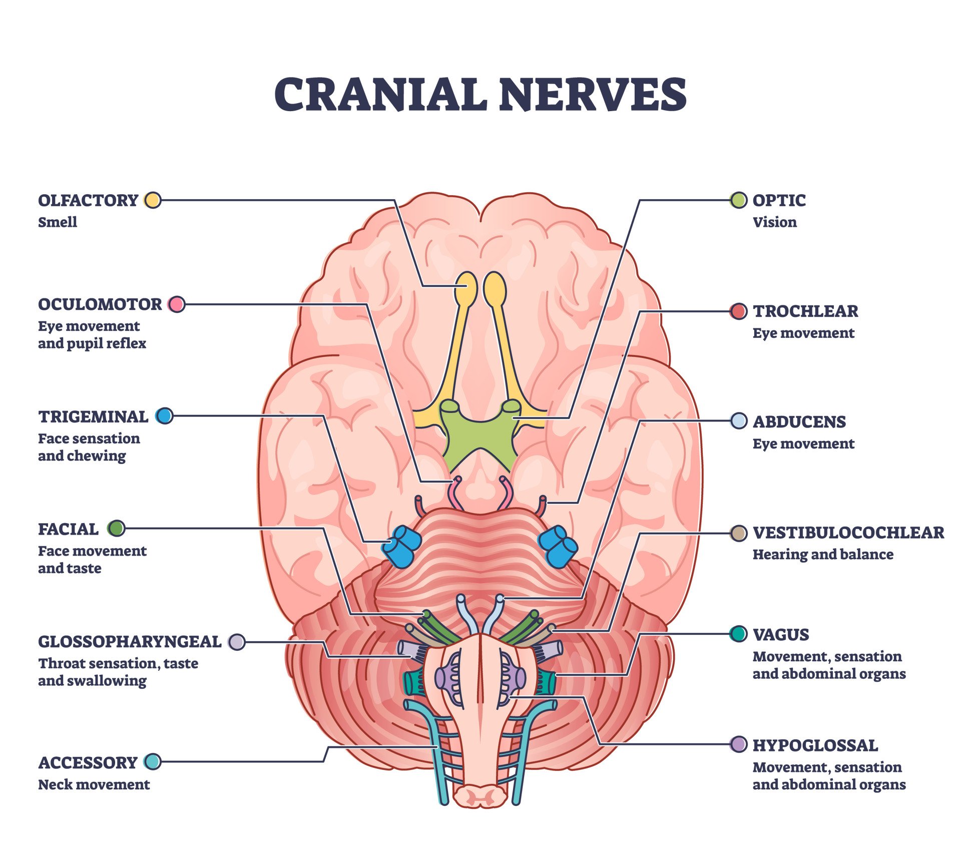
Mnemonic for Order of Cranial Nerves:
S ome S ay M arry M oney B ut M y B rother S ays B ig B rains M atter M ore
- Cranial I: Sensory
- Cranial II: Sensory
- Cranial III: Motor
- Cranial IV: Motor
- Cranial V: Both (sensory & motor)
- Cranial VI: Motor
- Cranial VII: Both (sensory & motor)
- Cranial VIII: Sensory
- Cranial IX: Both (sensory & motor)
- Cranial X: Both (sensory & motor)
- Cranial XI: Motor
- Cranial XII: Motor
Critical Evaluation of the Brain’s Anatomical Divisions
The forebrain, midbrain, and hindbrain scheme above is one of the most useful frameworks in biopsychology, but it has real limits. A balanced view weighs its strengths against these criticisms.
Divisions Are Heuristics, Not Hard Boundaries
This anatomical scheme is fundamentally developmental, tracing each structure back to where it arose in the embryo. It does not carve the brain neatly along functional lines.
Voluntary movement, for example, recruits the frontal cortex, basal ganglia, midbrain, cerebellum, and medullary tracts all at once. Mapping one function to one region oversimplifies how the brain actually works.
That is not the only wrinkle.
The scheme can also imply a tidiness the evidence does not support. The cerebellum, for instance, is not purely a motor coordinator. It also times short intervals and contributes to cognition, a role well outside the traditional “movement only” label.
Grouping and labelling structures by developmental region can obscure this multifunctionality just as easily as it can clarify the anatomy.
Function Is Distributed, Not Localised
Beyond the primary sensory and motor strips, most “higher” functions are not the property of one structure. They emerge from large-scale networks spanning several regions.
Even the thalamus, long described as a passive relay, turns out to be an active hub coordinating traffic across cortical areas (Sherman, 2016). Strong claims that a function has one single anatomical address deserve caution.
Modern network neuroscience makes the same point more broadly.
Researchers increasingly model the brain as a web of interacting hubs (Bassett & Sporns, 2017). Cognition is read off the pattern of connections between regions, not off any single structure in isolation.
This does not undo the developmental map. It simply says where a signal travels often matters more than which single box it starts in.
The “Triune Brain” Idea Is Contested
The medulla is often called the oldest part of the brain, and the neocortex the newest. This “older below, newer above” reading is popular, but it should be handled carefully.
That framing has a catch.
In its strongest form this becomes MacLean’s triune-brain model (MacLean, 1990). It pictures a reptilian core for instinct, a “paleomammalian” limbic layer for emotion, and a “neomammalian” cortex for reason, stacked in three evolutionary strata.
Comparative neuroscience has rejected this picture. The brain did not evolve by bolting new layers onto a preserved reptilian base, and “limbic” structures have homologues throughout the vertebrates (Cesario et al., 2020).
That reading is worth remembering next time “reptilian brain” comes up in conversation.
A network-level account fits the modern evidence better, while still keeping the developmental scheme’s real value for teaching and lesion localisation.
Why the Anatomy Still Matters
Despite these limits, the scheme does real explanatory work. Specific structures anchor major clinical syndromes: the substantia nigra and Parkinson’s disease, the hippocampus and amnesia, and the medulla and loss of vital function.
These tight, replicable links between structure and symptom show the divisions track real functional organisation. That is why the framework keeps generating testable predictions for diagnosis and treatment.
The anatomy earns its keep in the clinic.
The developmental route from three vesicles to five also gives the framework real predictive power. Every mature structure traces back to a known embryonic origin.
That is why a lesion in a given location reliably predicts a specific, recognisable pattern of symptoms, and why the scheme remains the standard teaching framework.
References
Purves, D., Augustine, G., Fitzpatrick, D., Katz, L., LaMantia, A., McNamara, J., & Williams, S. (2001). Neuroscience 2nd edition. sunderland (ma) sinauer associates. Types of Eye Movements and Their Functions.
Mayfield Brain and Spine (n.d.). Anatomy of the Brain. Retrieved July 28, 2021, from: https://mayfieldclinic.com/pe-anatbrain.htm
Robertson, S. (2018, August 23). What is Grey Matter? News Medical Life Sciences. https://www.news-medical.net/health/What-is-Grey-Matter.aspx
Guy-Evans, O. (2021, April 13). Temporal lobe: definition, functions, and location. Simply Psychology. https://www.simplypsychology.org/temporal-lobe.html
Guy-Evans, O. (2021, April 15). Parietal lobe: definition, functions, and location. Simply Psychology. https://www.simplypsychology.org/parietal-lobe.html
Guy-Evans, O. (2021, April 19). Occipital lobe: definition, functions, and location. Simply Psychology. https://www.simplypsychology.org/occipital-lobe.html
Guy-Evans, O. (2021, May 08). Frontal lobe function, location in brain, damage, more. Simply Psychology. https://www.simplypsychology.org/frontal-lobe.html
Guy-Evans, O. (2021, June 09). Gyri and sulci of the brain. Simply Psychology. https://www.simplypsychology.org/gyri-and-sulci-of-the-brain.html
Bassett, D. S., & Sporns, O. (2017). Network neuroscience. Nature Neuroscience, 20(3), 353–364.
Broca, P. (1861). Remarques sur le siège de la faculté du langage articulé, suivies d’une observation d’aphémie (perte de la parole). Bulletin de la Société Anatomique de Paris, 6, 330–357.
Cesario, J., Johnson, D. J., & Eisthen, H. L. (2020). Your brain is not an onion with a tiny reptile inside. Current Directions in Psychological Science, 29(3), 255–260.
Edlow, B. L., Olchanyi, M., Freeman, H. J., Li, J., Maffei, C., Snider, S. B., Zöllei, L., Iglesias, J. E., Augustinack, J., Bodien, Y. G., Haynes, R. L., Greve, D. N., Diamond, B. R., Stevens, A., Giacino, J. T., Destrieux, C., van der Kouwe, A., Brown, E. N., Folkerth, R. D., … Kinney, H. C. (2024). Multimodal MRI reveals brainstem connections that sustain wakefulness in human consciousness. Science Translational Medicine, 16(745), Article eadj4303. https://doi.org/10.1126/scitranslmed.adj4303
Ehringer, H., & Hornykiewicz, O. (1960). Verteilung von Noradrenalin und Dopamin (3-Hydroxytyramin) im Gehirn des Menschen und ihr Verhalten bei Erkrankungen des extrapyramidalen Systems. Klinische Wochenschrift, 38, 1236–1239.
Harlow, J. M. (1868). Recovery from the passage of an iron bar through the head. Publications of the Massachusetts Medical Society, 2(3), 327–347.
Hayman, L. A., Rexer, J. L., Pavol, M. A., Strite, D., & Meyers, C. A. (1998). Klüver-Bucy syndrome after bilateral selective damage of amygdala and its cortical connections. The Journal of Neuropsychiatry and Clinical Neurosciences, 10(3), 354–358.
Herculano-Houzel, S. (2009). The human brain in numbers: A linearly scaled-up primate brain. Frontiers in Human Neuroscience, 3, 31.
Klüver, H., & Bucy, P. C. (1939). Preliminary analysis of functions of the temporal lobes in monkeys. Archives of Neurology and Psychiatry, 42(6), 979–1000.
MacLean, P. D. (1990). The triune brain in evolution: Role in paleocerebral functions. Plenum Press.
Moruzzi, G., & Magoun, H. W. (1949). Brain stem reticular formation and activation of the EEG. Electroencephalography and Clinical Neurophysiology, 1(4), 455–473.
Papez, J. W. (1937). A proposed mechanism of emotion. Archives of Neurology and Psychiatry, 38(4), 725–743.
Scoville, W. B., & Milner, B. (1957). Loss of recent memory after bilateral hippocampal lesions. Journal of Neurology, Neurosurgery, and Psychiatry, 20(1), 11–21.
Sherman, S. M. (2016). Thalamus plays a central role in ongoing cortical functioning. Nature Neuroscience, 19(4), 533–541.



