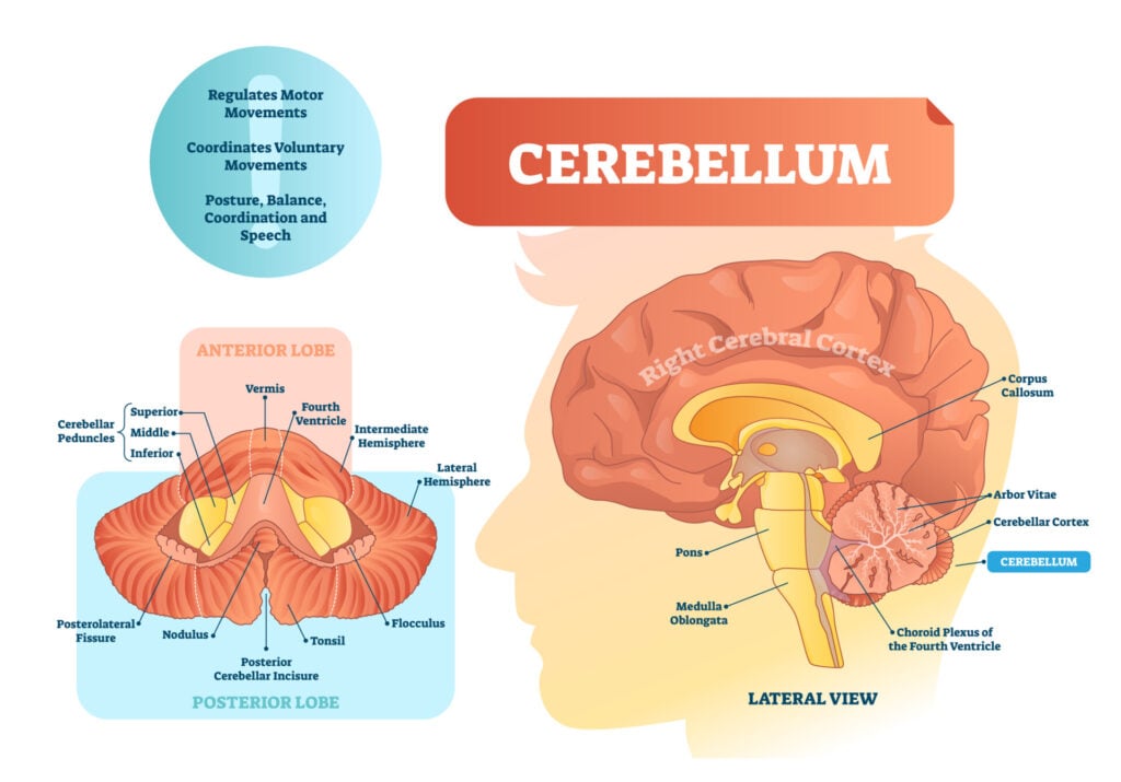
The cerebellum, which stands for ‘little brain,’ is a hindbrain structure that controls balance, coordination, movement, and motor skills, and it is thought to be important in processing some types of memory.

Key Takeaways
- The cerebellum is a brain structure located at the back of the head that helps coordinate movement, balance, and posture.
- It plays a crucial role in motor learning—helping us fine-tune skills like walking, typing, or riding a bike.
- Beyond movement, the cerebellum also supports cognitive functions like attention, language, and emotional regulation.
- Damage to the cerebellum can cause balance problems, tremors, speech difficulties, and even thinking or memory issues.
- Researchers have linked cerebellar differences to conditions such as ADHD, autism, anxiety disorders, and schizophrenia.
The cerebellum’s role is not only in physical coordination but also in the regulation of thought and emotion. It is also one of the few brain regions where adult neurogenesis has been observed (Ponti, Peretto & Bonfanti, 2008).
Did you know? Although the cerebellum only accounts for 10% of the overall brain mass, it contains over half of the brain's nerve cells.
Location
The cerebellum is located at the back of the brain, behind the brainstem, below the temporal and occipital lobes, and beneath the back of the cerebrum.
The cerebellum is also divided into two hemispheres, like the cerebral cortex. Unlike the cerebral hemispheres, each hemisphere of the cerebellum is associated with each side of the body.

Cerebellum functions
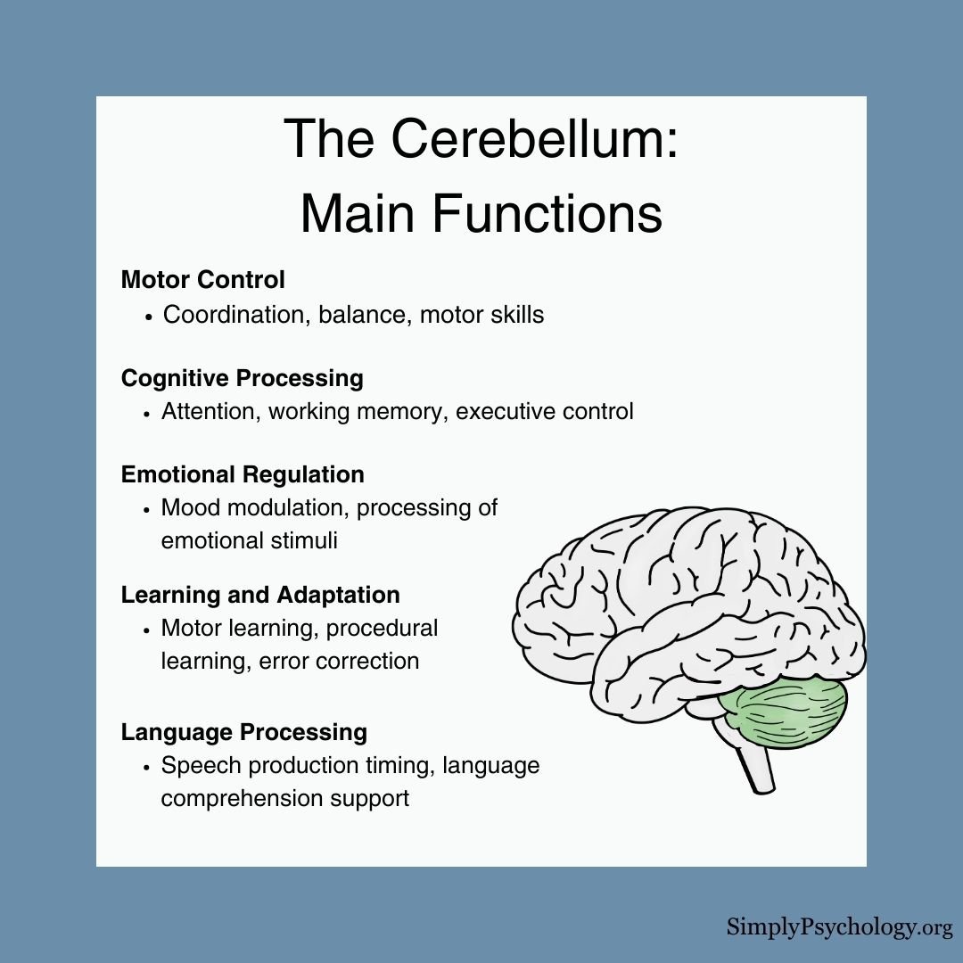
The cerebellum is essential not only for movement but also for various cognitive processes.
Motor Functions
Traditionally linked to movement, the cerebellum helps:
- Coordinate voluntary movements
- Maintain balance and posture
- Refine motor skills
- Support motor learning and adaptation
It receives sensory input about body position and planned movements, allowing it to fine-tune muscle actions—like coordinating eye movements—without conscious effort.
For example, when learning to ride a bike, the cerebellum adjusts and automates motor patterns over time.
Cognitive Functions
Research now shows the cerebellum also contributes to:
- Attention and executive control
- Language processing
- Spatial and temporal awareness
- Working memory
- Emotional regulation
In fact, much of the cerebellum connects to cerebral networks involved in thinking and emotion—not just motor control (Buckner, 2013; Buckner et al., 2013).

Structure
The cerebellum consists of the cerebellar cortex, the outer layer, containing folder brain tissue, filled with most of the cerebellum’s neurons. It plays a key role in processing and integrating information sent to the cerebellum.
There is also a fluid-filled ventricle and cerebellar nuclei, which is the innermost part, containing neurons that communicate information from the cerebellum to other areas of the brain.
Within the cerebellum, there are thought to be three anatomical lobes, which are divided by two fissures (large furrows)– the primary fissure and the posterolateral fissure:
- Anterior lobe (anterior meaning ‘to the front’): Primarily involved in the coordination of limb movements.
- Posterior lobe (posterior meaning ‘to the back’): The largest part and plays a significant role in planning, initiating, and timing movements.
- Flocculonodular lobe: The oldest part of the brain in terms of evolution. This part of the cerebellum is responsible for balance and spatial attention, as well as receiving visual input.
The cerebellum can also be divided into three functional areas:
- Cerebrocerebellum – this is the largest area of the cerebellum, responsible for planning movements and motor learning. It also works to regulate the coordination of muscle activation as well as eye movements.
- Spinocerebellum – this area functions in regulating body movements by allowing for error corrections.
- Vestibulocerebellum – this area is involved in controlling balance and flexes of the eyes.
Connections to other parts of the brain and body
Although the cerebellum is often described as a “little brain,” it functions more like a central hub—constantly exchanging information with other brain regions to coordinate movement, balance, and even cognition.
Input: Where the cerebellum gets its information
The cerebellum receives input from several key systems:
- Spinal cord – Sends information about body position and movement (proprioception).
- Vestibular system – Informs the cerebellum about head position and balance.
- Cerebral cortex – The motor and premotor areas send motor plans to the cerebellum via the middle cerebellar peduncle, allowing the cerebellum to refine upcoming movements.
- Sensory systems – Visual, auditory, and tactile input help the cerebellum adjust movements in real-time.
Integration: Processing information internally
Once information enters the cerebellum, it’s processed in two key areas:
- Cerebellar cortex – The outer layer where most neurons reside. It processes incoming signals and fine-tunes responses.
- Deep cerebellar nuclei – Including the dentate, emboliform, globose, and fastigial nuclei, these structures act as the cerebellum’s output centers, relaying instructions to other brain areas.
Neurotransmitters like GABA (an inhibitory neurotransmitter) and glutamate (an excitatory one) help regulate cerebellar signaling.
These chemicals are essential for balancing activation and inhibition across motor and cognitive circuits.
Output: Where the cerebellum sends its messages
Once processed, the cerebellum sends output through the superior cerebellar peduncle to the thalamus, which then relays signals to the motor cortex and other cortical areas.
This pathway helps adjust motor execution based on real-time sensory feedback.
The cerebellum also communicates with:
- Brainstem nuclei – To help control posture, eye movements, and reflexes.
- Basal ganglia – Although not directly connected, these two systems interact via shared loops with the cortex and thalamus, working together to initiate and refine movements.
- Prefrontal cortex – Involved in planning, attention, and working memory.
- Limbic system – Plays a role in emotional regulation and motivation, potentially explaining why cerebellar abnormalities are linked to mood disorders.
Related conditions
ADHD
Adults with ADHD show structural differences in the middle cerebellar peduncle, particularly reduced white matter organization.
These differences are linked to core symptoms like hyperactivity and inattention, suggesting that the cerebellum may play a role in the cognitive aspects of ADHD (Parkkinen et al., 2024).
This aligns with the cerebellum’s known involvement in attention and working memory—both areas of difficulty in ADHD.
Autism
Cerebellar disruptions, especially involving Purkinje cells, have been linked to autism-like behaviors in animal studies, such as social challenges and repetitive actions.
Abnormal connectivity between the cerebellum and cerebral cortex is also observed in autistic individuals (van der Heijden et al., 2021).
However, a large study of over 400 participants found no consistent anatomical differences in the cerebellum between autistic and non-autistic groups (Laidi et al., 2022), suggesting that functional or subtle structural changes may be more relevant than gross anatomical alterations.
Anxiety Disorders
Studies indicate that the cerebellum may contribute to anxiety-related responses.
Lesions in the cerebellum can reduce avoidance behaviors in animal models (Caulfield & Servatius, 2013), while increased cerebellar activity has been observed in humans with PTSD and generalized anxiety disorder (Abadie et al., 1999; Critchley et al., 2000).
Schizophrenia
Neuroimaging studies show reduced cerebellar volume and lower blood flow to the cerebellar cortex in individuals with schizophrenia, particularly during tasks involving working memory and attention (Nopoulos et al., 1999; Crespo-Facorro et al., 2007).
These findings support the cerebellum’s role in cognitive and emotional regulation, extending its relevance beyond motor coordination.
Damage
Cerebellar damage can lead to lasting problems with movement, coordination, and thinking, due to the loss of nerve cells.
Common Symptoms:
- Unsteady walking or balance issues
- Tremors (shaking)
- Slurred speech
- Jerky or inaccurate movements
- Difficulty with memory and concentration
Causes of Cerebellar Damage:
- Alcohol use – Intoxication temporarily affects coordination, while chronic misuse can cause lasting damage.
- Head injury – Especially trauma to the back of the head.
- Neurological conditions – Such as Parkinson’s disease, multiple sclerosis, or stroke.
- Infections or brain tumors – That affect the cerebellum.
- Toxins and medications – Including lead, mercury, or long-term use of benzodiazepines.
Taking steps to protect the brain—like avoiding substance misuse and preventing falls—can help reduce the risk of cerebellar damage.
References
Abadie, P., Boulenger, J. P., Benali, K., Barre, L., Zarifian, E., & Baron, J. C. (1999). Relationships between trait and state anxiety and the central benzodiazepine receptor: a PET study. European Journal of Neuroscience, 11 (4), 1470-1478.
Buckner, R. L. (2013). The cerebellum and cognitive function: 25 years of insight from anatomy and neuroimaging. Neuron, 80(3), 807-815.
Caulfield, M. D., & Servatius, R. J. (2013). Focusing on the possible role of the cerebellum in anxiety disorders. New Insights into Anxiety Disorders (Durbano F, Ed.). InTech, Rijeka, HR, 41-70.
Crespo-Facorro, B., Barbadillo, L., Pelayo-Terán, J. M., & Rodríguez-Sánchez, J. M. (2007). Neuropsychological functioning and brain structure in schizophrenia. International Review of Psychiatry, 19 (4), 325-336.
Critchley, H. D., Corfield, D. R., Chandler, M. P., Mathias, C. J., & Dolan, R. J. (2000). Cerebral correlates of autonomic cardiovascular arousal: a functional neuroimaging investigation in humans. The Journal of physiology, 523(1), 259-270.
Gowen, E., & Miall, R. C. (2007). The cerebellum and motor dysfunction in neuropsychiatric disorders. The Cerebellum, 6(3), 268-279.
Laidi, C., Floris, D. L., Tillmann, J., Elandaloussi, Y., Zabihi, M., Charman, T., … & Simonoff, E. (2022). Cerebellar atypicalities in autism?. Biological psychiatry, 92(8), 674-682.
Nopoulos, P. C., Ceilley, J. W., Gailis, E. A., & Andreasen, N. C. (1999). An MRI study of cerebellar vermis morphology in patients with schizophrenia: evidence in support of the cognitive dysmetria concept. Biological psychiatry, 46 (5), 703-711.
Parkkinen, S., Radua, J., Andrews, D. S., Murphy, D., Dell’Acqua, F., & Parlatini, V. (2024). Cerebellar network alterations in adult attention-deficit/hyperactivity disorder. Journal of Psychiatry and Neuroscience, 49(4), E233-E241.
Phillips, J. R., Hewedi, D. H., Eissa, A. M., & Moustafa, A. A. (2015). The cerebellum and psychiatric disorders. Frontiers in public health, 3, 66.
Ponti, G., Peretto, P., & Bonfanti, L. (2008). Genesis of neuronal and glial progenitors in the cerebellar cortex of peripuberal and adult rabbits. PLoS One, 3 (6), e2366.
Stoodley, C. J. (2016). The cerebellum and neurodevelopmental disorders. The Cerebellum, 15 (1), 34-37.
Further Information
D”Angelo, E. (2019). The cerebellum gets social. Science, 363(6424), 229-229.


