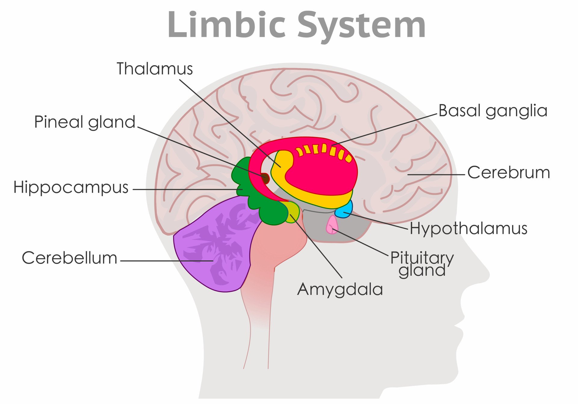
The question “Which part of the brain controls emotions?” reflects a common misconception—that emotions arise from a single, localized area of the brain.
In reality, emotional processing is distributed across a network of interconnected brain regions, each contributing to different aspects of emotional experience, regulation, and expression.
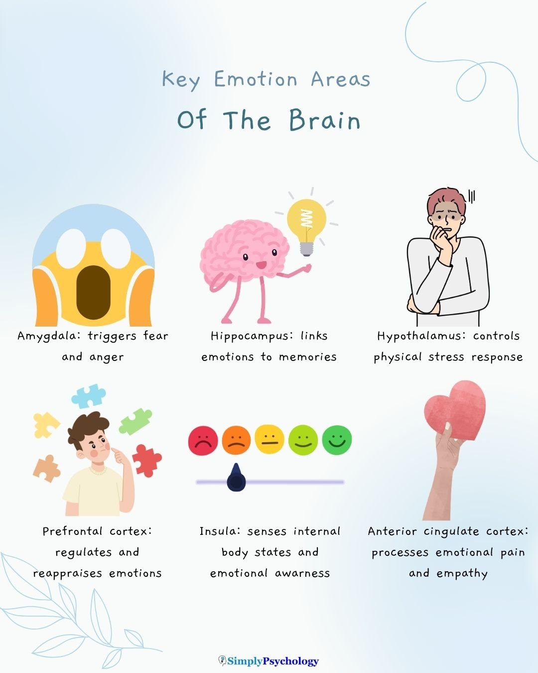
Rather than one “emotion center,” research points to a functional system involving structures such as the amygdala (threat detection), hippocampus (emotional memory), prefrontal cortex (regulation and reasoning), hypothalamus (physiological response), and insula (bodily awareness).
These areas work in concert to interpret stimuli, attach meaning, activate physiological responses, and guide behavior.

Key Takeaways
- No single emotion center: Emotions arise from a network of brain regions working together.
- Amygdala, hippocampus, hypothalamus, and PFC each play unique roles in emotion detection, memory, and regulation.
- Insula and ACC help us sense emotions in the body and navigate social experiences like empathy or shame.
- Teenage brains feel emotions more intensely due to a mature amygdala and still-developing prefrontal cortex.
- The brain can change: Practices like mindfulness and therapy can rewire emotional circuits over time.
Areas of the brain involved in emotions
Here are the key brain areas involved in controlling emotions:
Amygdala: The Brain’s Threat Detector
The amygdala is a crucial structure responsible for evaluating sensory information and quickly determining its emotional importance, particularly in processing fear and anger.
It plays a central role in tying emotional meaning to our memories and is important for the learning and triggering of emotions.
Research indicates that the amygdala’s central nucleus is particularly important in mediating fear responses.
Hyperactivity in the amygdala is frequently observed in anxiety disorders like specific phobia and social anxiety disorder, reflecting enhanced attention and emotional reactivity to threatening stimuli.
Elevated amygdala activity is also linked to depression, especially when processing negative emotional stimuli.
Hippocampus: Linking Emotion to Memory
The hippocampus serves as a “gateway” to memory, enabling us to form spatial memories and consolidate new ones.
It is an essential structure for learning and memory, integrating emotional experiences with cognitive processes.
Strong emotions can trigger the formation of powerful memories, with the hippocampus playing a role in encoding these emotionally arousing events at a deeper level.
Its structure and function are significantly linked to various mood and anxiety disorders.
Notably, individuals suffering from Post-Traumatic Stress Disorder (PTSD) often exhibit marked reductions in the volume of several parts of the hippocampus.
Hypothalamus: Controlling the Body’s Emotional Response
The hypothalamus is a critical component of the limbic system, involved in drives essential for individual survival.
It regulates fundamental homeostatic processes, including body temperature, appetite, blood pressure, and sexual motivation.
In emotional reactions, the hypothalamus plays a key role in activating the sympathetic nervous system.
This activation triggers physical responses, such as increased heart rate and blood pressure, which are central to the body’s “fight or flight” response.
It also coordinates reflexive changes in response to physical and psychological demands, providing a link between physiological systems and psychological stress.
Prefrontal Cortex: Regulating and Reframing Emotions
The prefrontal cortex (PFC) is responsible for higher-level cognitive functioning, including planning, decision-making, creative problem-solving, and emotion regulation.
It plays a crucial role in inhibiting impulsive reactions, particularly those originating from the amygdala.
The PFC’s ability to dampen amygdala activation allows for the suppression of negative emotions and supports reflection and empathy.
In anxiety and mood disorders, research indicates reduced activation in frontoparietal regions, including the PFC, during tasks involving emotional reappraisal and attention regulation.
Less activation in the prefrontal cortex, especially on the left side, is observed in depressed individuals.
Insula and Anterior Cingulate Cortex: Feeling Emotions in the Body
The insula and Anterior Cingulate Cortex (ACC) are important for integrating bodily sensations with emotional experience.
The insula, in particular, shows hyperactivity in anxiety disorders, reflecting enhanced attention and emotional reactivity, especially in interoceptive processing.
It’s also recognized as a critical brain area for sensations like limb ownership and is connected to emotion-related areas.
The ACC facilitates the inhibition of maladaptive behavioral responses and is involved in conflict monitoring and emotional pain perception.
Both the insula and ACC demonstrate altered neural processing during executive functioning and decision-making in anxiety disorders.

How these areas work together
Emotion is a complex interplay of various brain regions working together in intricate circuits, much like a relay race between instinct and reflection.
The amygdala, serving as the brain’s rapid threat detector, quickly evaluates sensory information for emotional importance, particularly fear and anger.
This immediate “bottom-up” response is then influenced by the prefrontal cortex (PFC), a “top-down” regulator.
This amygdala-PFC feedback loop allows the PFC to inhibit impulsive reactions and dampen amygdala activation, enabling the suppression and reframing of negative emotions.
Our conscious experience of emotions in the body is significantly mediated by the insula and the anterior cingulate cortex (ACC).
The insula integrates signals relating to internal bodily states like pain or hunger, which in turn give rise to emotions.
The ACC is crucial for conflict monitoring, emotional pain perception, and empathy. Together, the insula-ACC connection forms a vital part of the brain’s salience network, involved in directing attention and emotional awareness.
In response to trauma, the hippocampus contextualizes emotional experiences with cognitive processes.
Its strong link with the amygdala means that emotionally arousing events are encoded at a deeper level.
This interconnectedness allows these regions to collaboratively process and store the emotional dimensions of traumatic memories.
Emotions and the teenage brain
Teenage brains are a fascinating work in progress, often leading to intense emotional experiences and increased risk-taking.
This dynamic arises because the amygdala, the brain’s emotional core and threat detector, is largely developed and highly reactive during adolescence. It’s like a powerful, fully engaged emotional ‘accelerator’.
Meanwhile, the prefrontal cortex (PFC), crucial for judgment, impulse control, and planning, is still undergoing significant development, continuing to mature into early adulthood. This makes the PFC akin to underdeveloped ‘brakes.’
This developmental imbalance can result in intense mood swings, where emotions are felt strongly, and reactions might be impulsive.
For example, a minor disappointment might trigger an outsized emotional response, or the brain’s heightened sensitivity to reward can make risky behaviors seem more appealing, despite potential negative consequences.
It’s why decisions might seem more spontaneous and feelings more overwhelming, as the ‘accelerator’ is strong, but the ‘brakes’ are still under construction.
Complex Social Emotions: Empathy, Shame, and Embarrassment
Complex social emotions like empathy, shame, and embarrassment are not mere reflexes but involve sophisticated higher-level brain functions.
The medial prefrontal cortex (mPFC) is crucial for distinguishing between self and other perspectives, allowing for moral judgment and the understanding of intentions and beliefs.
The temporoparietal junction (TPJ) is also a key region in social cognition and mentalizing—the ability to infer others’ mental states.
Furthermore, the mirror neuron system, located in areas like the anterior cingulate cortex (ACC) and the insula, plays a significant role in empathy by enabling mimicry and the recognition of others’ actions and emotions.
These complex emotions are profoundly shaped by multifaceted factors beyond basic physiological responses.
Culture dictates appropriate emotional displays and even how emotions are perceived and categorized.
Memory integrates past experiences, influencing how we interpret and react emotionally to present social situations.
Relationships also heavily mould these emotions, as they are learned and negotiated within social contexts, such as family and community interactions.
For instance, shame may be considered a stigmatized emotion that many tend to hide rather than display in individualistic cultures.
Can we change how our brain handles emotions?
Yes, our brains can indeed change how they handle emotions, a process known as neuroplasticity. This means that the brain’s circuits can be rewired through various interventions.
- Strengthening the Prefrontal Cortex (PFC): The prefrontal cortex can be strengthened and its cortical thickness increased through practices such as mindfulness meditation. This enhanced PFC activity supports greater personal agency and allows for more thoughtful responses instead of automatic reactions.
- Reduced Amygdala Reactivity: As the prefrontal cortex becomes stronger, it gains a greater ability to dampen the activity of the amygdala, which is the brain’s threat detector. Studies using fMRI have shown that therapies, like labelling emotions, can reduce activity in the amygdala and limbic regions when processing negative emotional images or memories.
- Therapeutic Evidence: Neuroscience provides evidence that psychological therapies can produce actual changes in brain functioning. For example, Cognitive Behavioural Therapy (CBT) has been shown to result in significant “deactivations” in the amygdala, thalamus, and hippocampal regions. Mindfulness-based interventions also lead to structural and functional changes in the brain, improving emotional regulation.

