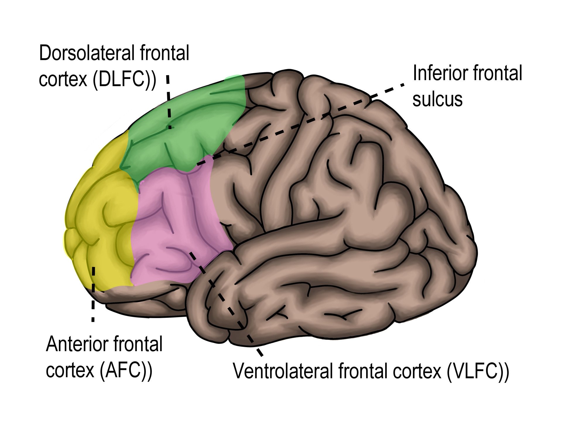
The dorsolateral prefrontal cortex (DLPFC) sits in the upper, outer part of the prefrontal cortex, at the front of the brain. It supports working memory, planning, attention, and self-control. Many core topics in psychology depend on it.
When you learn about executive functions, you’re really studying the capacities that the DLPFC helps support.
Researchers study the DLPFC with tasks like the Stroop test, the Wisconsin Card Sorting Test, and working memory tasks (e.g. n-back). The DLPFC is active in all of them. Changes in its functioning show how people think, adapt, and learn.
It also comes up in clinical psychology: many disorders (e.g. depression, ADHD, schizophrenia) involve differences in DLPFC function.
Knowing how the DLPFC is studied, what goes wrong, and how it can be strengthened or compensated for gives you a bridge between theory and application.
Key Takeaways
- Location: The DLPFC sits on the upper, outer surface of the prefrontal cortex, mainly Brodmann areas 9 and 46.
- Working Memory: Its neurons keep firing while information is held in mind, which makes it the engine of the brain’s mental workspace.
- Cognitive Control: It supports planning, attention, and flexible switching between rules. Stopping an action outright relies more on the right inferior frontal cortex.
- Cold Cognition: The DLPFC handles the “cold” side of control. Valuing options and feeling their consequences rely more on ventromedial and orbitofrontal regions.
- Late Maturation: It is among the last cortical regions to mature, reaching an adult profile only in late adolescence or beyond.
- Clinical Links: DLPFC underactivity features in depression, ADHD, and schizophrenia, but the pattern is probabilistic, not universal.
- Modern Evidence: Meta-analyses link a shared frontoparietal control network to many disorders, and rTMS aimed at the left DLPFC beats sham in depression.

Location and Structure of the DLPFC
The dorsolateral prefrontal cortex (DLPFC) is a part of the frontal lobe of the brain. It sits toward the top and side (dorsolateral = dorsal + lateral) of the prefrontal cortex.
You can find it on the middle frontal gyrus, which is one of the ridges on the brain’s surface in the frontal lobe.
The Brodmann areas system numbers regions of the cortex. The numbering is based on how the cells are organized. The DLPFC mainly covers Brodmann Area 9 and Area 46.
Area 9 sits toward the top (dorsal). Area 46 sits more toward the side (lateral). The two areas are close neighbours and often work together.
The DLPFC connects to many other brain regions. Sensory areas feed it input. It also links with memory-related areas like the hippocampus, and with subcortical structures like parts of the thalamus and basal ganglia.
This network of connections lets the DLPFC integrate incoming information, keep things in mind, plan actions, and control behaviour.
Laterality matters. Verbal tasks (language work) tend to activate more of the left DLPFC, while spatial and visual tasks often use more of the right DLPFC.
DLPFC vs Other Prefrontal Regions
The prefrontal cortex is a family of subregions, each tied to a different facet of executive function. The table shows where the DLPFC fits among them.
| Region | Location | Main role |
|---|---|---|
| Dorsolateral (DLPFC) | Upper, outer surface; Brodmann areas 9 and 46 | Working memory, planning, reasoning, set-shifting, top-down attention control |
| Ventromedial (vmPFC) | Lower, inner surface; medial parts of areas 10, 11, 12 and 25 | Value, emotion, and social decision-making |
| Orbitofrontal (OFC) | Underside above the eye sockets; areas 11, 13 and 47 | Reward valuation and flexible responding when outcomes change |
| Anterior cingulate (ACC) | Medial strip over the corpus callosum; areas 24 and 32 | Conflict monitoring and error detection |
| Frontopolar | Extreme front tip; rostral area 10 | Abstract “meta” operations, such as holding a main goal while pursuing a sub-goal |
The boundaries are not razor-sharp, and the functions overlap. Even so, the DLPFC is the region most closely tied to the “cold,” computational side of executive control.
Functions of the DLPFC
The dorsolateral prefrontal cortex (DLPFC) is one of the brain’s key regions for executive functions and higher-order cognition.
That means it helps with thinking tasks where you have to plan, hold information in mind, make decisions, and control your behaviour.
Researchers distinguish three core executive functions (Diamond, 2013):
- Inhibitory control: suppressing automatic but inappropriate responses, such as ignoring a distraction.
- Working memory: holding and updating information in mind.
- Cognitive flexibility: switching between rules, tasks, or perspectives.
Higher-order skills such as planning and problem-solving are built from these three. The three are separable but related.
Miyake and colleagues (2000) tested a battery of frontal lobe tasks. They found that shifting, updating, and inhibition were moderately correlated yet clearly distinct. They called this the “unity and diversity” of executive functions.
What links them? According to Miller and Cohen (2001), the prefrontal cortex actively maintains patterns of activity that represent goals. It then sends bias signals that steer processing elsewhere in the brain.
Working memory is that maintenance. Flexibility is updating the goal, and inhibition is biasing away competing responses.
One caution applies. Stopping an action outright depends on the right inferior frontal cortex, not the DLPFC (Aron et al., 2004).
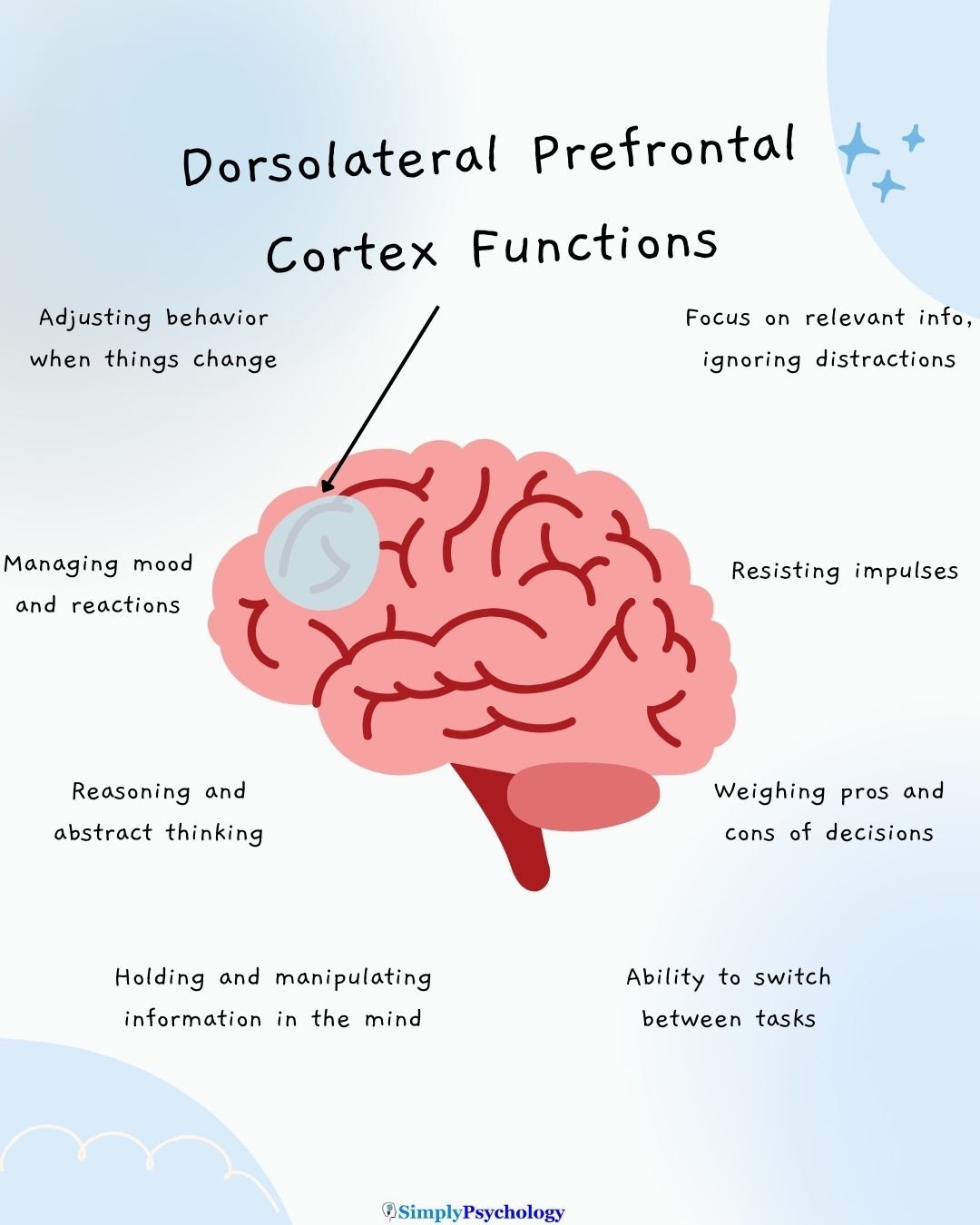
Working Memory and Attention
Working memory is like a mental scratchpad: the ability to hold and work with information in your mind for short periods.
The DLPFC is especially important for manipulating information, not just remembering it. That means rearranging items, comparing them, or updating what’s relevant.
For example, if you remember numbers and then have to sort them in your head, the DLPFC helps with that.
The evidence for this role stretches back almost a century. Jacobsen (1936) found that monkeys with frontal lesions could still see, move, and learn simple discriminations, but failed once a delay separated the hidden food from their choice.
Fuster and Alexander (1971) later recorded prefrontal neurons that changed their firing during that delay. Funahashi, Bruce, and Goldman-Rakic (1989) then showed how precise this firing can be.
- Aim: To test whether the delay-period firing of dorsolateral prefrontal neurons is tied to a specific remembered location.
- Method: Monkeys fixated a central spot while a cue flashed at one of eight locations. After a delay of 1–6 seconds, they made an eye movement to where the cue had been.
- Results: Many neurons near the principal sulcus fired throughout the delay. Each fired most for cues in its own preferred part of the visual field, forming a “memory field.”
- Conclusion: Individual DLPFC neurons hold a remembered location online through sustained, spatially tuned firing. This became the textbook cellular account of the brain’s mental workspace.
Attention is also guided by the DLPFC. It helps you focus on what’s important and ignore irrelevant distractions.
It also helps you shift your focus when needed, perhaps from listening in class to taking notes and then back again. This ability to flexibly shift and maintain attention is essential to learning and problem-solving.
Decision-making and Problem-solving
The DLPFC contributes to decision-making by helping you evaluate different options and think about consequences.
When you weigh pros and cons, predict what might happen, or choose between short-term reward and long-term benefit, the DLPFC is active.
This is the “cold” side of decision-making. It means holding options in mind and reasoning about them. Valuing those options and feeling their consequences relies more on the ventromedial and orbitofrontal cortex.
The Iowa Gambling Task shows the difference. Patients with ventromedial damage, though intellectually intact, keep choosing options with large immediate rewards but bigger long-term losses (Bechara et al., 1994).
Problem-solving often involves planning steps, dealing with unexpected obstacles, and adjusting strategies.
The DLPFC supports flexible thinking. If your first idea doesn’t work, it helps you switch to a new approach. It also helps you stay goal-oriented, keeping in mind what you want to achieve while resisting distractions.
The classic test of this flexibility is the Wisconsin Card Sorting Test. Participants sort cards by colour, shape, or number. They must work out the rule from feedback, and the rule changes without warning.
- Aim: To test whether the frontal lobes are specifically needed for shifting cognitive set, by comparing patients with frontal and non-frontal lesions.
- Method: Patients with surgical lesions in different brain regions sorted cards by colour, form, or number. The correct rule was worked out from right/wrong feedback and changed periodically without warning.
- Results: Patients with dorsolateral frontal damage perseverated after the rule changed, sorting by the old rule despite repeated feedback. Patients with lesions elsewhere did not show this pattern.
- Conclusion: The dorsolateral frontal cortex is critical for cognitive flexibility and for inhibiting an outdated response set. Perseveration became a hallmark sign of prefrontal dysfunction.
Emotional Regulation and Self-Control
Emotional regulation means being able to manage your feelings so that they’re appropriate for the situation.
The DLPFC plays a big part here, helping suppress or adjust emotional reactions when necessary (for example, cooling down anger or resisting impulses).
One strategy is cognitive reappraisal. It means reinterpreting an upsetting situation to change its emotional impact. Reappraisal recruits lateral and medial prefrontal regions that dampen reactivity in the amygdala, which helps generate fear and threat responses.
This control can fail under pressure. High levels of catecholamines, a family of chemical messengers, can take the prefrontal cortex offline, shifting control back to the amygdala and to habit (Arnsten, 2009). That is why stress erodes self-control.
It helps you think before acting, rather than acting purely out of emotion.
Self-control is closely related: it’s the ability to resist temptations, delay gratification (choosing a bigger reward later rather than a smaller one now), or ignore impulses.
Studies show that greater activity in the DLPFC is associated with better self-control in tasks (e.g., resisting unhealthy food choices) and that disrupting DLPFC activity makes self-control harder.
The marshmallow test shows how lasting this link can be. Four decades later, adults who had struggled to delay gratification as preschoolers still showed poorer impulse control.
When suppressing responses to tempting cues, they also showed reduced prefrontal recruitment and exaggerated activity in the ventral striatum, a reward region (Casey et al., 2011).
Development and Plasticity of the DLPFC
The DLPFC isn’t fully mature at birth – in fact, it keeps developing through adolescence and into early adulthood.
During the teen years, the structure of the DLPFC changes. Its cortex tends to thin. This thinning is thought to reflect pruning, the removal of unused neural connections, which leaves the remaining ones more efficient.
Also, white matter (nerve fibres that help different brain areas communicate) increases in the prefrontal cortex during this period, helping signals travel faster.
These physical changes coincide with improvements in executive functions. Adolescents become better at planning, focusing attention, resisting impulsive responses, and switching tasks.
As the DLPFC matures, these high-level thinking skills become more reliable and flexible.
The best-known evidence for this late maturation comes from a longitudinal imaging study.
- Aim: To map the order in which regions of the cerebral cortex mature in living children.
- Method: Thirteen healthy children had repeated structural MRI scans every two years for 8–10 years, from roughly ages 4 to 21. The researchers built a four-dimensional map of grey-matter change.
- Results: Primary sensory and motor cortices matured first, and higher-order association cortices matured last. The dorsolateral prefrontal cortex was among the very latest, not reaching an adult profile until late adolescence and beyond.
- Conclusion: The prefrontal cortex is the last cortex to come fully online, maturing after the areas whose outputs it must integrate.
Maturation is gradual and varies between individuals. There is no single birthday when the DLPFC is “finished.”
Neuroplasticity means the brain can change with experience, and the DLPFC is plastic. Learning new high-demand cognitive tasks (like working memory training) can increase activity in the DLPFC, recruit more neurons, or alter how neurons fire in response to a task.
Also, therapies, education, or structured training can strengthen connections or improve control over behaviour and emotion via the DLPFC.
The prefrontal cortex also changes with age. A meta-analysis pooled fMRI studies of the n-back working memory task, in which people spot items repeated from a few steps earlier.
It compared young (about 24), middle-aged (about 38), and older (about 67) adults (Yaple et al., 2019). Parietal and cingulate activation appeared at every age.
Prefrontal activation was robust in young adults, weaker in middle age, and essentially absent in older adults. Working memory leans heavily on prefrontal integrity, and that contribution declines across adulthood.
Clinical Relevance of the DLPFC
When it is impaired or underactive, many psychological disorders show deficits in these areas. Problems can include:
- Difficulty keeping thoughts in mind
- Poor planning
- Trouble shifting attention
- Weak self-control
These impairments can seriously affect a person’s daily functioning — school, work, social life.
Dopamine helps explain why. Prefrontal function depends on dopamine and noradrenaline staying within a narrow optimal range, and too much or too little of either impairs it (Arnsten, 2009). That dependence is why disorders of dopamine signalling, such as schizophrenia and ADHD, so often involve the prefrontal cortex.
Below are some specific disorders where DLPFC dysfunction plays a role.
Depression and Mood Disorders
In major depressive disorder (MDD), studies often find that the left DLPFC is underactive. This underactivity is linked with symptoms like negative thinking, low motivation, and difficulty initiating behaviour.
When treatments like transcranial magnetic stimulation (TMS) successfully increase DLPFC activity, people’s depression symptoms often reduce.
Trial evidence supports a causal role. A network meta-analysis of randomised trials compared several active repetitive TMS (rTMS) protocols with sham stimulation. Those aimed at the left DLPFC reduced depressive symptoms significantly more (Brunoni et al., 2017).
Koenigs and Grafman (2009) describe a division of labour. The DLPFC tends to be underactive in the cognitive and psychomotor features of depression, such as poor concentration, indecision, and blunted drive. The ventromedial prefrontal cortex is implicated more in its affective and self-referential features.
This suggests that part of depression’s cognitive symptoms may come from a failure in the DLPFC’s ability to regulate mood and control negative thought patterns.
ADHD and Executive Dysfunction
Attention-Deficit Hyperactivity Disorder (ADHD) is another disorder where the DLPFC often shows reduced function.
People with ADHD may struggle with working memory deficits, which means they find it harder to hold information in mind and use it.
They may also have difficulty with impulse control and resisting distractions. Imaging studies point the same way. Networks involving the DLPFC are less active or less well-connected in ADHD compared to neurotypical individuals.
These impairments help explain symptoms like forgetfulness, interrupting others, and trouble finishing tasks, which are common in ADHD.
A large imaging study adds a developmental angle. Shaw et al. (2007) analysed 824 scans from children with and without ADHD. Cortical maturation followed the same regional sequence in both groups but ran later in ADHD.
The median age of reaching peak cortical thickness was 10.5 years in ADHD, compared with 7.5 years in typically developing children. That is a three-year gap.
The delay was greatest in prefrontal regions that govern attention and motor planning. This fits the way many people’s ADHD traits change as they get older.
Schizophrenia and Other Conditions
In schizophrenia, DLPFC dysfunction is associated with more severe cognitive deficits, such as problems with memory, planning, and organizing thoughts.
The DLPFC tends to show reduced activity or abnormal connectivity with other brain regions like the cerebellum.
The classic evidence is hypofrontality: the DLPFC fails to activate when a task demands it.
- Aim: To test whether the DLPFC is physiologically dysfunctional in schizophrenia specifically when a prefrontal task demands it.
- Method: Twenty medication-free patients with chronic schizophrenia and 25 controls had regional cerebral blood flow measured. Measurements came at rest, during number matching, and during the Wisconsin Card Sorting Test.
- Results: During card sorting, controls increased DLPFC blood flow but patients did not. In patients, the degree of DLPFC activation correlated with test performance.
- Conclusion: Schizophrenia involves a task-dependent failure to activate the DLPFC, called hypofrontality. This linked cognitive difficulties to a specific prefrontal physiology.
These changes are linked with disorganized thinking, difficulties in following conversations or tasks, and sometimes psychosis (hallucinations or delusions).
Other conditions involve the wider prefrontal cortex.
In addiction, Goldstein and Volkow (2011) argue that prefrontal disruption underlies loss of control over drug use, compulsive seeking, and harmful choices that persist despite mounting costs. Addiction is thus partly a failure of prefrontal control over a hijacked reward system.
Critical Evaluation of DLPFC Research
The case for the DLPFC’s role in executive function is strong, but it has limits worth knowing.
Strengths: Converging Evidence
The prefrontal evidence base spans single neurons, lesioned patients, developmental imaging, and meta-analysis. Very different methods point to the same conclusion:
- Single neurons: recordings reveal memory fields in the monkey DLPFC (Funahashi et al., 1989).
- Lesions: damage to dorsolateral frontal cortex causes perseveration (Milner, 1963).
- Developmental imaging: the DLPFC matures late (Gogtay et al., 2004).
- Meta-analysis: n-back fMRI studies tie prefrontal engagement to working memory performance (Yaple et al., 2019).
No single method is decisive. Lesions are imprecise, animal work assumes a mapping onto humans, and imaging is correlational. Their convergence is what makes the account credible.
It makes the DLPFC a model case of a control region whose role can be pinned down empirically, not merely asserted. It is among the most secure structure-to-function mappings in biopsychology.
Limitations: Networks, Not a Single Seat
The DLPFC is not a single seat of function. Functions localise to subregions and distributed networks.
Working memory, flexibility, value, and inhibition depend on different prefrontal regions wired into different networks (Aron et al., 2004; Koenigs & Grafman, 2009). Treating the DLPFC as the seat of self-control is therefore a reductionist error.
It also invites reverse inference: assuming that a region performs a task’s function because it activates during the task.
Much of the strongest evidence is indirect. Lesions vary between patients and damage grey and white matter together. Monkey recordings assume that the findings map onto humans. Imaging correlations cannot show that a prefrontal difference causes behaviour.
Disorder findings such as hypofrontality in schizophrenia or delayed maturation in ADHD are robust but probabilistic. Not every patient shows the pattern.
Contemporary Research
Recent work asks whether the single-disorder findings above are really separate stories. It also tests whether stimulating the DLPFC changes symptoms.
- Aim: To test whether disrupted cognitive-control circuitry is shared across psychiatric disorders rather than specific to each diagnosis.
- Method: McTeague and colleagues (2017) pooled neuropsychological, grey-matter, and fMRI findings from studies of schizophrenia, mood, anxiety, and substance-use disorders.
- Results: All three evidence types converged on the frontoparietal multiple-demand network, anchored in dorsolateral prefrontal and parietal cortex, and its link with the salience network. This held regardless of diagnosis.
- Conclusion: Cognitive-control difficulties across disorders may reflect one shared, transdiagnostic circuit rather than entirely separate pathologies.
As a large, multi-modal meta-analysis, this outranks any single-disorder imaging study. It does not overturn the disorder-specific findings. It shows what they share.
A second line of work tests causation directly. In a network meta-analysis, active rTMS aimed at the left DLPFC beat sham stimulation for depression (Brunoni et al., 2017). That moves the field from “the DLPFC is underactive in depression” toward “raising DLPFC activity has a causal antidepressant effect.”
Rival View: Adaptive Coding
The idea of fixed prefrontal subregions has a genuine rival. Duncan (2001) proposed adaptive coding: prefrontal neurons flexibly change what they represent from moment to moment. They carry whichever inputs, rules, or rewards are currently relevant.
On this view, the tidy map of subregions summarises average tendencies across many neurons and tasks. It does not describe what any single neuron is for.
The two accounts are not wholly incompatible. The frontoparietal circuit that McTeague et al. (2017) found disrupted overlaps heavily with DLPFC territory. That supports a domain-general reading of this map.
Ventromedial and orbitofrontal regions, by contrast, still respond more selectively to value and emotion than adaptive coding predicts.
The balanced reading is that the subdivision scheme captures real, useful average tendencies, while adaptive coding is a needed corrective against reading it as a literal map.
References
Arnsten, A. F. T. (2009). Stress signalling pathways that impair prefrontal cortex structure and function. Nature Reviews Neuroscience, 10(6), 410–422. https://doi.org/10.1038/nrn2648
Aron, A. R., Robbins, T. W., & Poldrack, R. A. (2004). Inhibition and the right inferior frontal cortex. Trends in Cognitive Sciences, 8(4), 170–177. https://doi.org/10.1016/j.tics.2004.02.010
Asgharian Asl, F., Abbaszade, S., Derakhshani, H., Vaghef, L., & Asgharian Asl, A. (2024). Unilateral vs. Bilateral DLPFC rTMS: Comparative effects on depression, visual-spatial memory, inhibitory control and cognitive flexibility in major depressive disorder. Frontiers in Psychiatry, 15, 1400414. https://doi.org/10.3389/fpsyt.2024.1400414
Bechara, A., Damasio, A. R., Damasio, H., & Anderson, S. W. (1994). Insensitivity to future consequences following damage to human prefrontal cortex. Cognition, 50(1–3), 7–15. https://doi.org/10.1016/0010-0277(94)90018-3
Benschop, L., Vanhollebeke, G., Li, J., Leahy, R. M., Vanderhasselt, M., & Baeken, C. (2022). Reduced subgenual cingulate–dorsolateral prefrontal connectivity as an electrophysiological marker for depression. Scientific Reports, 12, Article 16903. https://doi.org/10.1038/s41598-022-20274-9
Brunoni, A. R., Chaimani, A., Moffa, A. H., Razza, L. B., Gattaz, W. F., Daskalakis, Z. J., & Carvalho, A. F. (2017). Repetitive transcranial magnetic stimulation for the acute treatment of major depressive episodes: A systematic review with network meta-analysis. JAMA Psychiatry, 74(2), 143–152. https://doi.org/10.1001/jamapsychiatry.2016.3644
Casey, B. J., Somerville, L. H., Gotlib, I. H., Ayduk, O., Franklin, N. T., Askren, M. K., Jonides, J., Berman, M. G., Wilson, N. L., Teslovich, T., Glover, G., Zayas, V., Mischel, W., & Shoda, Y. (2011). Behavioral and neural correlates of delay of gratification 40 years later. Proceedings of the National Academy of Sciences, 108(36), 14998–15003. https://doi.org/10.1073/pnas.1108561108
Diamond, A. (2013). Executive functions. Annual Review of Psychology, 64, 135–168. https://doi.org/10.1146/annurev-psych-113011-143750
Duncan, J. (2001). An adaptive coding model of neural function in prefrontal cortex. Nature Reviews Neuroscience, 2(11), 820–829. https://doi.org/10.1038/35097575
Funahashi, S., Bruce, C. J., & Goldman-Rakic, P. S. (1989). Mnemonic coding of visual space in the monkey’s dorsolateral prefrontal cortex. Journal of Neurophysiology, 61(2), 331–349. https://doi.org/10.1152/jn.1989.61.2.331
Fuster, J. M., & Alexander, G. E. (1971). Neuron activity related to short-term memory. Science, 173(3997), 652–654. https://doi.org/10.1126/science.173.3997.652
Gogtay, N., Giedd, J. N., Lusk, L., Hayashi, K. M., Greenstein, D., Vaituzis, A. C., Nugent, T. F., Herman, D. H., Clasen, L. S., Toga, A. W., Rapoport, J. L., & Thompson, P. M. (2004). Dynamic mapping of human cortical development during childhood through early adulthood. Proceedings of the National Academy of Sciences, 101(21), 8174–8179. https://doi.org/10.1073/pnas.0402680101
Goldstein, R. Z., & Volkow, N. D. (2011). Dysfunction of the prefrontal cortex in addiction: Neuroimaging findings and clinical implications. Nature Reviews Neuroscience, 12(11), 652–669. https://doi.org/10.1038/nrn3119
Jacobsen, C. F. (1936). Studies of cerebral function in primates. I. The functions of the frontal association areas in monkeys. Comparative Psychology Monographs, 13(3), 1–60.
Koenigs, M., & Grafman, J. (2009). The functional neuroanatomy of depression: Distinct roles for ventromedial and dorsolateral prefrontal cortex. Behavioural Brain Research, 201(2), 239–243. https://doi.org/10.1016/j.bbr.2009.03.004
Kumari, V., Peters, E. R., Fannon, D., Antonova, E., Premkumar, P., Anilkumar, A. P., Williams, S. C., & Kuipers, E. (2009). Dorsolateral Prefrontal Cortex Activity Predicts Responsiveness to Cognitive–Behavioral Therapy in Schizophrenia. Biological Psychiatry, 66(6), 594–602. https://doi.org/10.1016/j.biopsych.2009.04.036
McTeague, L. M., Huemer, J., Carreon, D. M., Jiang, Y., Eickhoff, S. B., & Etkin, A. (2017). Identification of common neural circuit disruptions in cognitive control across psychiatric disorders. American Journal of Psychiatry, 174(7), 676–685. https://doi.org/10.1176/appi.ajp.2017.16040400
Miller, E. K., & Cohen, J. D. (2001). An integrative theory of prefrontal cortex function. Annual Review of Neuroscience, 24, 167–202. https://doi.org/10.1146/annurev.neuro.24.1.167
Milner, B. (1963). Effects of different brain lesions on card sorting: The role of the frontal lobes. Archives of Neurology, 9(1), 90–100. https://doi.org/10.1001/archneur.1963.00460070100010
Miyake, A., Friedman, N. P., Emerson, M. J., Witzki, A. H., Howerter, A., & Wager, T. D. (2000). The unity and diversity of executive functions and their contributions to complex “frontal lobe” tasks: A latent variable analysis. Cognitive Psychology, 41(1), 49–100. https://doi.org/10.1006/cogp.1999.0734
Shaw, P., Eckstrand, K., Sharp, W., Blumenthal, J., Lerch, J. P., Greenstein, D., Clasen, L., Evans, A., Giedd, J., & Rapoport, J. L. (2007). Attention-deficit/hyperactivity disorder is characterized by a delay in cortical maturation. Proceedings of the National Academy of Sciences, 104(49), 19649–19654. https://doi.org/10.1073/pnas.0707741104
Smucny, J., Dienel, S. J., Lewis, D. A., & Carter, C. S. (2021). Mechanisms underlying dorsolateral prefrontal cortex contributions to cognitive dysfunction in schizophrenia. Neuropsychopharmacology, 47(1), 292–308. https://doi.org/10.1038/s41386-021-01089-0
Weinberger, D. R., Berman, K. F., & Zec, R. F. (1986). Physiologic dysfunction of dorsolateral prefrontal cortex in schizophrenia. I. Regional cerebral blood flow evidence. Archives of General Psychiatry, 43(2), 114–124. https://doi.org/10.1001/archpsyc.1986.01800020020004
Yaple, Z. A., Stevens, W. D., & Arsalidou, M. (2019). Meta-analyses of the n-back working memory task: fMRI evidence of age-related changes in prefrontal cortex involvement across the adult lifespan. NeuroImage, 196, 16–31. https://doi.org/10.1016/j.neuroimage.2019.03.074