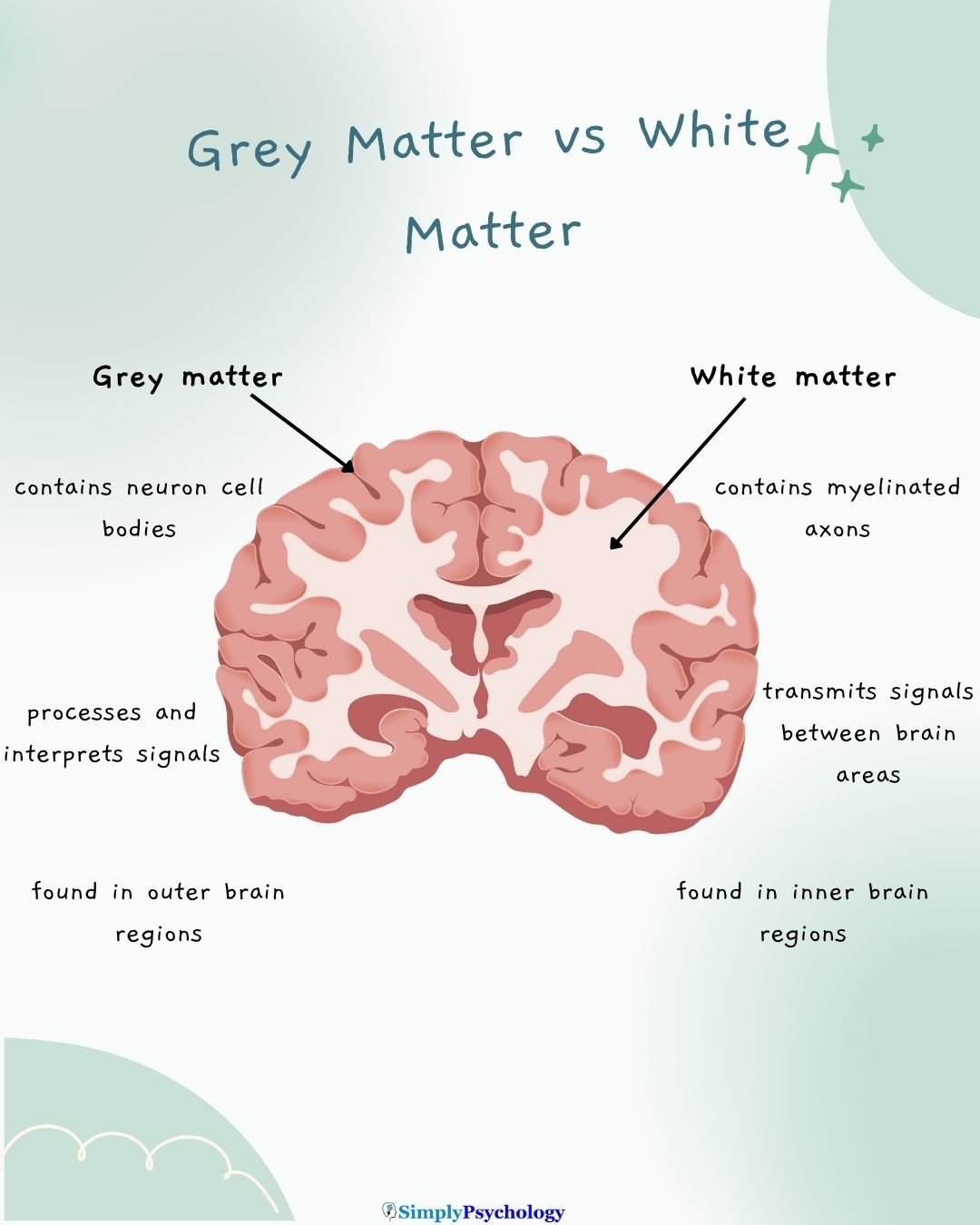
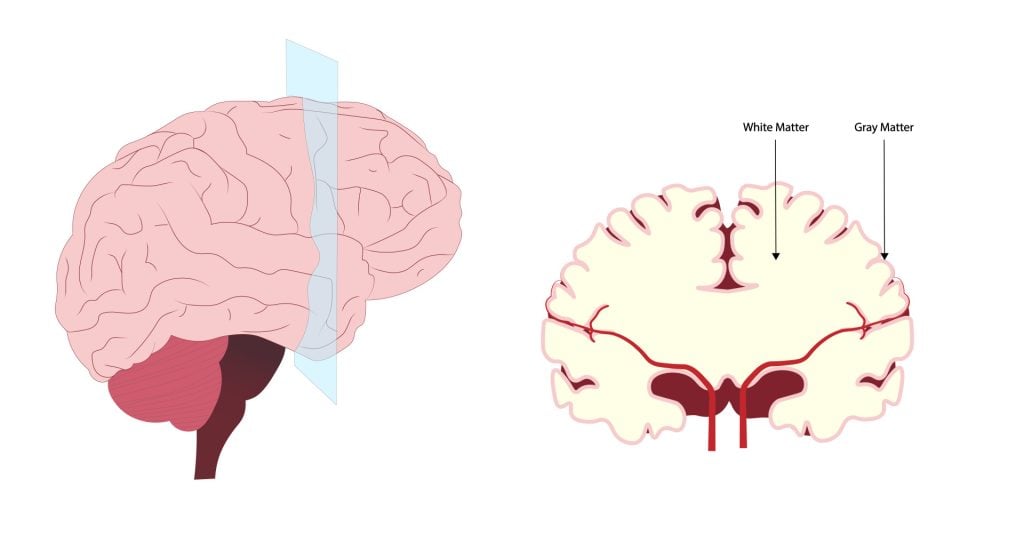
The central nervous system (CNS) is made up of white matter and grey matter.
White matter, comprising about half of the brain, consists of bundles of myelinated axons (nerve fibers).
Located in the deeper parts of the brain, white matter acts as the brain’s communication network, connecting different areas of grey matter and facilitating coordinated brain function.

Where is white matter found?
Its white appearance comes from the myelin sheath, a fatty substance surrounding the axons.
White matter is found in several key areas of the central nervous system:
- Brain:
- Located in the deeper tissues, beneath the grey matter
- Present in both the cerebrum and cerebellum
- Spinal Cord:
- Surrounds the grey matter, which is located in the center
This distribution allows white matter to effectively connect different regions of grey matter throughout the central nervous system, facilitating communication between various parts of the brain and between the brain and spinal cord.
What White Matter Consists of
White matter consists of millions of bundles of axons, glial cells, and nodes of Ranvier.
Axons are portions of the nerve cell (neuron) that carry electrical signals between different parts of the CNS.
Axons are covered in a fatty insulating substance called myelin sheath. Myelin is white which is what gives white matter its characteristic color.

Myelin sheath is made by the types of non-neuronal cells that are also present in the white matter, called oligodendrocytes.
These types of cells are known as glial cells, which are cells that provide support or protection to the neurons.
Oligodendrocytes form the myelin and wrap this around the axons up to 150 times. All the layers of myelin are tightly compressed around the axon to ensure it is protected.
On the axon, there are gaps in the myelin sheath called nodes of Ranvier. These unmyelinated gaps cause the impulses traveling along the length of the axon to leap from node to node.
When it does this, the signal increases in velocity so it can reach its destination quicker than traveling down the axon without nodes.
Function
White matter’s primary function is to transmit signals between different brain regions. It therefore plays a crucial role in a variety of brain functions:
- Neural Communication:
- Facilitates rapid transmission of signals between different brain regions
- Enables coordination and integration of information across the brain
- Learning and Skill Acquisition:
- Structural changes in white matter correlate with learning complex tasks
- Fields (2010) found significant increases in white matter volume, organization, and connectivity in adults learning to read
- Klingberg et al. (2000) observed that decreased reading ability correlated with decreased white matter diffusion in the temporoparietal area
- Cognitive Function:
- White matter structure is associated with various cognitive abilities
- Schmithorst et al. (2005) found a correlation between greater axon organization in frontal and occipital-parietal areas and higher IQ scores
- Brain Plasticity and Development:
- Ongoing myelination until the late 20s aligns with the period of cortical synaptic restructuring
- Teicher et al. (2004) found that children who experienced abuse or neglect had a 17% smaller corpus callosum (the largest white matter structure in the brain), suggesting early experiences affect white matter development
White Matter vs. Grey Matter
Below are some of the key differences between white matter and grey matter:
| White Matter | Grey Matter |
| White in color | Grey in colour |
| Located deep in the brain; outer portion of the spinal cord | Located in outer layer of brain (cortex); central portion of spinal cord |
| Composed mainly of myelinated axons and oligodendrocytes | Composed mainly of neuron cell bodies, dendrites, and unmyelinated axons |
| Primary function is to transmit signals between different brain regions | Primary function is to process and analyze information |
| Fast signal transmission | Slower signal transmission |
| Shows structural changes with learning and experience | Undergoes pruning and reorganization during development |
| Appears bright on T1-weighted MRI images | Appears dark on T1-weighted MRI images |

Associated Disorders
White matter plays a vital role in how the brain communicates, develops, and functions. When this tissue is damaged or disrupted, it can contribute to a wide range of neurological and psychiatric conditions—some common, others more complex.
Multiple Sclerosis and Demyelination
Multiple sclerosis (MS) is one of the most well-known disorders affecting white matter. In MS, the immune system mistakenly attacks the myelin sheath, leading to demyelination—a breakdown of the protective covering around axons. This disrupts signal transmission and can cause:
- Muscle weakness
- Coordination problems
- Fatigue
- Vision disturbances
Over time, persistent demyelination may also damage the axons themselves, resulting in irreversible neuronal loss.
White Matter Disease and Small Vessel Damage
White matter disease—often identified as white matter hyperintensities on MRI scans—is commonly linked to aging and cerebral small vessel disease.
These bright spots typically indicate areas of poor blood flow or inflammation. They are associated with:
- Memory problems
- Slower thinking
- Higher risk of stroke and vascular dementia
Managing vascular health (e.g., controlling blood pressure) can help reduce progression.
Psychiatric and Developmental Conditions
Disruptions in white matter structure have been observed in several psychiatric disorders:
- Depression: Reduced white matter integrity, especially in tracts connecting the thalamus and frontal lobe, has been linked to both current depression and genetic vulnerability.
- Schizophrenia: White matter abnormalities often emerge during adolescence, especially in the forebrain, when myelination is still developing.
- ADHD, OCD, PTSD, and autism spectrum disorder have all shown atypical white matter development in key brain pathways.
These findings highlight the importance of white matter in regulating attention, emotion, and executive functioning.
Alzheimer’s Disease and Cognitive Decline
In Alzheimer’s disease, white matter changes can appear even before symptoms of memory loss. Researchers have found that:
- White matter lesions are associated with early mild cognitive impairment (MCI)
- Disruptions in white matter tracts may impair communication between memory-related regions like the hippocampus and prefrontal cortex
This suggests that preserving white matter health could help delay or reduce cognitive decline.
Research insights
A 2024 brain imaging study found that people with depression have reduced white matter integrity, especially in pathways involving the thalamus.
Even individuals without depression but at high genetic risk showed similar white matter changes.
In multiple sclerosis (a disease attacking white matter), a 2023 clinical analysis revealed that an old antihistamine drug (clemastine) increased myelin in patients’ brain white matter.
This hints at a potential new therapy to repair white matter damage.
Physical activity appears beneficial for white matter. A 2022 study of older adults reported that higher fitness levels and intense exercise were linked to stronger white matter microstructure, suggesting that regular exercise helps maintain healthy “brain wiring.”
In 2023, the FDA approved an AI-based MRI software that automatically maps white matter microstructure in the brain.
This technology provides a detailed, automated view of white matter and can quickly flag subtle abnormalities in white matter tracts.
How to Strengthen White Matter
While there’s no cure for white matter disease, several strategies may support white matter health and slow decline.
Exercise Regularly
Aerobic and resistance training improve blood flow and brain connectivity. Studies show higher fitness levels are linked to stronger white matter microstructure, especially in older adults.
Learn New Skills
Challenging the brain—through language learning, music, or complex tasks—can promote white matter plasticity and reinforce communication between regions.
Practice Mindfulness
Mindfulness and meditation may reduce stress-related inflammation and improve white matter integrity in areas tied to focus and emotion.
Protect Against Injury
Wearing helmets and avoiding excess alcohol can reduce the risk of falls and head injuries, both of which may damage white matter.
Support Vascular Health
Controlling blood pressure, quitting smoking, and eating a heart-healthy diet can slow the progression of white matter lesions and lower stroke risk.
References
Fields, R. D. (2010). Neuroscience. Change in the brain’s white matter. Science (New York, NY), 330(6005), 768-769.
Klingberg, T., Hedehus, M., Temple, E., Salz, T., Gabrieli, J. D., Moseley, M. E., & Poldrack, R. A. (2000). Microstructure of temporo-parietal white matter as a basis for reading ability: evidence from diffusion tensor magnetic resonance imaging. Neuron, 25 (2), 493-500.
Newman, T. (2017, August 16). White matter: The brain’s flexible but underrated superhighway. Medical News Today https://www.medicalnewstoday.com/articles/318966
Pani, J., Eikenes, L., Reitlo, L. S., Stensvold, D., Wisløff, U., & Håberg, A. K. (2022). Effects of a 5-Year Exercise Intervention on White Matter Microstructural Organization in Older Adults. A Generation 100 Substudy. Frontiers in Aging Neuroscience, 14, 859383. https://doi.org/10.3389/fnagi.2022.859383
Schmithorst, V. J., Wilke, M., Dardzinski, B. J., & Holland, S. K. (2005). Cognitive functions correlate with white matter architecture in a normal pediatric population: a diffusion tensor MRI study. Human brain mapping, 26 (2), 139-147.
Teicher, M. H., Dumont, N. L., Ito, Y., Vaituzis, C., Giedd, J. N., & Andersen, S. L. (2004). Childhood neglect is associated with reduced corpus callosum area. Biological psychiatry, 56 (2), 80-85.
Yamazaki, R., & Ohno, N. (2025). The potential of repurposing clemastine to promote remyelination. Frontiers in Cellular Neuroscience, 19, 1582902. https://doi.org/10.3389/fncel.2025.1582902


