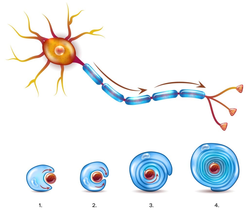
The myelin sheath is a fatty, protective coating that wraps around the axons of many neurons. Much like insulation on electrical wires, this sheath allows nerve signals to travel faster and more efficiently.
Without myelin, communication between the brain and body would slow down, affecting everything from movement to thought.
This sheath isn’t continuous. Instead, it has small gaps called nodes of Ranvier that are essential for speeding up signal transmission.

Key Takeaways
- Myelin is a protective coating that wraps around nerve fibers, helping messages travel quickly between the brain and body.
- It speeds up signals by letting them “jump” between small gaps (called nodes of Ranvier), reaching speeds up to 120 meters per second.
- It keeps the brain working well by helping nerves save energy and adapt to learning and new experiences.
- Damage to myelin can cause problems like numbness, blurry vision, or weak muscles—common in conditions like multiple sclerosis.
- Myelin grows over time, starting before birth and continuing into adulthood, following the brain’s growth and development.

How Myelin Supports Efficient Signaling
Myelin plays a critical role in ensuring that nerve impulses are transmitted quickly, accurately, and with minimal energy loss. It does this through several complementary mechanisms:
- Saltatory conduction: Myelin enables electrical impulses to “jump” between the nodes of Ranvier, dramatically increasing conduction speed—up to 120 meters per second.
- Signal containment: The sheath prevents electrical signals from leaking out of the axon, preserving signal strength and direction.
- Ion regulation: Myelin helps control sodium and potassium flow, which is essential for nerve impulses. Paranodal junctions (specialized contact points between myelin and axons that help keep signal-related proteins in the right places) ensure these ions are properly separated, preventing signal disruption.
- Potassium clearance: During signal transmission, potassium levels rise around the axon. Myelin contains channels that help remove this excess potassium, while nearby astrocytes absorb and transport it out of the nervous system.
- Energy efficiency: By concentrating ion channels at specific points and reducing the need for continuous ion exchange, myelin minimizes the energy required for repeated signal transmission.
Together, these features allow neurons to transmit signals rapidly and reliably, which is vital for everything from reflexes to complex thought.

How Myelin Supports Brain Health
While its role in speeding up communication is well known, recent research reveals that myelin is more than just insulation.
- It adapts with experience: Neural activity can change the way myelin forms, helping the brain strengthen or weaken specific connections over time.
- It provides metabolic support: Oligodendrocytes—the cells that produce myelin in the brain—can supply energy to axons, helping them function for longer periods without damage.
These discoveries show that myelin is a dynamic part of the brain, involved in both learning and long-term neural health.
History
In 1854, Rudolf Virchow introduced the term “myelin”, deriving it from the Greek word for marrow, to describe a structure richly present in the brain.
Since 1949, the predominant view has been that myelin’s sole function is to optimize nerve conduction speed and minimize energy use in axons.
Recent research
However, recent findings challenge this perspective, suggesting that myelin and the cells forming it can undergo changes based on neural activity, thereby influencing how neural circuits function.
Additionally, evidence is growing that these cells offer metabolic support to neurons through their myelin sheaths.
This expanded understanding of myelin and its relationship with neurons sheds light not only on its role in normal physiological functions but also in the pathology of various neurological and psychiatric disorders.

Myelination: Formation and Development
Formation Process
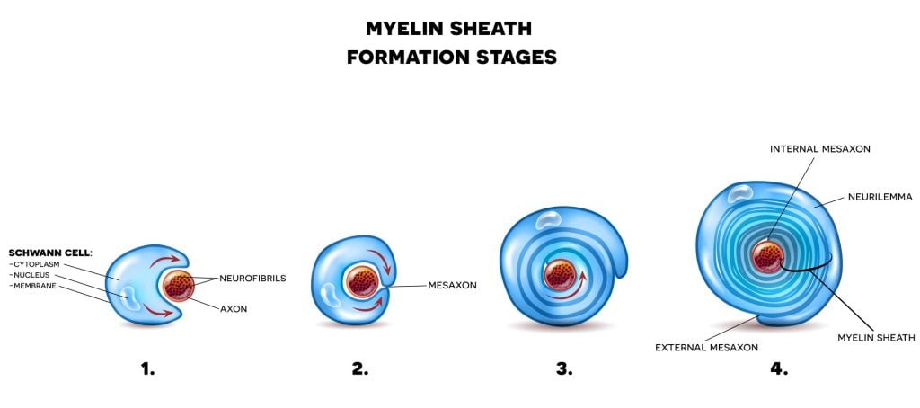
Myelin sheath is a lipid-rich, white substance that forms a protective layer around nerve cell axons throughout the nervous system.
Two specialized cell types are responsible for producing this vital structure: oligodendrocytes in the central nervous system (brain and spinal cord) and Schwann cells in the peripheral nervous system.

These cells create the myelin sheath by wrapping multiple layers of their plasma membrane tightly around the axon, forming concentric layers of insulation.
While Schwann cells can only myelinate one axon segment at a time, oligodendrocytes can simultaneously myelinate multiple axon segments through their star-shaped structure.
The resulting sheath isn’t continuous along the axon; instead, it has small gaps called nodes of Ranvier that occur every 0.2 to 2 millimeters along its length.
When axons lack this myelin covering, they are considered “unmyelinated” and conduct electrical signals more slowly than their myelinated counterparts.

When Does Myelination Happen?
Myelination begins in the womb and continues into adulthood:
- Infancy (0–2 years): Rapid myelination supports motor and sensory development.
- Childhood (5–7 years): Key connections between brain areas like the thalamus and prefrontal cortex mature.
- Adulthood (20s–50s): Myelination of higher-order brain regions continues, supporting decision-making and complex thinking.
- Later life: While myelin may thin with age, it remains vital for healthy brain function.
This timeline helps explain why different cognitive and motor skills develop and peak at different stages of life.
What Happens When Myelin Is Damaged?
Damage to the myelin sheath disrupts nerve signal transmission, leading to a range of neurological symptoms. This is known as demyelination.
Common causes include:
- Autoimmune diseases (e.g., multiple sclerosis)
- Infections or injuries
- Genetic conditions
Symptoms of Myelin Damage
- Muscle weakness or spasms
- Numbness or tingling
- Vision problems
- Poor coordination or balance
- Pain, dizziness, or sensory changes
While some damage can be repaired naturally or with treatment, the extent of recovery depends on the underlying cause and severity.
Myelin and Nervous System Efficiency
Beyond its structural role, myelin actively contributes to signal reliability and energy balance:
- Maintains signal integrity: Myelin ensures that potassium levels don’t rise too high during signal transmission.
- Works with astrocytes: These neighboring brain cells absorb excess potassium and transport it out of the nervous system.
- Supports saltatory conduction: The nodes of Ranvier contain ion channels that allow impulses to “jump” along the axon, conserving energy.
This combination of physical insulation and cellular teamwork keeps your nervous system functioning smoothly.

References
Cech, D. J., & Martin, S. T. (2011). Functional Movement Development Across the Life Span-E-Book . Elsevier Health Sciences.
Fields, R. D. (2008). White matter in learning, cognition and psychiatric disorders. Trends in neurosciences, 31(7), 361-370.
Fünfschilling, Ursula, Lotti M. Supplie, Don Mahad, Susann Boretius, Aiman S. Saab, Julia Edgar, Bastian G. Brinkmann et al. “Glycolytic oligodendrocytes maintain myelin and long-term axonal integrity.” Nature 485, no. 7399 (2012): 517-521.
Huxley, A. F., & Stämpfli, R. (1949). Evidence for saltatory conduction in peripheral myelinated nerve fibres. The Journal of physiology, 108(3), 315.
Moore, S., Meschkat, M., Ruhwedel, T., Trevisiol, A., Tzvetanova, I. D., Battefeld, A., Kusch, K., Kole, M. H. P., Strenzke, N., Möbius, W., de Hoz, L. & Nave, K. A. (2020). A role of oligodendrocytes in information processing. Nature communications, 11 (1), 1-15.
Kirkwood, C. (2015, March 24). Myelin: An Overview . Brain Facts. https://www.brainfacts.org/brain-anatomy-and-function/anatomy/2015/myelin
Ratini, M. (2019, August 7). What Is a Myelin Sheath? WebMD. https://www.webmd.com/multiple-sclerosis/myelin-sheath-facts
Osika, A. (2020, October 29). The myelin sheath and myelination . Kenhub. https://www.kenhub.com/en/library/anatomy/the-myelin-sheath-and-myelination
Stadelmann, C., Timmler, S., Barrantes-Freer, A., & Simons, M. (2019). Myelin in the central nervous system: structure, function, and pathology. Physiological reviews, 99(3), 1381-1431.


