The peripheral nervous system (PNS) consists of all nerves outside the brain and spinal cord, which together make up the central nervous system (CNS). It acts as a communication relay between the CNS and the rest of the body.
It enables movement, sensation, and involuntary functions like heart rate and digestion.

What does the PNS do?
The PNS has many essential functions throughout the body:
- Sensation: Sensory information is carried from the eyes, ears, nose, tongue, and skin to the brain. For example, when you smell food, this information is carried from the nose to the brain.
- Movement: The PNS carries signals that allow for voluntary movements such as walking, and maintaining posture, and for reflex movements such as automatically moving a hand away from a hot surface.
- Involuntary processes: Vital, unconscious processes such as heartbeat and breathing are controlled by the PNS.
- Digestion: The PNS sends messages to the digestive system to keep food moving along, for example, by helping to produce saliva when food is presented.
The functions of the PNS are controlled by different substructures which will be explained in the next section.
Parts and Divisions of the Peripheral Nervous System
The peripheral nervous system (PNS) is divided into two main branches, each with distinct roles:
- The somatic nervous system (SNS), which controls voluntary, conscious movement
- The autonomic nervous system (ANS), which regulates involuntary organ activity
These subdivisions help regulate both voluntary and involuntary body functions.

Somatic Nervous System
The somatic nervous system is responsible for conscious, voluntary movements, like walking, talking, or typing.
It includes two types of nerves:
- Sensory neurons (afferent neurons): Carry information from sensory organs (skin, eyes, ears, etc.) to the central nervous system (CNS).
Example: When you touch a hot stove, sensory neurons alert the brain. - Motor neurons (efferent neurons): Transmit signals from the CNS to skeletal muscles, enabling movement.
Example: Your brain sends motor commands that help you lift a cup or run.
This system also controls reflex actions through a structure called the reflex arc, which allows for fast, automatic responses without involving the brain.
Autonomic Nervous System
The autonomic nervous system manages involuntary processes: functions you don’t consciously control, like heartbeat, digestion, or breathing. It is further divided into three parts:
- Sympathetic nervous system (fight or flight):
Prepares the body for stressful situations. It increases heart rate, opens airways, dilates pupils, and slows digestion. - Parasympathetic nervous system (rest and digest):
Calms the body after stress. It reduces heart rate, promotes digestion, and restores energy balance. - Enteric nervous system (the gut’s “second brain”):
A large web of neurons in the gut wall that can keep digestion moving even when cut off from the rest of the nervous system.
Together, the SNS and ANS allow the PNS to handle both physical movement and automatic life-sustaining processes.

The Spinal Reflex Arc
Reflexes are the nervous system’s fastest responses. A spinal reflex arc can trigger a protective movement before the brain even registers what happened.
How the Reflex Arc Works
A sensory neuron carries the signal from skin or muscle in along the dorsal root, into the spinal cord’s grey matter.
Grey matter is the butterfly-shaped core of cell bodies and synapses running down the centre of the cord. It is surrounded by the white-matter tracts that carry traffic up and down to the brain.
The path is short.
Within the grey matter the signal crosses one or more synapses. A motor neuron then carries the resulting command back out along the ventral root to the muscle, which contracts.
The whole loop finishes inside the cord. That is what makes the reflex so fast: the brain only learns what happened afterwards.
This anatomy is old and simple, but it is exactly what makes a fast, protective reflex possible before conscious thought can intervene.
The Knee-Jerk Reflex: A Landmark Study
The simplest reflex arc crosses just one synapse. Its clearest example is the knee-jerk, tested by tapping the tendon below the kneecap.
- Aim: Liddell and Sherrington (1924) set out to determine the neural pathway behind the tonic stretch reflex, the sustained contraction produced when a muscle is passively stretched.
- Method: Working with decerebrate cats, in which the forebrain is surgically disconnected from the brainstem, the researchers stretched specific limb muscles and measured how quickly the reflex contraction followed.
- Results: A sustained stretch produced a correspondingly sustained reflex contraction in the very same muscle, and the short delay showed the pathway crossed only a single synapse.
- Conclusion: The knee-jerk is produced by the simplest possible circuit, a single synapse linking a sensory neuron directly to a motor neuron, with no interneuron in between.
The decerebrate preparation isolated the reflex from brain influence, but it also means the finding says nothing about how an intact, conscious brain modifies the same reflex.
The Withdrawal Reflex
Not all reflexes are this simple.
Withdrawing a hand from something hot crosses at least two synapses.
The sensory neuron first excites an interneuron, which then excites the motor neuron that pulls the hand away.
This costs a little time compared with the knee-jerk. But it buys flexibility: an interneuron can combine signals from several sources and coordinate more than one muscle group at once.
The knee-jerk shows the fastest, simplest option: one synapse, nothing to coordinate. The withdrawal reflex trades some of that speed for the ability to integrate several signals at once and organise a more complex response.
Both patterns matter throughout the body. Which reflex pattern a given task needs depends on how much coordination the movement demands.
Clinical Uses of Reflex Testing
Because each reflex is generated at a known spinal level, tapping a tendon and grading the response is one of the fastest tools in a neurological exam.
A reduced or absent reflex at one level, paired with normal reflexes below it, can help locate a problem in the spinal cord or a peripheral nerve.
The Babinski sign is a classic example. In adults, the big toe should curl down, not extend upward, when the sole is stroked.
An upward-extending toe in an adult signals that descending pathways from the brain have stopped suppressing this primitive reflex. It is a reminder that spinal reflexes are shaped by the brain, not produced by the cord in isolation.
Reflex testing also matters after injury. The pattern of reflexes that remains below the level of damage helps show how complete a spinal cord injury is.
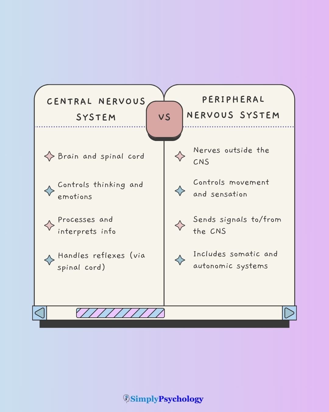
Central nervous system vs peripheral nervous system
The central nervous system (CNS) and peripheral nervous system (PNS) serve distinct but complementary roles.
The CNS, consisting of the brain and spinal cord, acts as the body’s command center and is protected by bone and protective membranes.
The PNS comprises nerves branching throughout the body, lacking bony protection and thus more vulnerable to injury.
Neither system works in isolation.
While the CNS processes and makes decisions, the PNS functions as a messenger network through sensory and motor neurons.
CNS neurons also have limited regenerative capacity, making injuries often permanent.
PNS neurons, however, can regenerate under certain conditions, allowing for potential recovery from peripheral nerve damage.
Nerves in the Peripheral Nervous System
The PNS is made up of nerve cells (neurons) that carry messages back and forth between the CNS and the muscles, organs, and senses in the body’s periphery.

Within the PNS, some nerves are attached to the spinal cord (spinal nerves), and others are attached directly to the brain (cranial nerves).
Spinal nerves
There are 31 pairs of spinal nerves, which emerge from the spinal cord and branch out to serve different areas of the body. These nerves carry:
- Sensory signals from the skin, muscles, and organs to the spinal cord and brain
- Motor commands from the brain and spinal cord to the muscles and glands
Each spinal nerve is a mixed nerve: for most of its length, it carries both sensory and motor fibres together. This mixing is undone only at the spinal cord itself, where the nerve splits into a dorsal root, carrying sensory fibres, and a ventral root, carrying motor fibres.

Types of spinal nerves (grouped by region):
- 8 cervical nerves, which serve the chest, head, neck, shoulders, arms, and hands (called C1-C8).
- 12 thoracic nerves, which serve the back, abdominal muscles, and intercostal muscles (called T1 – T12).
- 5 lumbar nerves, which serve the lower abdomen, thighs, and legs (called L1-L5).
- 5 sacral nerves, which serve the legs, feet, and genital areas (called S1-S5).
- 1 coccygeal pair of nerves (called Co1).
These nerves exit the spinal column through openings in the vertebrae and form a vast network that links every part of the body to the nervous system.
Example: The Sciatic Nerve
The sciatic nerve, which extends from the lower spine down to the toes, is the longest nerve in the human body.
It plays a major role in leg movement and is commonly associated with conditions like sciatica.
Cranial nerves
Unlike spinal nerves, cranial nerves connect directly to the brain, bypassing the spinal cord.
There are 12 pairs of cranial nerves, and most are involved in sensory and motor functions of the head and neck.
Functions of Cranial Nerves:
- Transmit sensory input from the eyes, ears, nose, tongue, and face
- Send motor signals to muscles involved in facial expression, chewing, swallowing, and speech

For example, when eating:
- Cranial nerves help you chew and swallow (motor function)
- They also relay taste signals back to the brain (sensory function)
There are 12 pairs of cranial nerves attached to the brain:
- Olfactory nerves are sensory nerves related to the sense of smell.
- Optic nerves are sensory nerves related to the sense of sight.
- Oculomotor, trochlear, and abducens nerves are motor nerves responsible for regulating voluntary eye movements.
- Vestibulocochlear nerves are sensory nerves related to the sense of hearing, linked with sound, orientation, and balance.
- Glossopharyngeal nerve: A sensory and motor nerve responsible for taste and swallowing.
- Hypoglossal nerve: A motor nerve responsible for tongue movements.
- Vagus nerves are both sensory and motor nerves responsible for movements of the lower head, throat, neck, chest, and abdomen, as well as autonomic functions such as breathing and heart rate.
- Accessory nerve: A motor nerve responsible for movement of the head, neck, and shoulders.
- Facial nerves are sensory and motor nerves related to the taste buds and movements of the face (facial expressions).
- Trigeminal nerves are sensory and motor nerves that carry signals from the eyes, teeth, and face, as well as impulses from the lower jaw and muscles involved with chewing.
Why the Peripheral Nervous System Matters
The peripheral nervous system (PNS) is not just a conduit for signals between the brain and body; it’s a dynamic network essential for survival, adaptation, and overall well-being.
1. It Powers Everyday Actions
The PNS lets you walk, grip a pencil, feel textures, or react quickly to danger.
These responses happen through sensory and motor nerves working in sync with the brain and spinal cord.
2. It Keeps Your Body in Balance
The autonomic division of the PNS regulates involuntary processes like heart rate, blood pressure, and digestion.
This helps maintain homeostasis, your body’s internal balance, even during stress or illness.
The ANS produces these effects two ways at once: direct nerve signals to organs, and hormone release from glands.
The hypothalamus coordinates both routes. That is how a stressful thought in the brain becomes a bodily change like a faster heartbeat.
3. It Can Regenerate After Injury
Unlike the central nervous system, the PNS has some ability to repair itself.
Cells like Schwann cells help guide the healing of damaged nerves, improving chances of recovery after injury.
PNS nerves are wrapped by Schwann cells.
CNS neurons, by contrast, are wrapped by a different cell type called oligodendrocytes. This difference is why a severed peripheral nerve can sometimes regrow toward its target, while a severed spinal cord or brain pathway generally cannot.
4. It Reflects Your Overall Health
Tingling, numbness, or muscle weakness may indicate larger issues like diabetes, autoimmune disorders, or nutritional deficiencies.
Tracking PNS symptoms can support early diagnosis of systemic problems.
5. It Includes the “Second Brain”
The enteric nervous system, part of the PNS, controls digestion independently of the brain.
It’s so influential that scientists call it the “second brain.”
It even plays a role in mood regulation and immune function.
Critical Evaluation
The CNS/PNS/somatic/autonomic map is one of the most useful frameworks in biopsychology, and it earns that status honestly.
A balanced view, though, has to weigh its real explanatory power against a few ways the tidy diagram oversimplifies the underlying biology.
The Voluntary/Involuntary Boundary Isn’t Fixed
The somatic system is defined by voluntary control, yet it also runs the fully automatic reflex arc described above. The autonomic system is defined by involuntary control, yet its balance can be shifted, with training, through biofeedback and slow breathing.
Neither label is an absolute rule. Each describes a system’s typical mode, not a hard limit on what it can do.
Biofeedback makes this concrete. A person watches their own heart rate or skin conductance displayed in real time, and learns, with practice, to shift their own balance toward calm.
Slow, deliberate breathing works on the same principle, nudging the body toward parasympathetic dominance on purpose. Control here is real, even though the system runs on its own the rest of the time.
The Reflex Arc Isn’t Purely Bottom-Up
The reflex arc completes its loop without consulting the brain, but that does not make it independent of the brain. Descending pathways from the cortex and brainstem constantly adjust spinal reflex circuits to match the body’s ongoing task, rather than leaving them as a fixed loop (Grillner, 1975).
The adult Babinski sign shows this clearly.
When the sole is stroked, an upward-extending big toe means the brain’s normal suppression of an infant reflex has broken down (Van Gijn, 1978).
This is exactly why a reflex test is diagnostically useful in the first place. An abnormal reflex is not just a spinal-cord fact; it is evidence about what the brain is or is not doing to it.
A purely bottom-up account is therefore only a first approximation. The cord and the brain are always working together.
The Enteric Nervous System’s Independence
The clearest challenge to a strict top-down model is the enteric nervous system. It is a mesh of hundreds of millions of neurons embedded in the gut wall, using much of the same neurochemistry as the brain. It is sometimes called the body’s “second brain.”
It can keep coordinating digestion even when its links to the rest of the nervous system are cut (Gershon, 1998). Sympathetic and parasympathetic input still modulate it day to day, and a strong stress response can override it, so it is not wholly autonomous.
Yet it is not fully independent. But its very existence shows that at least part of the periphery is doing genuine local computation, not simply relaying commands issued elsewhere.
More recent clinical research extends this: enteric neuron dysfunction is now linked to disorders once thought of as purely brain-based. Parkinson’s disease pathology, for example, is increasingly understood to appear in gut neurons years before it appears in the brain (Rao & Gershon, 2016).
Contemporary Research
The classical reflex circuit is not just historical curiosity. It is now an active constraint on modern attempts to restore movement after spinal cord injury.
Formento and colleagues (2018) combined computer modelling of the spinal circuitry with direct testing in people with chronic spinal cord injury. They found that continuous epidural stimulation, the protocol that restores stepping in animal studies, blocks natural proprioceptive signals and disrupts the very reciprocal-inhibition circuits Liddell and Sherrington described.
Burst and precisely patterned stimulation avoided this problem. It left room for natural signals to keep flowing and produced much more robust voluntary leg control.
This mechanistic finding is backed by an independent clinical result. Precisely patterned epidural stimulation, timed to a person’s own attempted movement and combined with rehabilitation training, restored voluntary stepping in people with spinal cord injury (Wagner et al., 2018).
Some of the improvement lasted even after the stimulation itself was switched off.
Key Takeaways
- PNS Scope: All nerves outside the brain and spinal cord, twelve pairs of cranial nerves and thirty-one pairs of spinal nerves, connecting the CNS to the rest of the body.
- Two Main Branches: The somatic nervous system controls voluntary movement, while the autonomic nervous system regulates involuntary processes like heart rate and digestion.
- Three ANS Divisions: Sympathetic (fight or flight), parasympathetic (rest and digest), and enteric (the gut’s own “second brain”) work together to maintain balance.
- Reflex Arc: A protective reflex, like the knee-jerk, can be organised entirely within the spinal cord, faster than a decision made by the brain.
- Regeneration: PNS nerves can often repair themselves after injury, unlike the more fragile neurons of the brain and spinal cord.
- Modern Evidence: Recent spinal cord injury research shows that restoring movement depends on preserving the reflex circuit’s natural wiring, not simply overriding it with continuous stimulation.
Sources
Dorland, W. A. N. (2011). Dorland’s Illustrated Medical Dictionary E-Book. Elsevier Health Sciences.
Eyesenck, M. W. (2012). Simply Psychology. New York: Taylor & Francis.
Formento, E., Minassian, K., Wagner, F., Mignardot, J. B., Le Goff-Mignardot, C. G., Rowald, A., Bloch, J., Micera, S., Capogrosso, M., & Courtine, G. (2018). Electrical spinal cord stimulation must preserve proprioception to enable locomotion in humans with spinal cord injury. Nature Neuroscience, 21(12), 1728–1741. https://doi.org/10.1038/s41593-018-0262-6.
Gershon, M. D. (1998). The second brain: A groundbreaking new understanding of nervous disorders of the stomach and intestine. HarperCollins.
Goldstein, D. S. (2010). Adrenal responses to stress. Cellular and Molecular Neurobiology, 30 (8), 1433-1440.
Grillner, S. (1975). Locomotion in vertebrates: Central mechanisms and reflex interaction. Physiological Reviews, 55(2), 247–304. https://doi.org/10.1152/physrev.1975.55.2.247.
Liddell, E. G. T., & Sherrington, C. S. (1924). Reflexes in response to stretch (myotatic reflexes). Proceedings of the Royal Society of London. Series B, 96(675), 212–242. https://doi.org/10.1098/rspb.1924.0023.
Martin, G. N., Carlson, N. R., & Buskist, W. (2009). Psychology, 4th European edition. Harlow: Pearson Education, 723-725.
Rao, M., & Gershon, M. D. (2016). The bowel and beyond: The enteric nervous system in neurological disorders. Nature Reviews Gastroenterology & Hepatology, 13(9), 517–528. https://doi.org/10.1038/nrgastro.2016.107.
Van Gijn, J. (1978). The Babinski sign and the pyramidal syndrome. Journal of Neurology, Neurosurgery & Psychiatry, 41(10), 865–873. https://doi.org/10.1136/jnnp.41.10.865.
Wagner, F. B., Mignardot, J.-B., Le Goff-Mignardot, C. G., Demesmaeker, R., Komi, S., Capogrosso, M., Rowald, A., Seáñez, I., Caban, M., Pirondini, E., Vat, M., McCracken, L. A., Heimgartner, R., Fodor, I., Watrin, A., Seguin, P., Paoles, E., Van Den Keybus, K., Eberle, G., … Courtine, G. (2018). Targeted neurotechnology restores walking in humans with spinal cord injury. Nature, 563(7729), 65–71. https://doi.org/10.1038/s41586-018-0649-2.


