Sensory neurons carry incoming information from the body’s sensory receptors toward the central nervous system (brain and spinal cord). Motor neurons carry outgoing commands away from the central nervous system to the muscles and glands.

Key Takeaways
- Sensory neurons carry information toward the central nervous system (CNS), while motor neurons send commands away from the CNS to muscles or glands.
- Sensory neurons are afferent and found in dorsal root ganglia, whereas motor neurons are efferent and located in the spinal cord or brain.
- Structurally, sensory neurons are usually unipolar, and motor neurons are typically multipolar.
- Both types work together in reflex arcs, allowing the body to detect and respond to stimuli rapidly.
- Understanding these neurons helps explain how the brain and body communicate to sense the world and produce behavior.
Sensory vs. motor neurons: Differences
Below are the main differences between sensory and motor neurons:
| Feature | Sensory neurons | Motor neurons |
|---|---|---|
| Also called | Afferent neurons | Efferent neurons |
| Direction of signal | Toward the CNS | Away from the CNS |
| Carries information | From receptors in the skin, eyes, ears and internal organs to the CNS | From the CNS to effectors (muscles and glands) |
| Cell body location | Dorsal root ganglia, just outside the spinal cord | Inside the CNS (spinal cord’s ventral horn or the brain) |
| Typical structure | Usually unipolar: one process that splits into two branches | Multipolar: many processes leave the cell body |
| Role in a reflex | Detect the stimulus and signal the CNS | Receive the signal and trigger the response |
Function
Sensory neurons transmit information from sensory receptors (in the skin, eyes, ears, tongue, etc.) to the CNS, enabling perception of stimuli.
Motor neurons transmit commands from the CNS to effectors (muscles or glands), enabling responses or actions.
In other words, sensory neurons inform the brain/spinal cord of what’s happening, while motor neurons direct the body on how to react.
Touching a hot surface shows both jobs. Sensory neurons signal the CNS about the heat they detect, and motor neurons then drive the hand to move away.
Direction of Signal (Afferent vs. Efferent)
Sensory input travels toward the central nervous system along afferent pathways, whereas motor impulses travel away from the CNS along efferent pathways.
A helpful shorthand is “afferent arrives, efferent exits” the CNS.
Between the two sits a third class, the relay neuron or interneuron. It connects sensory and motor neurons inside the CNS. It is also by far the most numerous type of neuron there.
Location of Cell Bodies
The cell bodies of sensory neurons are often located in clusters just outside the spinal cord. These clusters, called dorsal root ganglia, are part of the peripheral nervous system. Sensory neurons relay signals from the periphery into the CNS.
In contrast, the cell bodies of motor neurons reside within the central nervous system, for example in the spinal cord’s ventral horn or in the brain. Their long axons extend out to muscles or glands.
Structure
Structurally, many sensory neurons are unipolar. They have a single long process that splits into two branches, one connecting to the sensory receptor and one entering the CNS.
This allows sensory neurons to quickly transmit signals from the body into the spinal cord.
Not every sensory neuron is unipolar. Bipolar neurons, with one axon and one dendrite, serve the retina and the olfactory epithelium (the smell-sensing lining of the nose).
Motor neurons, on the other hand, are typically multipolar. They have one long axon (the fiber that carries signals away from the cell body) and many branched dendrites (the fibers that receive signals). This suits them to integrating inputs. They also control outputs to multiple muscle fibers.
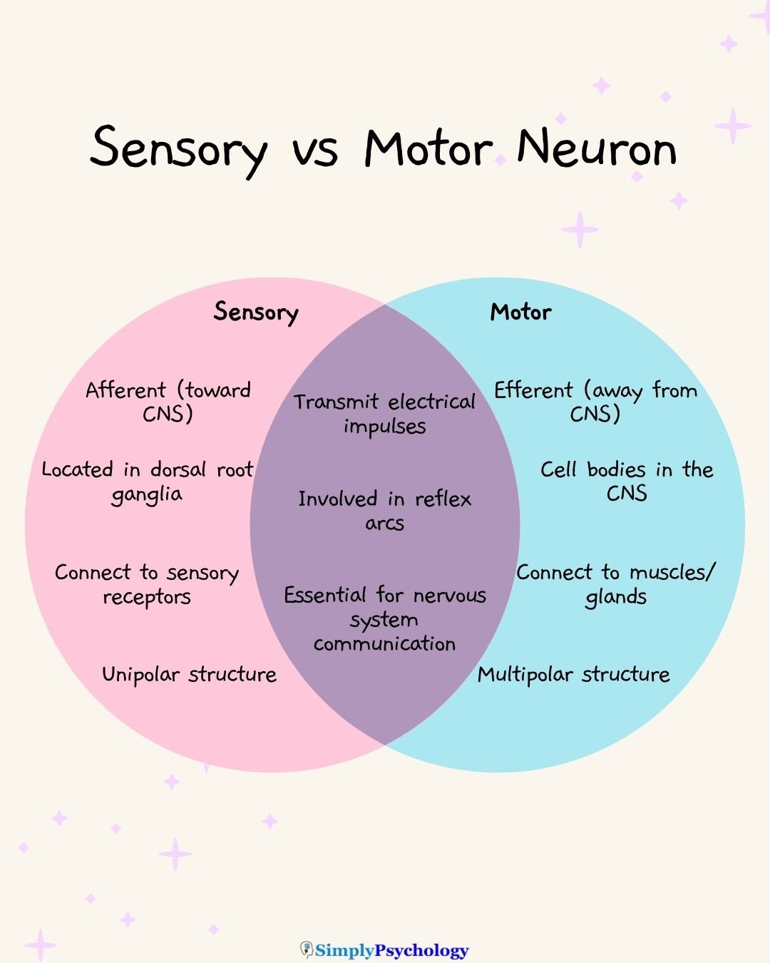
Mnemonic: “SAME DAVE” stands for Sensory Afferent, Motor Efferent; Dorsal Afferent, Ventral Efferent. Sensory neurons are afferent and enter the spinal cord on the dorsal side. Motor neurons are efferent and exit on the ventral side.
What Are Sensory Neurons?
Sensory neurons are specialized nerve cells that carry information from sensory receptors to the central nervous system (CNS).
Sensory neurons respond to three broad kinds of stimulus:
- Physical: touch, temperature, sound and light.
- Chemical: taste and smell.
- Internal: signals such as blood pressure or joint position.
When these impulses reach the brain, they are translated into sensations such as vision, hearing, taste and touch.

You’ll find sensory neurons in receptors located throughout the body: in the skin, eyes, ears, tongue, and internal organs.
Their cell bodies are grouped in clusters called dorsal root ganglia, part of the peripheral nervous system (PNS). From here, their axons enter the CNS. They deliver information for processing.
Importantly, not all sensory input reaches conscious awareness. Some sensory neurons stop at the spinal cord, forming part of a reflex arc that allows for quick, automatic responses.
This setup ensures the body can react rapidly to danger. You can pull your hand from a hot surface before the brain fully registers the pain.
Structurally, most sensory neurons are unipolar, with a single process that splits to connect the sensory receptor and the spinal cord. This design allows for fast, efficient signal transmission.
Do sensory neurons have dendrites? Yes. Sensory neurons typically have long dendrites, the branches that receive signals, and short axons, the fibers that send them.
In short, sensory neurons are the input system of the nervous system, constantly relaying data from the body and environment to the CNS for perception or reflexive action.
What Are Motor Neurons?
Motor neurons are nerve cells that carry commands from the central nervous system (CNS) to muscles or glands, enabling movement and physiological responses.
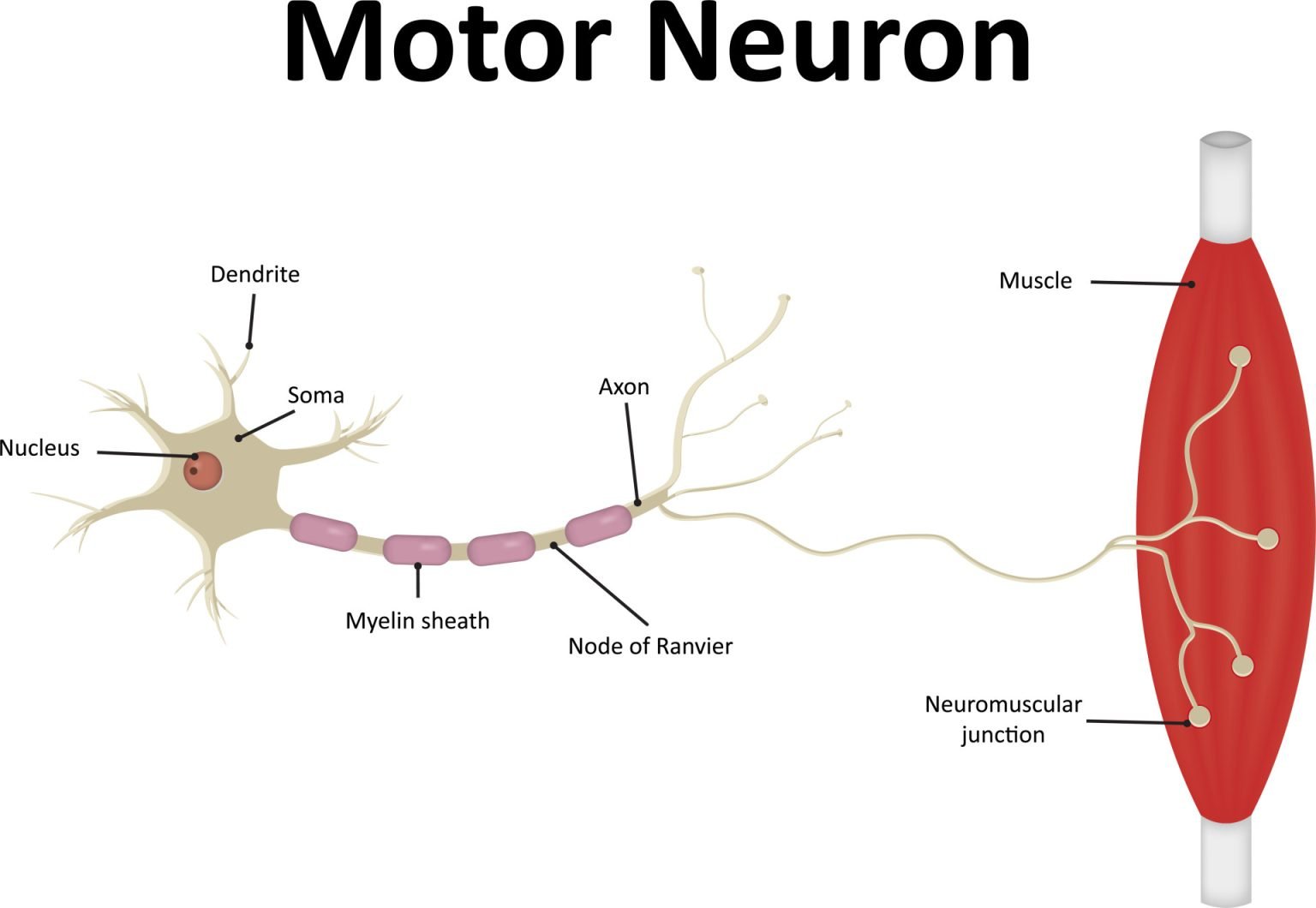
Motor neurons fall into two groups:
- Upper motor neurons: run between the brain and the spinal cord, passing commands to lower motor neurons.
- Lower motor neurons: run from the spinal cord out to the muscles and connect to them directly.
Motor neuron cell bodies sit within the spinal cord or the motor areas of the brain. Their axons then extend into the peripheral nervous system (PNS). There they reach effectors, the tissues that carry out a response: skeletal muscle, smooth muscle and glands.
When activated, motor neurons release neurotransmitters at the neuromuscular junction, the point where nerve meets muscle. The transmitter is acetylcholine. It triggers the muscle to contract.
Toxins show how much depends on this step. Curare blocks acetylcholine at the junction and causes paralysis. Botulinum toxin blocks acetylcholine release, and it is used both cosmetically and therapeutically.
Motor neurons play a key role in both voluntary movements (e.g., walking, speaking) and involuntary reflexes.
For example, pulling your hand away from something hot involves motor neurons. They receive the signal via the spinal cord. Then they immediately activate your arm muscles.
Structurally, they are typically multipolar, with many dendrites for receiving input and a single long axon for sending signals to distant targets. This allows motor neurons to integrate complex information before initiating an action.
In essence, motor neurons are the output system of the nervous system. They turn CNS decisions into action in the body.
How Sensory and Motor Neurons Work Together (Sensory-Motor Integration)
Sensory and motor neurons do not operate in isolation. In fact, they are two halves of a complete circuit.
The classic example of their cooperation is a reflex arc or any simple stimulus-response pathway.
Typically, a sensory neuron will detect a change or stimulus and send a signal into the CNS.
There, the signal is often passed to an interneuron, also known as a relay neuron. The interneuron processes the information. It quickly relays it to a motor neuron, which produces a response.
Interneurons are the third category of neuron, found entirely within the CNS, that serve as the connectors and processors between input and output.
When you touch something hot, a withdrawal reflex pulls your hand away before you consciously feel the pain. The loop runs through the spinal cord rather than the brain:
- Detect: Heat receptors in the skin excite a sensory neuron, which sends an electrical impulse along its axon toward the spinal cord.
- Relay: At the first synapse (the tiny gap between two neurons), the sensory neuron releases glutamate onto an interneuron in the spinal cord’s gray matter.
- Integrate: If the combined input pushes the interneuron past its firing threshold, it fires and excites a motor neuron at the next synapse.
- Act: The motor neuron carries the impulse to the flexor muscle of the arm and releases acetylcholine at the neuromuscular junction, so the muscle contracts.
- Perceive: Only afterwards do pathways carry the signal up to the brain, where the pain is consciously registered.
In total, the signal crosses two synapses, and an interneuron combines the inputs on the way. This quick loop lets the body respond instantly to danger.
Some reflexes, like the knee-jerk reaction, involve a direct connection between sensory and motor neurons for maximum speed.
More commonly, interneurons serve as intermediaries, helping coordinate appropriate responses.
Regardless of complexity, reflexes follow the same basic pattern:
sensory input → central processing → motor output.
This rapid communication between neurons allows the nervous system to protect the body and maintain functional control without conscious effort.
According to Charles Sherrington’s studies of spinal reflexes, signals cross a reflex arc more slowly than they run along an uninterrupted axon. He reasoned that the delay arises where one neuron meets the next. He named that junction the synapse.

