Interneurons (also known as relay neurons) are specialized nerve cells that primarily serve as connectors within the central nervous system (CNS). Their unique role is to facilitate communication between other neurons.
Their main job is to connect other neurons. Usually, they link sensory neurons (which detect stimuli) with motor neurons (which trigger movement). Others form brain networks for complex processing.
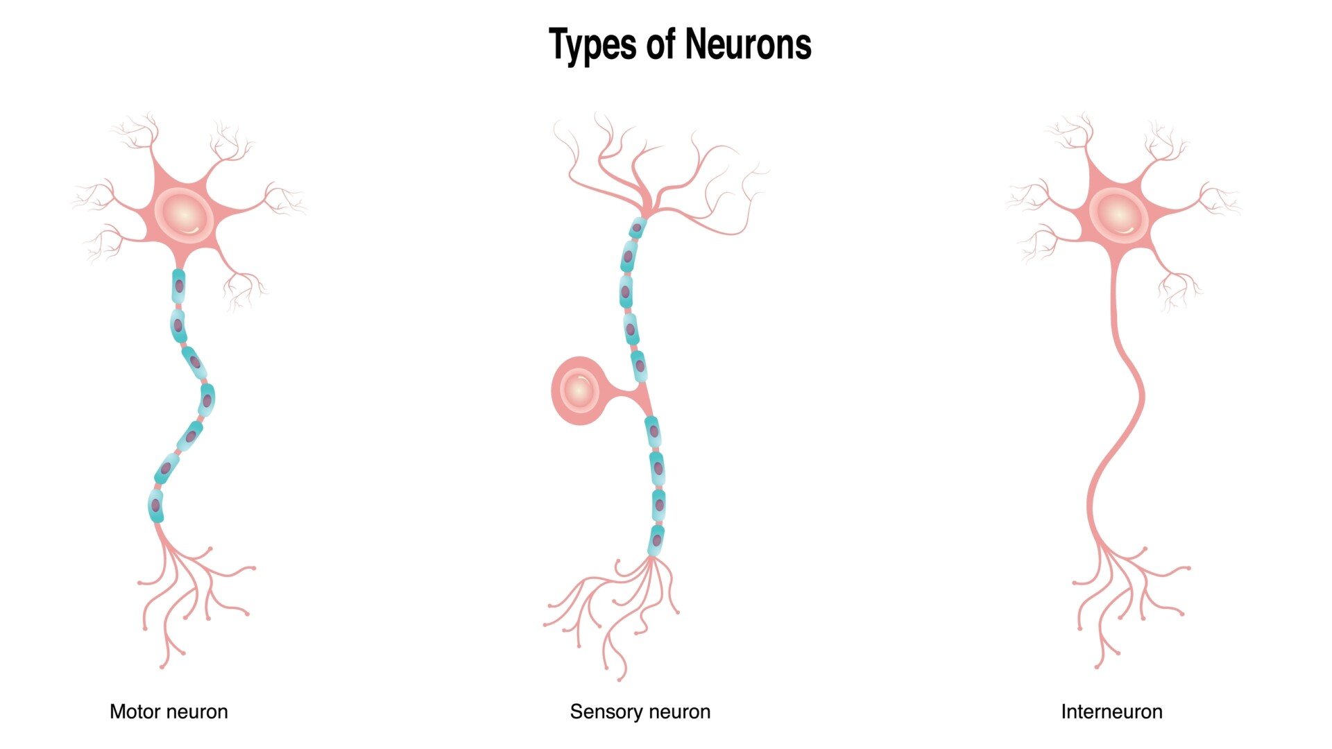
They are incredibly numerous, making up around 20–30% of all neurons in the cerebral cortex. Most of the rest are excitatory pyramidal cells, which project over long distances.
Interneurons keep brain activity balanced and coordinated.
In a spinal reflex, interneurons connect incoming sensory neurons to motor neurons. Sensory neurons bring information into the CNS, and motor neurons send commands out. The response is immediate and automatic, with no direct brain involvement.
Key Takeaways
- Definition: Interneurons connect other neurons entirely within the central nervous system, linking sensory input to motor output and processing information in between.
- Functions: They relay signals in reflexes, integrate information, modulate whether signals excite or inhibit, and support learning, memory and decision-making.
- Inhibition: Most interneurons release GABA or glycine, which inhibit other neurons. This keeps brain activity balanced and prevents overstimulation.
- Classification: Neuroscientists group them by connectivity, neurotransmitter, molecular marker (parvalbumin, somatostatin, VIP) and shape (basket, chandelier, Martinotti).
- Prevalence: Interneurons make up around 20–30% of cortical neurons. Most of the rest are excitatory pyramidal cells.
- Development: They are born in the ganglionic eminences, migrate into the cortex, and help open and close critical periods.
- Clinical Links: Disrupted interneuron function is linked to epilepsy and schizophrenia. Much of the human evidence is correlational.
What do interneurons do?
Below are some of the main functions of interneurons:
Relaying signals
Interneurons are vital for relaying messages, especially in quick, automatic responses like reflex arcs.
For example, in a withdrawal reflex, sensory neurons bring information (e.g., from touching a hot stove) into the spinal cord, where interneurons directly connect them to motor neurons.
This allows immediate action without conscious brain involvement, saving crucial seconds. Not every reflex needs an interneuron, though. The knee-jerk reflex uses a single synapse and none.
Interneurons also coordinate the whole body. When a bare foot steps on something sharp, three things happen in sequence:
- Excitatory interneurons trigger the flexor muscles that lift the injured leg.
- Inhibitory interneurons relax the opposing extensor muscles, so the leg can bend freely.
- A commissural interneuron crosses the spinal cord’s midline and straightens the other leg to take the body’s weight.
This is the crossed-extensor reflex. It needs no instruction from the brain.
As Sherrington (1906) argued, the spinal cord does not merely relay signals. It organises coherent whole-body movement from a purely local stimulus.
Similarly, in the visual system, rods and cones are connected to retinal ganglion cells via interneurons.
Processing information
Within the brain, interneurons are deeply involved in integrating sensory input and higher-level signals.
They process vast streams of information from our senses, emotions, and memories, helping the brain to form coherent thoughts, perceptions, and plans.
This integrative function is fundamental to how we make sense of our environment and interpret complex information.
One way interneurons do this is lateral inhibition. A strongly active neuron uses interneurons to suppress its neighbours, which exaggerates the contrast between active and quiet pathways.
Touch shows the effect. The fingertips tell one touch point from two far better than the back does. Lateral inhibition sharpens the signals from their densely packed receptors.
The retina uses the same trick. Horizontal and amacrine cells, both interneurons, sharpen the boundaries between light and dark regions before the signal reaches the brain.
Modulation
Interneurons play a significant role in modulating neural signals by determining whether a message is excitatory or inhibitory.
An excitatory signal makes a receiving neuron more likely to fire, while an inhibitory signal decreases this likelihood.
The balance is delicate. It is mediated by neurotransmitters such as glutamate (excitatory) and GABA (inhibitory). That allows precise control of brain activity, preventing overstimulation and ensuring appropriate responses.
Cognitive Control
These connector cells are fundamental to higher cognitive functions such as learning, memory, and decision-making.
They coordinate the interactions between neurons that form new memories and support problem-solving. They also underpin executive control, such as planning and judgment, particularly in the frontal and prefrontal cortex.
Two cell types matter here. Fast-spiking parvalbumin interneurons pace gamma-band oscillations, rhythmic activity linked to attention and working memory (Cardin et al., 2009). Vasoactive intestinal peptide (VIP) interneurons inhibit other inhibitory cells. This disinhibition boosts one circuit while leaving others untouched, which helps during attention.
Circuit Motifs
Beyond individual cells, interneurons form recurring wiring patterns called circuit motifs. These turn basic excitation and inhibition into useful computations.
- Feedforward inhibition: An incoming signal excites an interneuron and the target together. Inhibition arrives just after excitation, sharpening response timing and preventing runaway excitation.
- Feedback inhibition: An active neuron excites an interneuron through a branch of its own axon, and that interneuron inhibits it. Spinal Renshaw cells brake motor neuron firing this way (Eccles et al., 1954).
- Lateral inhibition: An active pathway suppresses its neighbours, sharpening contrast, as in two-point touch discrimination and the retina.
- Disinhibition: Inhibiting an inhibitory cell raises activity downstream. VIP interneurons use it to boost one pathway on demand.
Interneuron circuits can even generate rhythm. In the spinal cord, central pattern generators produce the alternating flexor and extensor activity of walking without a fresh brain command for each step.
Two properties combine to do this. In lamprey spinal cord experiments, activating NMDA receptors on interneurons alone produced locomotor-like rhythmic bursting (Wallén & Grillner, 1987). Reciprocal inhibition between flexor and extensor interneuron pools then makes the two alternate when both receive steady excitation.
Types of Interneurons
Interneurons are incredibly diverse. The most basic split is where each one sends its output.
- Local-circuit interneurons: Short axons keep their influence within a small patch of tissue, such as one cortical column or spinal segment.
- Relay (projection) interneurons: Longer axons carry output to distant parts of the CNS, sometimes across the midline to the opposite side of the body.
These schemes overlap rather than compete. One cell can be a GABAergic, parvalbumin-expressing, basket-shaped, local-circuit interneuron. Beyond connectivity, interneurons can also be grouped by:
1. Neurotransmitter Type
Most are GABAergic, meaning they use GABA to inhibit other neurons. GABAergic interneurons are the main source of inhibition in the cerebral cortex. They make their targets less likely to fire.
Interneurons in the spinal cord and brainstem often use glycine, another inhibitory transmitter, to organise reflex circuits.
A smaller group is excitatory, releasing glutamate or acetylcholine. These cells pass signals onward instead of damping them. In a reflex arc, one may relay a sensory signal straight to a motor neuron.
Quick Guide: What Are These Neurotransmitters?
- GABA (Gamma-Aminobutyric Acid): The brain’s main inhibitory neurotransmitter. It helps calm activity and prevent overstimulation.
- Glycine: Another inhibitory neurotransmitter, mainly active in the spinal cord and brainstem.
- Acetylcholine: Often involved in attention and learning. It can excite or inhibit neurons depending on the receptor.
- Glutamate: The brain’s main excitatory neurotransmitter. It plays a key role in learning, memory, and brain development.
2. Molecular Markers
In the cortex, most fall into three major groups:
- Parvalbumin (PV): Fast-spiking cells that tightly control timing. They contact the cell body or axon initial segment, so they decide whether and when targets fire.
- Somatostatin (SST): Cells that target dendrites and regulate incoming signals, filtering each input before the neuron combines them.
- Vasoactive intestinal peptide (VIP): Cells that inhibit other interneurons, mainly SST cells, creating a “disinhibitory” effect that boosts activity in excitatory neurons.
This scheme was consolidated by a consensus paper from more than 40 cortical-circuit researchers (DeFelipe et al., 2013). They argued that no single criterion reliably predicts a cell’s function, so marker, connectivity and firing pattern should be combined.
3. Shape and Structure
Some classic examples:
- Basket cells: Wrap branching axons around the cell bodies of many neighbours, delivering fast, powerful inhibition to a whole local population.
- Chandelier cells: Target axon initial segments, where electrical signals begin, giving them an almost veto-like influence over whether a neuron fires.
- Martinotti cells: Send axons up through the cortical layers to the dendrites of pyramidal neurons in the outermost layer.
Shape and chemistry often line up. Most basket cells express parvalbumin, and Martinotti cells typically express somatostatin. Labels such as “fast-spiking basket cell” and “parvalbumin interneuron” therefore often describe overlapping populations, though the overlap is incomplete (Markram et al., 2004).
Development and Plasticity
Unlike excitatory neurons, which are born in the cortex, interneurons are generated elsewhere, in the ganglionic eminences of the developing forebrain. They then migrate a long way into the cortex.
This sideways journey is called tangential migration: the young cells move parallel to the cortical surface, not radially outward. Marín and Rubenstein (2001) reviewed the chemical signals that guide it. Their account established a developmental programme separate from the one that builds excitatory cortex.
Once in place, they:
- Integrate into circuits by forming synapses with excitatory neurons and other interneurons.
- Mature gradually, often helping open or close critical periods for learning.
Even in adulthood, interneurons remain plastic. Their connections can strengthen or weaken in response to experience, helping the brain adapt to new learning or injury.
Fast-spiking parvalbumin interneurons are one trigger that opens a critical period, a window of heightened plasticity in sensory cortex. Their later maturation helps close it again.
Davis et al. (2015) showed how far adult plasticity can be pushed.
- Aim: To test whether transplanted embryonic interneurons could reopen critical-period plasticity in adult mouse visual cortex. The team also asked whether this reverses amblyopia caused by early visual deprivation.
- Method: Embryonic inhibitory precursors from the medial ganglionic eminence were transplanted into adult mouse visual cortex. Some mice had been made amblyopic by depriving one eye of vision early in life.
- Results: The cells matured and reinstated ocular-dominance plasticity in adults. In amblyopic mice, transplantation recovered visual cortical responses and performance on a behavioural visual acuity test.
- Conclusion: Adding interneurons at the right developmental stage can reopen plasticity in adulthood and reverse an established sensory impairment.
This is a rare causal demonstration, but it used an invasive procedure in mice, so human treatment remains distant.
Interneurons and Mental Health
Disruption in interneuron function can lead to circuit imbalances linked to several psychological and neurological conditions:
- Schizophrenia: Reduced activity in parvalbumin (PV) interneurons, especially in the prefrontal cortex, is associated with impaired cognitive function and disrupted gamma oscillations. These deficits may contribute to issues with attention and working memory.
- Autism Spectrum Disorder (ASD): Researchers propose that reduced GABA signaling involving interneurons contributes to the sensory overload some autistic people report. This is one contributing thread, not a full explanation.
- Epilepsy: Interneuron loss or dysfunction reduces inhibition in seizure-prone areas, leading to hyperexcitability.
- Other Disorders: Interneurons are also implicated in anxiety, depression, Alzheimer’s disease, and Parkinson’s disease. In these conditions, disrupted inhibitory balance may affect fear regulation, mood, memory, and motor control.
Because interneurons are key to neural balance and coordination, they are central to research on brain disorders and treatment development.
Interneurons vs Sensory and Motor Neurons
Below is how to know the difference between these types of neurons:
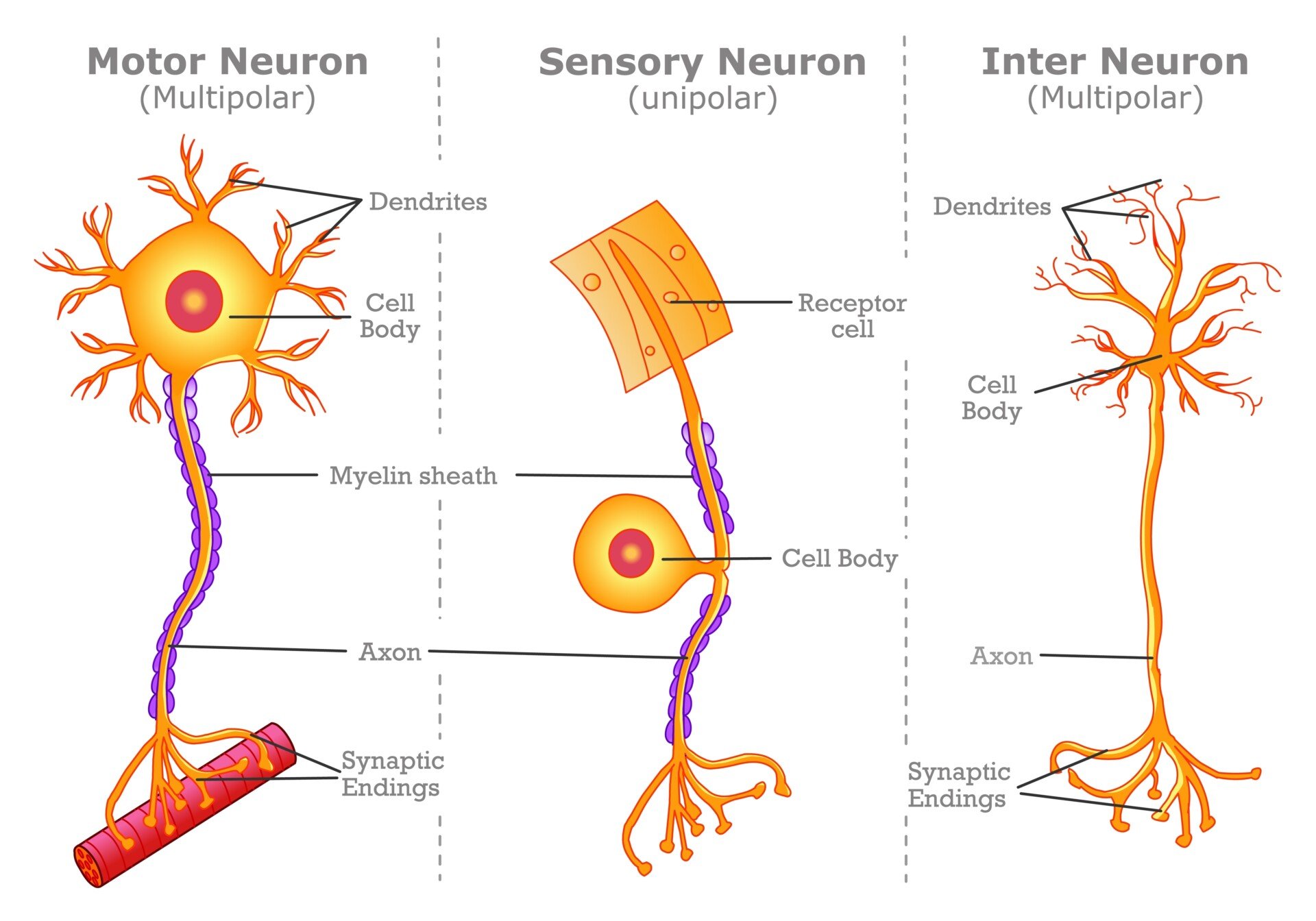
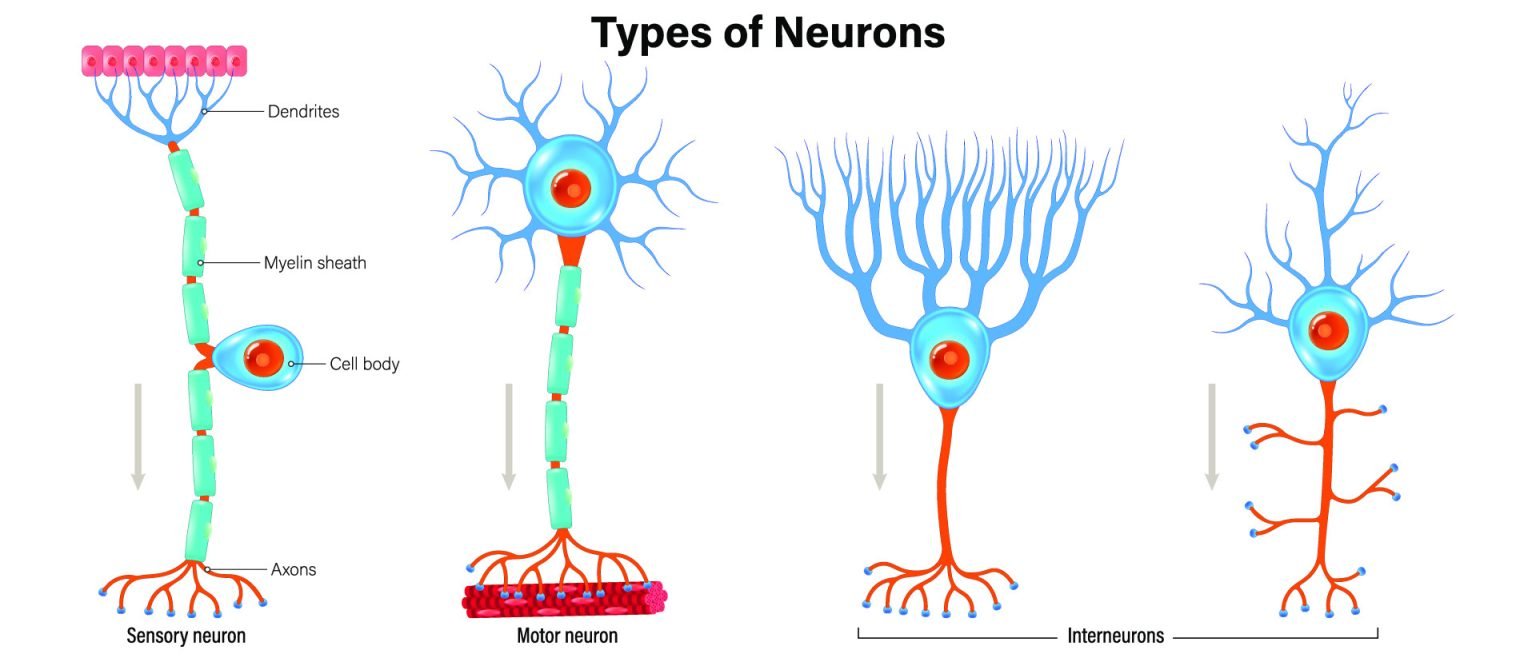
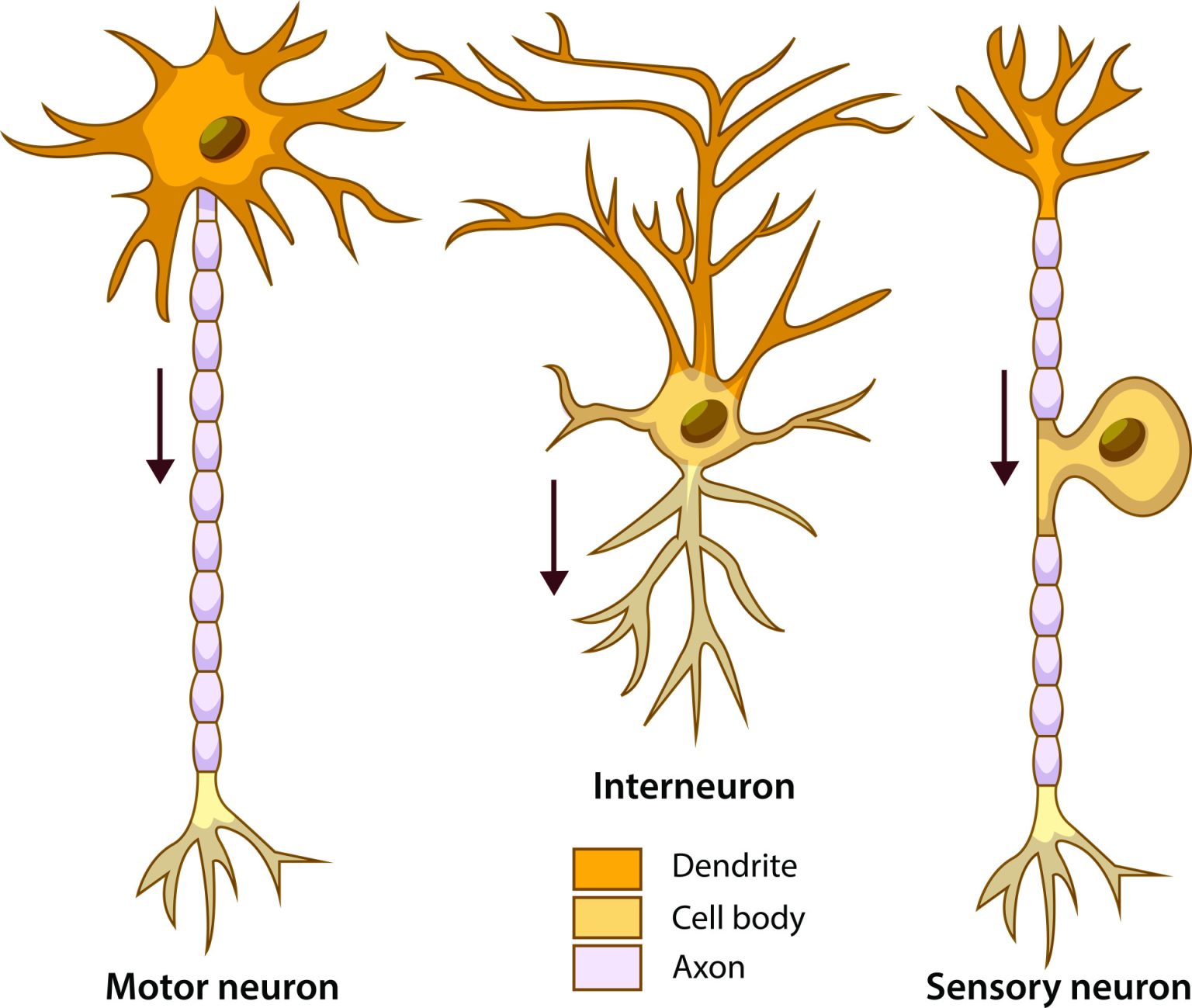
- Sensory Neurons (Afferent): These neurons carry sensory information from the body’s periphery (like receptor cells in our five senses) to the central nervous system (CNS), which includes the brain and spinal cord. They “arrive” with information.
- Motor Neurons (Efferent): These neurons transmit commands from the CNS to the muscles and glands, dictating how the body should respond. They cause an “exit” of commands.
- Interneurons: These are primarily found within the CNS (brain and spinal cord) and act as connectors. They link sensory neurons to motor neurons, and facilitate communication between other neurons for processing and modulation of signals.
Each type plays a distinct role in the nervous system’s communication network.
Interneurons go by several names. They are also called connector neurons and association neurons, and UK exam specifications often call them relay neurons. Do not confuse this with relay interneurons, a narrower subtype whose long axons project to distant parts of the CNS.

Critical Evaluation of Interneuron Research
The strongest human evidence on interneurons comes from schizophrenia research, and each type of evidence has its own limits.
Contemporary Research
Fewer Parvalbumin Cells in Schizophrenia
Kaar et al. (2019) pooled the available postmortem data to resolve conflicting individual findings.
- Aim: To resolve conflicting postmortem findings on parvalbumin interneuron density and parvalbumin gene expression (mRNA) in the prefrontal cortex in schizophrenia.
- Method: A meta-analysis pooled 274 people with schizophrenia and 275 comparison subjects. Hedges’ g was calculated separately for cell density and mRNA, with postmortem interval and publication year tested as covariates.
- Results: Parvalbumin cell density was reduced in frontal cortex (g = −0.27, p = .03). The mRNA reduction (g = −0.44) was not significant (p = .12).
- Conclusion: The data tentatively support a deficient GABAergic system in schizophrenia, though further region- and layer-specific studies are needed.
The result is mixed. The authors report it openly. Postmortem interval affected the mRNA estimate, so that measure is less secure.
Chung et al. (2016) looked for a cellular explanation. Using fluorescent immunohistochemistry and confocal microscopy on postmortem prefrontal cortex, they found that parvalbumin interneurons in schizophrenia carried 18% fewer excitatory synaptic inputs. The deficit was absent in monkeys given antipsychotic medication long term.
Pacing Gamma Rhythms
Why should a parvalbumin deficit matter for thinking? Cardin et al. (2009) used optogenetics, which switches genetically targeted cells on with light, to drive fast-spiking interneurons in mouse cortex. Doing so amplified gamma oscillations and sharpened the cortex’s response to sensory input. Activating pyramidal neurons did not.
Gamma disruption is a well-documented feature of schizophrenia. A deficit in the cells that pace the rhythm is therefore a compelling candidate mechanism for its cognitive symptoms.
Mapping Interneuron Subtypes
The newest work resolves interneurons into finer subtypes. Enwright et al. (2026) used single-nucleus RNA sequencing on postmortem prefrontal cortex from 19 people with no known psychiatric disorder.
The result is a detailed map. It holds 37 transcriptionally distinct subtypes across five major classes, including 11 somatostatin and 9 parvalbumin subtypes. Chandelier cells proved distinct from parvalbumin basket cells at the level of gene expression.
The authors offer the work as a reference map for future schizophrenia research. It is a starting point. They do not claim that any one subtype is yet the source of dysfunction.
Limitations of the Evidence
Four limits temper how far these findings can be pushed.
- Simplified Scheme: “Interneuron” covers cells that differ widely in shape, transmitter and firing, and some types resist clean classification (DeFelipe et al., 2013; Markram et al., 2004).
- Postmortem Correlation: Postmortem studies compare brains after death, often after years of antipsychotic exposure. They cannot show that interneuron loss causes symptoms.
- Animal Models: Cardin et al. (2009) and Davis et al. (2015) used invasive techniques in mice. Whether the same effects occur in the human cortex remains an inference.
- Static Snapshots: Postmortem tissue cannot separate a developmental deficit from the effects of illness, medication or the dying process.
Progress is most likely to come from pairing single-cell profiling of interneuron subtypes with recordings from living human tissue, for example during clinically necessary epilepsy surgery.
References
Cardin, J. A., Carlén, M., Meletis, K., Knoblich, U., Zhang, F., Deisseroth, K., Tsai, L.-H., & Moore, C. I. (2009). Driving fast-spiking cells induces gamma rhythm and controls sensory responses. Nature, 459(7247), 663–667. https://doi.org/10.1038/nature08002
Chung, D. W., Fish, K. N., & Lewis, D. A. (2016). Pathological basis for deficient excitatory drive to cortical parvalbumin interneurons in schizophrenia. American Journal of Psychiatry, 173(11), 1131–1139. https://doi.org/10.1176/appi.ajp.2016.16010025
Davis, M. F., Figueroa Velez, D. X., Guevarra, R. P., Yang, M. C., Habeeb, M., Carathedathu, M. C., & Gandhi, S. P. (2015). Inhibitory neuron transplantation into adult visual cortex creates a new critical period that rescues impaired vision. Neuron, 86(4), 1055–1066. https://doi.org/10.1016/j.neuron.2015.03.062
DeFelipe, J., López-Cruz, P. L., Benavides-Piccione, R., Bielza, C., Larrañaga, P., Anderson, S. A., Burkhalter, A., Cauli, B., Fairén, A., Feldmeyer, D., Fishell, G., Fitzpatrick, D., Freund, T. F., González-Burgos, G., Hestrin, S., Hill, S., Hof, P. R., Huang, Z. J., Jones, E. G., … Ascoli, G. A. (2013). New insights into the classification and nomenclature of cortical GABAergic interneurons. Nature Reviews Neuroscience, 14(3), 202–216. https://doi.org/10.1038/nrn3444
Eccles, J. C., Fatt, P., & Koketsu, K. (1954). Cholinergic and inhibitory synapses in a pathway from motor-axon collaterals to motoneurones. The Journal of Physiology, 126(3), 524–562. https://doi.org/10.1113/jphysiol.1954.sp005226
Enwright, J. F., Tamburino, A. M., Tumkaya, T., Lovatt, D., Pan, J., Gunaratna, R., Fagegaltier, D., Wang, X., Marino, M. J., Fish, K., Arion, D., Gonzalez-Burgos, G., & Lewis, D. A. (2026). Transcriptional profiles of somatostatin and parvalbumin interneuron subtypes in the human dorsolateral prefrontal cortex: Implications for schizophrenia. American Journal of Psychiatry, 183(8), 576–591. https://doi.org/10.1176/appi.ajp.20250355
Kaar, S. J., Angelescu, I., Reis Marques, T., & Howes, O. D. (2019). Pre-frontal parvalbumin interneurons in schizophrenia: A meta-analysis of post-mortem studies. Journal of Neural Transmission, 126(12), 1637–1651. https://doi.org/10.1007/s00702-019-02080-2
Marín, O., & Rubenstein, J. L. R. (2001). A long, remarkable journey: Tangential migration in the telencephalon. Nature Reviews Neuroscience, 2(11), 780–790. https://doi.org/10.1038/35097509
Markram, H., Toledo-Rodriguez, M., Wang, Y., Gupta, A., Silberberg, G., & Wu, C. (2004). Interneurons of the neocortical inhibitory system. Nature Reviews Neuroscience, 5(10), 793–807. https://doi.org/10.1038/nrn1519
Sherrington, C. S. (1906). The integrative action of the nervous system. Constable.
Wallén, P., & Grillner, S. (1987). N-methyl-D-aspartate receptor-induced, inherent oscillatory activity in neurons active during fictive locomotion in the lamprey. The Journal of Neuroscience, 7(9), 2745–2755. https://doi.org/10.1523/jneurosci.07-09-02745.1987

