The autonomic nervous system (ANS) is part of the peripheral nervous system. It automatically regulates the body’s internal organs, including heart rate, digestion, breathing, and pupil size.
It has three divisions: the sympathetic (“fight or flight”), the parasympathetic (“rest and digest”), and the enteric system, which governs the gut. Together they maintain homeostasis, the body’s stable internal balance.

The autonomic nervous system differs from the somatic nervous system (another branch of the peripheral nervous system), which is associated with controlling voluntary body movements.
Although most of the functions of the ANS are automatic, they can, however, work in conjunction with the somatic nervous system.
Divisions of the Autonomic Nervous System
There are three branches to the ANS; the sympathetic nervous system, the parasympathetic nervous system, and the enteric nervous system:
- Sympathetic: The “fight-or-flight” system; mobilizes energy, increases heart rate, and redirects blood to muscles during stress.
- Parasympathetic: The “rest-and-digest” system; conserves energy, slows heart rate, and promotes digestion during recovery.
- Enteric: The “second brain”; an independent network of neurons embedded in the gastrointestinal tract that manages digestion and nutrient absorption.

Sympathetic Nervous System
The sympathetic nervous system is involved in responses that help us deal with stress.
It slows bodily processes that are less important in emergencies, such as digestion.
For instance, if the temperature of a room is too hot, the sympathetic system will encourage the body to sweat in response to this change.
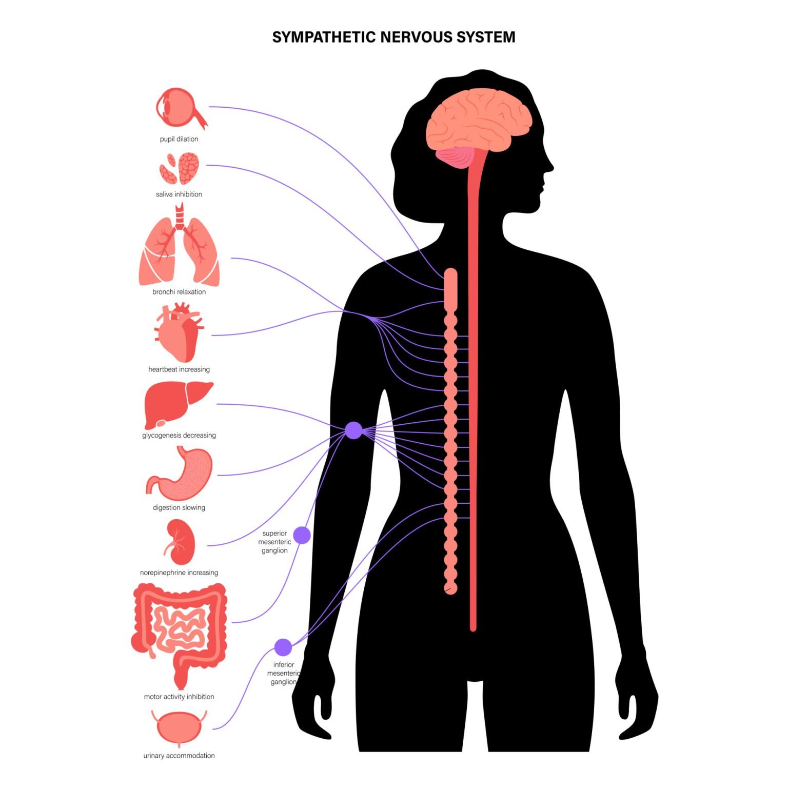
When activated, the SNS triggers a cascade of physiological changes:
- Heart and Lungs: Increases heart rate and blood pressure; dilates bronchi to increase air intake.
- Vision: Dilates pupils to enhance vision.
- Digestion: Inhibits non-vital functions such as gastrointestinal activity and salivation (causing a dry mouth).
- Energy Mobilisation: Stimulates the liver to release glucose and the adrenal glands to release adrenaline (epinephrine), providing energy for immediate action.
The primary neurotransmitter used by the SNS at the target organ is norepinephrine (noradrenaline)
Fight or Flight Response
The most noticeable function of the sympathetic branch is during the fight-or-flight response.
The sympathetic system activates and releases epinephrine (adrenaline) during threatening or stressful conditions, providing an automatic response.
Sarah walks home alone at night. She hears footsteps behind her. Her heart races, pupils dilate, and she starts to sweat. This is her sympathetic nervous system preparing her to react: either to flee or defend herself.
Aim: Cannon asked whether fear and rage trigger one coordinated bodily reaction.
Method: Cannon provoked fear and rage in cats and dogs. He then used surgery and drugs (cutting sympathetic nerves, removing adrenal glands, injecting adrenaline) to isolate the autonomic changes.
Results: The same stereotyped changes (racing heart, mobilized blood sugar, halted digestion) appeared across different threats and species. Denervating an organ or removing the adrenal glands abolished its part in the reaction. The pattern still appeared in calm animals given adrenaline alone.
Conclusion: Fear and rage provoke one generalized sympathetic-adrenal mobilization, the fight-or-flight response, which restores homeostasis once the emergency passes (Cannon, 1929, 1932).
This is the founding demonstration linking the sympathetic nervous system to emotional arousal. Its evidence base, though, is entirely non-human.
This fast adrenaline surge is only half the response. In parallel, the hypothalamus triggers the pituitary gland, which signals the adrenal cortex to release cortisol. This second pathway takes minutes rather than seconds, but it sustains the body’s readiness for longer.
Selye’s General Adaptation Syndrome (1956) describes this two-stage pattern: the fast adrenaline surge is the alarm reaction, and the slower cortisol response is the resistance stage that follows.
Although the sympathetic nervous system was evolutionarily used in life-threatening situations, modern-day life, such as work stressors and relationship problems, can also trigger this response.
Similarly, people with anxiety disorders and phobias can show the same autonomic responses to situations that pose no real threat.
Parasympathetic Nervous System
The parasympathetic nervous system acts to calm the body and conserve energy, often called the rest-and-digest system. It becomes dominant once an emergency has passed: pupils constrict, heart rate returns to its resting rhythm, and sweating stops.
Without it, the body would stay constantly alert, draining energy and driving chronic stress. Neither system, in fact, is ever fully switched off.
In practice, both systems are active to some degree at all times. An organ’s resting state reflects the ratio of sympathetic to parasympathetic drive, not one system simply switching the other off.
Anatomically, it is known as the Craniosacral system, as its preganglionic fibres emerge with cranial nerves (III, VII, IX, and X) and from the sacral spinal cord.
The primary neurotransmitter for the parasympathetic system is acetylcholine

Sympathetic vs. Parasympathetic Nervous System
| Organ/System | Sympathetic Nervous System (“Fight or Flight”) | Parasympathetic Nervous System (“Rest and Digest”) |
|---|---|---|
| Heart | Increases heart rate | Decreases heart rate |
| Lungs | Dilates bronchi (more air in) | Constricts bronchi (returns to normal) |
| Pupils | Dilates pupils (better vision in danger) | Constricts pupils (normal vision) |
| Digestive System | Slows digestion | Stimulates digestion |
| Bladder | Relaxes bladder (inhibits urination) | Contracts bladder (promotes urination) |
| Salivary Glands | Inhibits saliva production | Stimulates saliva production |
| Sweat Glands | Activates sweating | No significant effect |
| Liver | Stimulates glucose release | Promotes glucose storage |
| Adrenal Glands | Stimulates adrenaline release | No direct stimulation |
| Reproductive Organs | Decreases function | Stimulates arousal |
Enteric Nervous System
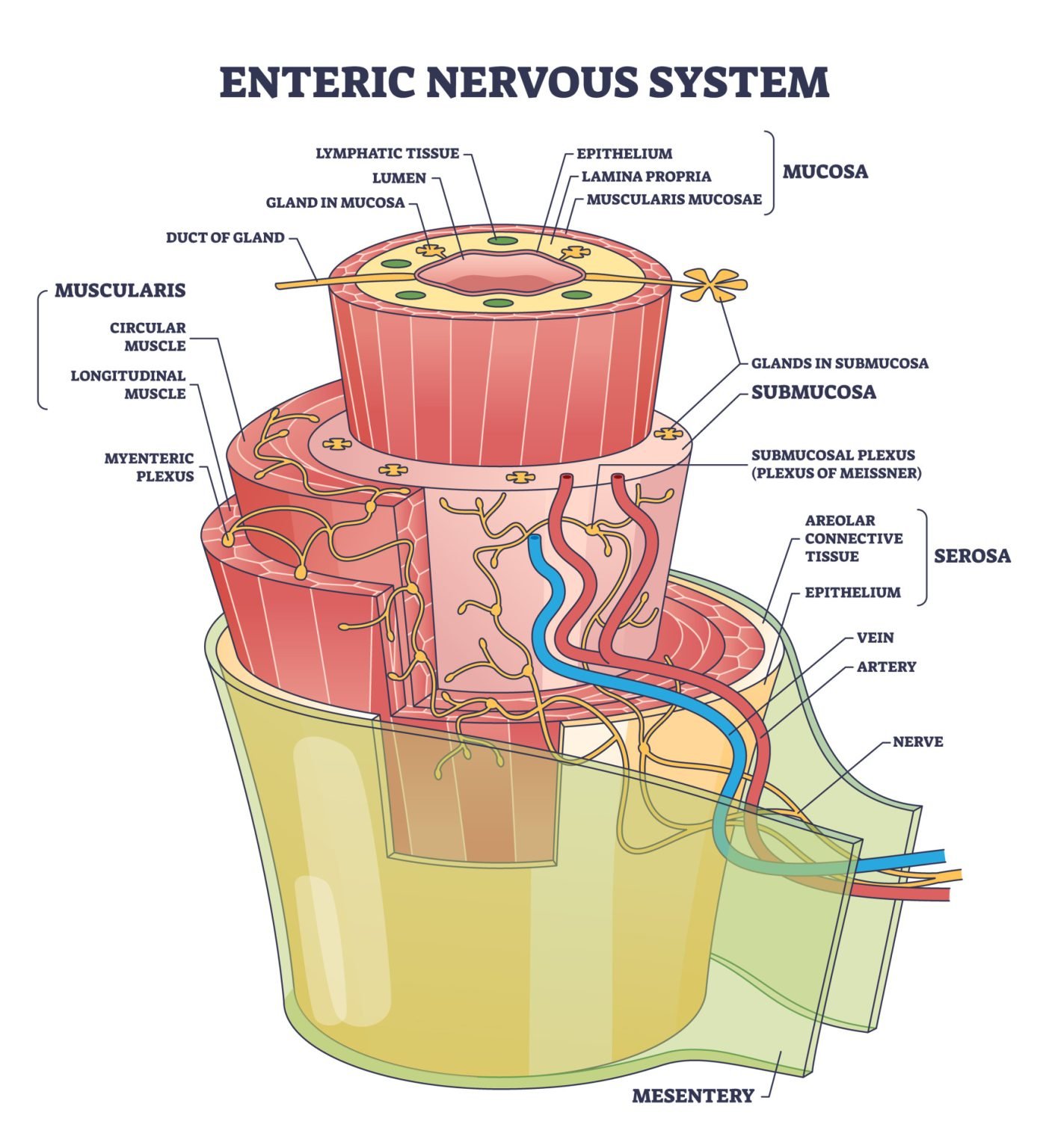
The Enteric Nervous System (ENS) is a specialized branch of the Autonomic Nervous System (ANS) that governs the gastrointestinal (GI) tract.
It is unique because it can operate autonomously, meaning it can manage digestion even if its connection to the brain is severed.
Key Biological Features
- Independent Circuitry: While it interacts with the Sympathetic and Parasympathetic branches, the ENS contains its own complete reflex arcs that allow it to function without input from the Central Nervous System (CNS).
- Anatomical Location: It consists of a vast network of roughly 200–600 million neurons embedded directly within the walls of the gastrointestinal tract, stretching from the esophagus to the anus (Gershon, 1998).
- Complex Neurochemistry: The ENS uses over 30 neurotransmitters identical to those found in the brain, including acetylcholine, dopamine, and serotonin. Notably, about 95% of the body’s serotonin is found in the gut.
Primary Functions
- Blood Flow & Defense: The submucosal (Meissner’s) plexus modulates local blood flow and secretion, and regulates immune responses to protect against ingested pathogens.
- Motility: The myenteric (Auerbach’s) plexus drives peristalsis, the rhythmic muscle contractions that move food through the digestive tract.
- Secretion: Controlling the release of digestive enzymes, acids, and mucus necessary for breaking down food.
The ENS is bidirectional. It doesn’t just take orders from the brain; it sends a massive amount of sensory information back to the CNS via the vagus nerve. This is the body’s longest cranial nerve.
This explains why emotional states (like anxiety) can immediately affect digestive comfort (“butterflies” in the stomach).
Neurotransmitters
The autonomic nervous system (ANS) relies on neurotransmitters to regulate involuntary functions such as heart rate, digestion, and respiration.
Three primary chemicals dictate whether an organ is stimulated or inhibited:
- Acetylcholine (ACh): The universal “start” signal at the first synapse (ganglion) for both branches. It is also the primary inhibitory signal for the heart in the parasympathetic branch.
- Norepinephrine (Noradrenaline): The primary excitatory signal for the sympathetic branch at the target organ.
- Epinephrine (Adrenaline): Released as a hormone into the blood to create a widespread, long-lasting “full body” stress response.
| ANS Branch | First Synapse (Ganglion) | Target Organ Synapse | Main Biological Effect |
| Sympathetic | Acetylcholine | Norepinephrine | Increases Heart Rate; Dilates Bronchioles |
| Parasympathetic | Acetylcholine | Acetylcholine | Decreases Heart Rate; Stimulates Digestion |
Receptors: Why the Same Neurotransmitter Has Different Effects
Acetylcholine can excite one organ and calm another because its effect depends on the receptor it binds, not the molecule itself. At every autonomic ganglion, it acts on fast nicotinic receptors that reliably fire the next neuron.
At parasympathetic target organs, acetylcholine instead binds slower muscarinic receptors. Noradrenaline acts on adrenergic receptors at most sympathetic targets.
This receptor map is where many drugs work. Beta-blockers occupy cardiac β₁ receptors to slow a stress-driven heart, while anticholinergic drugs block muscarinic receptors to reduce parasympathetic effects such as salivation.
The Sympathetic “Fight or Flight” Circuit
The sympathetic nervous system is designed for speed and wide distribution.
- Stage 1 (Preganglionic): Short neurons exit the spinal cord and release Acetylcholine at a ganglion.
- Stage 2 (Postganglionic): Long neurons travel to the heart, lungs, or muscles and release Norepinephrine.
- The Adrenal Boost: The system triggers the adrenal medulla, the inner part of the adrenal gland, to release Epinephrine and Norepinephrine into the blood. This hormonal “rush” lasts longer than a simple nerve impulse.
The Parasympathetic “Rest and Recover” Circuit
The parasympathetic system is more localized and uses only one type of neurotransmitter.
- Stage 1 (Preganglionic): Long neurons exit the brainstem (Vagus nerve) or lower spine and release Acetylcholine.
- Stage 2 (Postganglionic): Short neurons right next to the organ release Acetylcholine again.
- Cholinergic Fibers: Because both stages use ACh, these fibers are called cholinergic.
Dysfunction of the Autonomic Nervous System
ANS Dysfunction (or dysautonomia) is the failure of the involuntary systems to maintain homeostasis.
It occurs when the delicate balance between mobilization (Sympathetic) and restoration (Parasympathetic) is disrupted, often leading to a high allostatic load: the “wear and tear” on the body.
The ANS and Emotional Stress
Chronic stress, anxiety, and trauma can disrupt normal autonomic regulation by overactivating the sympathetic nervous system. This keeps the body in a prolonged “fight or flight” state, raising heart rate and cortisol while impairing digestion and sleep.
One reason the response lingers is psychological, not physical. Simply thinking about a past threat, or imagining a future one, can keep the sympathetic system switched on with no real danger present. Researchers call this perseverative cognition (Brosschot et al., 2006).
Over time, this imbalance can lead to exhaustion and health issues.
Researchers believe an overactive autonomic nervous system may be the root cause of panic attacks. Poor regulation of the brain’s locus coeruleus, which triggers the fight-or-flight response, may overstimulate the limbic system, leading to chronic anxiety and recurrent panic attacks.
ANS Development and Aging
The autonomic nervous system develops gradually after birth and becomes more stable into later childhood and into adulthood.
However, with aging, the ANS may become less responsive.
Older adults often show reduced heart rate variability, slower pupillary responses, and impaired thermoregulation, making them more vulnerable to fainting, temperature extremes, and stress-related health issues.
These changes reflect a natural decline in autonomic flexibility, which can also affect resilience to illness and recovery.
Autonomic Neuropathy and Other ANS Disorders
Autonomic neuropathy refers to the damage of autonomic nerves. These are disorders that can affect the sympathetic nerves, parasympathetic nerves, or both.
The features of autonomic neuropathy include having a fixed heart rate, constipation, abnormal sweating, decreased pupil size, and absent or delayed light reflexes (Bankenahally & Krovvidi, 2016).
There are a number of other disorders which can be the result of ANS dysfunction:
- Acute autonomic paralysis – associated with spinal cord injury, resulting in acute and uncontrolled hypertension.
- Multiple system atrophy – a rare condition that causes gradual damage to the nerve cells.
- Pure autonomic failure – dysfunction of many processes controlled by the ANS.
- Postural orthostatic tachycardia syndrome (POTS) – standing triggers an excessive sympathetic heart-rate rise, causing dizziness, fainting, and fatigue.
- Familial dysautonomia – also known as Riley-Day syndrome. This is an inherited condition where the nerve fibers do not function properly, so these individuals have trouble feeling pain, temperature, pressure, and positioning their arms and legs.
Taking Care of the Autonomic Nervous System
While many autonomic processes happen automatically, lifestyle choices can influence how well the system functions.
- Manage stress: Chronic stress overactivates the sympathetic system and disrupts autonomic balance.
- Practice relaxation techniques: Deep breathing, mindfulness meditation, and yoga activate the parasympathetic system and promote calm.
- Exercise regularly: Physical activity improves heart rate variability and supports overall autonomic tone (the resting balance between the sympathetic and parasympathetic systems).
- Support gut health: Good sleep, hydration, and a fiber-rich diet support the enteric nervous system.
- Limit stimulants: Reducing caffeine and alcohol may help those prone to sensitivity or dysregulation.
- Seek professional support: Biofeedback, vagal nerve stimulation, or therapy can help manage anxiety or trauma-related ANS imbalance.
Taking care of the autonomic nervous system means maintaining habits that support physical and emotional resilience.
Critical Evaluation of the Autonomic Nervous System Model
The sympathetic-parasympathetic model captures a real psychological truth: much of stress and emotion has a physical basis in autonomic arousal. It maps cleanly onto anatomy, neurotransmitters, and clinical practice, and Cannon’s fight-or-flight account remains a well-supported description of the acute stress response.
Contemporary psychophysiology still draws on this basic framework to study stress, emotion regulation, and clinical disorders.
The model has real limitations, though, worth weighing against its strengths.
- Oversimplified opposition: The two branches are not always strict opposites. Some organs receive input from only one branch, and some responses need both systems working together.
- “Automatic” is misleading: Breathing, biofeedback, and relaxation training can voluntarily shift autonomic activity, so the system is not as fixed as its name implies.
- Individual variation: Autonomic tone differs between people and by sex. The classic model was based mainly on male samples; females may show an additional “tend-and-befriend” response (Taylor et al., 2000).
- Correlation, not causation: Autonomic arousal reliably accompanies emotion, but the same bodily pattern doesn’t reveal which emotion is present, or prove that arousal causes it.
- Animal-model evidence: Cannon’s and Selye’s foundational studies used cats, dogs, and rats, not humans. Neither alone shows how closely animal autonomic physiology maps onto the more cognitively mediated human stress response.
- Inconsistent measurement: Much of the modern evidence rests on heart-rate variability. But studies differ in which index, recording length, and posture they use, and these choices change the result (Laborde et al., 2017).
Contemporary Research
Research since 2015 has extended this picture, from how heart-rate variability indexes self-regulation to whether the ANS itself can be directly targeted for treatment.
Heart-Rate Variability and Self-Regulation
Heart-rate variability (HRV), the beat-to-beat variation in heart rate, is a widely studied marker of parasympathetic function.
A meta-analysis pooling 123 studies and over 14,000 participants tested whether HRV predicts self-regulation.
It found that higher HRV was reliably, though only modestly, linked to better emotion regulation and effortful control (Holzman & Bridgett, 2017).
Age moderated the strength of the link.
A 2025 study of 70 university students found a similar pattern: HRV measured early in a semester predicted lower test anxiety by its end (Grabo et al., 2025).
That is one small, not-yet-replicated study.
ANS Dysregulation and Mental Health
A second strand asks whether blunted autonomic flexibility marks psychopathology itself.
One meta-analysis screened over 3,600 reports and retained 37 studies covering 2,347 participants.
It found a small but real link between reduced heart-rate variability and psychological difficulties, strongest for externalizing problems such as conduct difficulties (Beauchaine et al., 2019).
Women showed greater reactivity than men.
Consumer wearables now put an HRV score in front of millions of users.
But bench-testing seven popular devices against a medical-grade ECG found errors ranging from about 4% to over 100% (Stone et al., 2021).
Targeting the Vagus Nerve
Vagus-nerve stimulation (VNS), delivered surgically or through the ear, is an approved treatment for some epilepsy and treatment-resistant depression.
Researchers are now investigating it as a way to boost parasympathetic tone in anxiety and inflammation.
Porges’ polyvagal theory proposes that the vagus nerve has two branches: a newer branch supporting calm social engagement, and an older branch driving shutdown or freezing.
The theory is influential in trauma therapy.
But a detailed re-examination of the comparative-anatomy evidence disputes its central claim that the two branches act independently (Grossman, 2023).
It is best treated as a framework, not an established fact.
Together, these findings do not undermine the ANS framework. They refine it.
The sympathetic and parasympathetic systems remain psychology’s best-supported map of the body’s stress and recovery machinery. New research adds nuance to how automatic, universal, and easily measured it really is.
Which division of the autonomic nervous system returns the body to a relaxed condition after an emergency?
The parasympathetic division of the autonomic nervous system is responsible for returning the body to a relaxed and restorative state after an emergency or stress.
It counteracts the effects of the sympathetic division, which initiates the “fight or flight” response during emergencies. The parasympathetic system promotes “rest and digest” functions, restoring balance and conserving energy.
Which division of the ans can function independently without being stimulated by the central nervous system?
The enteric division of the autonomic nervous system (ANS) can function independently without being stimulated by the central nervous system. It primarily manages the functions of the gastrointestinal tract, including digestion and motility, and can operate autonomously but also communicates with the central nervous system.
Which division of the autonomic nervous system prepares the body for action in a stressful situation?
The sympathetic division of the autonomic nervous system prepares the body for action in stressful situations, often referred to as the “fight or flight” response. It increases heart rate, dilates airways, and redirects blood flow to muscles, among other responses, to ready the body for immediate action.
Related articles:
Neurotransmitters: Types, Function and Examples
Peripheral Nervous System (PNS): Parts and Function
Sympathetic Nervous System: Functions & Examples
Parasympathetic Nervous System (PSNS) Functions & Division

References
Bankenahally, R., & Krovvidi, H. (2016). Autonomic nervous system: anatomy, physiology, and relevance in anaesthesia and critical care medicine. BJA Education, 16(11), 381–387. https://doi.org/10.1093/bjaed/mkw011
Beauchaine, T. P., Bell, Z., Knapton, E., McDonough-Caplan, H., Shader, T., & Zisner, A. (2019). Respiratory sinus arrhythmia reactivity across empirically based structural dimensions of psychopathology: A meta-analysis. Psychophysiology, 56(5), Article e13329. https://doi.org/10.1111/psyp.13329
Brosschot, J. F., Gerin, W., & Thayer, J. F. (2006). The perseverative cognition hypothesis: A review of worry, prolonged stress-related physiological activation, and health. Journal of Psychosomatic Research, 60(2), 113–124. https://doi.org/10.1016/j.jpsychores.2005.06.074
Cannon, W. B. (1929). Bodily changes in pain, hunger, fear and rage: An account of recent researches into the function of emotional excitement (2nd ed.). Appleton.
Cannon, W. B. (1932). The wisdom of the body. W. W. Norton.
Gershon, M. D. (1998). The second brain: A groundbreaking new understanding of nervous disorders of the stomach and intestine. HarperCollins.
Grabo, L. M., Schulz, A., & Bellingrath, S. (2025). Vagally-mediated heart rate variability longitudinally predicts test anxiety in university students. Anxiety, Stress, & Coping, 38(4), 409–422. https://doi.org/10.1080/10615806.2025.2460230
Grossman, P. (2023). Fundamental challenges and likely refutations of the five basic premises of the polyvagal theory. Biological Psychology, 180, Article 108589. https://doi.org/10.1016/j.biopsycho.2023.108589
Holzman, J. B., & Bridgett, D. J. (2017). Heart rate variability indices as bio-markers of top-down self-regulatory mechanisms: A meta-analytic review. Neuroscience & Biobehavioral Reviews, 74, 233–255. https://doi.org/10.1016/j.neubiorev.2016.12.032
Laborde, S., Mosley, E., & Thayer, J. F. (2017). Heart rate variability and cardiac vagal tone in psychophysiological research: Recommendations for experiment planning, data analysis, and data reporting. Frontiers in Psychology, 8, Article 213. https://doi.org/10.3389/fpsyg.2017.00213
Selye, H. (1956). The stress of life. McGraw-Hill.
Stone, J. D., Ulman, H. K., Tran, K., Thompson, A. G., Halter, M. D., Ramadan, J. H., Stephenson, M., Finomore, V. S., Galster, S. M., Rezai, A. R., & Hagen, J. A. (2021). Assessing the accuracy of popular commercial technologies that measure resting heart rate and heart rate variability. Frontiers in Sports and Active Living, 3, Article 585870. https://doi.org/10.3389/fspor.2021.585870
Taylor, S. E., Klein, L. C., Lewis, B. P., Gruenewald, T. L., Gurung, R. A. R., & Updegraff, J. A. (2000). Biobehavioral responses to stress in females: Tend-and-befriend, not fight-or-flight. Psychological Review, 107(3), 411–429. https://doi.org/10.1037/0033-295X.107.3.411

