The parasympathetic nervous system (PSNS) is one of two main divisions of the autonomic nervous system. It controls involuntary functions such as heartbeat, digestion, and breathing.
Known as the rest and digest system, the PSNS slows down bodily activity after stress, helping your body return to a calm, balanced state.
It works in opposition to the sympathetic nervous system (SNS), which is responsible for the “fight or flight” response.

Key Takeaways
- Rest and Digest: The parasympathetic system conserves energy and returns the body to calm after stress, opposing the sympathetic “fight or flight” branch.
- Craniosacral Outflow: Its fibers leave the central nervous system through cranial nerves III, VII, IX, and X and sacral segments S2 to S4.
- Vagus Nerve: The longest cranial nerve supplies the heart, lungs, and most of the gut and is the main route for slowing the heart.
- Acetylcholine: Both synapses release acetylcholine, which acts on nicotinic receptors at ganglia and muscarinic receptors at target organs.
- Autonomic Balance: Both branches are always partly active, so organ states reflect their balance, not an on-off switch.
- Vagal Tone: Heart-rate variability indexes vagal control of the heart, though imperfectly and mostly through correlational evidence.
- Trainable System: HRV biofeedback produced large reductions in self-reported stress and anxiety (Goessl et al., 2017).
How the PSNS Works
The PSNSoperates automatically, without conscious effort. It sends signals from the brain and lower spinal cord to major organs to slow things down, promote digestion, and encourage recovery.
Key components of the PSNS include:
- Cranial nerves, especially the vagus nerve, which makes up about 75% of the parasympathetic system and reaches organs like the heart, lungs, and stomach.
- Spinal nerves from the sacral region (S2-S4) that support bladder and reproductive functions.
- Neurotransmitter: The PSNS primarily uses acetylcholine, a chemical messenger that slows heart rate, stimulates digestion, and promotes glandular secretions.
Craniosacral Outflow and the Vagus Nerve
The parasympathetic system leaves the central nervous system from two widely separated regions. This is called craniosacral outflow.
The sympathetic system, by contrast, leaves from the continuous middle stretch of the spinal cord, a thoracolumbar outflow. The physiologist John Langley introduced the term “parasympathetic” and this scheme (Langley, 1921).
The cranial half serves the head, chest, and gut, while the sacral half serves the pelvic organs.
Cranial Nerves and the Vagus Nerve
Four of the twelve cranial nerves carry the cranial half of the outflow:
- Oculomotor (III): constricts the pupil and adjusts the lens for near focus.
- Facial (VII): drives tears from the lacrimal glands and saliva from several salivary glands.
- Glossopharyngeal (IX): supplies the parotid salivary gland.
- Vagus (X): supplies the heart, lungs, and almost the whole digestive tract.
The vagus is by far the largest contributor. Its name comes from the Latin for “wandering”, and it is the longest cranial nerve.
It leaves the brainstem and winds through the neck and chest into the abdomen. On the way, it branches to the heart, lungs, stomach, pancreas, liver, gallbladder, and most of the intestines.
It is also the main route for slowing the heart. A continuous “vagal brake” holds resting heart rate well below the heart’s own pacemaker rate.
Roughly four-fifths of its fibers run the other way, from body to brain, carrying information about the heart, lungs, and gut. It is a two-way conduit, not just an outgoing control cable.
Sacral Nerves and Ganglia
The sacral half arises from spinal segments S2 to S4, whose fibers form the pelvic splanchnic nerves. They supply what the vagus does not reach: the descending colon and rectum, the bladder, and the reproductive organs.
Every autonomic pathway uses two neurons in series. They relay at a ganglion, a cluster of nerve-cell bodies outside the central nervous system.
In the parasympathetic system, the ganglion sits on or beside the target organ, so the first neuron’s axon is long and the second’s very short.
Because these ganglia are scattered across individual organs and not linked together, parasympathetic action is slower, more localized, and more targeted. Restoration, unlike alarm, can afford to be selective. The gut can speed up digestion without the heart slowing at the same time.
Acetylcholine and Its Receptors
The parasympathetic system is cholinergic throughout: it signals with the neurotransmitter acetylcholine (ACh) at both of its synapses.
The first neuron releases ACh in the ganglion, and the second releases it again onto the organ.
This is where it parts company with the sympathetic system. Sympathetic pathways use ACh at the ganglion but release noradrenaline at most target organs. So acetylcholine marks “rest and digest” and noradrenaline marks “fight or flight.”
The receptor, not the molecule, sets a transmitter’s effect. Dale (1914) separated two receptor families in pharmacological studies of choline esters. They differ in speed and location:
- Nicotinic receptors: fast, found at every autonomic ganglion, and named because nicotine mimics ACh there.
- Muscarinic receptors: slower, found at parasympathetic target organs, and named after the mushroom toxin muscarine.
Several muscarinic subtypes, distributed differently across organs, let the same molecule slow the heart, contract the gut, and constrict the pupil.
Drugs exploit this map. Muscarinic blockers such as atropine speed a slow heart and dilate the pupil, which is why related eye drops widen the pupil for an examination.
Loewi’s Frog-Heart Experiment (1921)
The first direct proof that nerves signal chemically came from the vagus and a frog heart.
- Aim: To test whether nerves signal organs electrically or by releasing a chemical, using the vagus slowing the heart as the test case.
- Method: Loewi stimulated the vagus of an isolated, beating frog heart, which slowed it. He then moved the fluid bathing that heart to a second frog heart whose vagus had not been stimulated.
- Results: The fluid slowed the second heart too, while fluid from stimulating the accelerator nerve sped it up. The slowing substance was named “Vagusstoff” and later identified as acetylcholine.
- Conclusion: Nerves communicate by releasing chemical neurotransmitters, not by direct electrical conduction. The vagus slows the heart by secreting acetylcholine.
Every later account of the cholinergic parasympathetic system rests on this demonstration (Loewi, 1921). It earned a Nobel Prize.
Key functions of the PSNS
The parasympathetic nervous system regulates everyday functions that keep your body healthy and balanced. Key functions include:
- Slowing heart rate and breathing to resting levels
- Constricting pupils to reduce light intake in relaxed conditions
- Stimulating digestion through increased salivation and intestinal movement
- Promoting urination by contracting the bladder
- Facilitating bowel movements via intestinal contractions
- Producing tears to lubricate and protect eye tissue
- Releasing bile to help digest fats
Imagine you've just finished giving an important presentation at work. As you sit back down at your desk, you notice your racing heart begins to slow. Your tense muscles start to relax. Your churning stomach settles enough that you're actually feeling hungry.

These calming sensations are your parasympathetic nervous system at work, helping your body return to its normal, relaxed state after the stressful event has passed.

Why the PSNS is important
Chronic stress keeps the sympathetic system in overdrive, which can lead to high blood pressure, digestive issues, sleep problems, and anxiety.
The parasympathetic system helps counterbalance this stress, protecting both physical and mental health.
For example, after a tense meeting or a near accident, the PSNS kicks in to slow your heartbeat, relax your muscles, and bring your body back to baseline.
Over time, a well-functioning PSNS helps:
- Improve digestion
- Lower stress levels
- Support cardiovascular health
- Promote restorative sleep
Parasympathetic vs. sympathetic nervous system
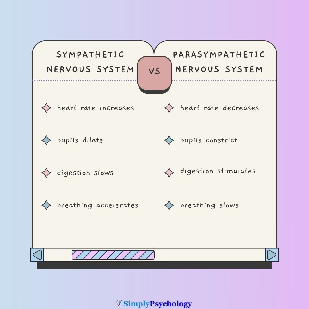
Think of the parasympathetic and sympathetic systems as a brake and gas pedal.
The sympathetic nervous system gets you ready for action (increased heart rate, energy mobilization), while the parasympathetic system helps you slow down and recover.
The two systems are not an on-off pair. Both are always partly active, and each organ’s state reflects the balance between them, known as autonomic tone.
Health depends on how flexibly the body shifts that balance: ramping up sympathetic activity to meet a challenge, then recruiting the parasympathetic system to recover.
| Function | Sympathetic (Fight or Flight) | Parasympathetic (Rest and Digest) |
|---|---|---|
| Heart rate | Increases | Decreases |
| Pupils | Dilate | Constrict |
| Digestion | Slows | Stimulates |
| Breathing | Accelerates | Slows |
| Salivation | Inhibits | Stimulates |
| Bladder | Relaxes | Contracts |
Vagal Tone and Heart-Rate Variability
At the heart, the parasympathetic share of autonomic tone is called vagal tone. It is a key measure in psychophysiology.
The vagus does not slow the heart to a steady rate. It adjusts the heart beat by beat, lengthening and shortening the gaps between beats in time with breathing.
This variation is called heart-rate variability (HRV). It is the small, ongoing difference in the time between one heartbeat and the next.
Its most informative component is respiratory sinus arrhythmia (RSA): the heart speeds up on inhalation and slows on exhalation. RSA tracks the vagal brake, breath by breath.
Because the vagus drives most of this variation, higher HRV is widely used as a non-invasive index of vagal control of the heart (Grossman & Taylor, 2007). The respiration-linked, high-frequency part matters most.
Higher resting vagal tone signals a flexible, well-regulated autonomic system.
Thayer and Lane (2000) proposed that it reflects a neurovisceral integration network linking the prefrontal cortex, limbic structures, and the heart.
On this account, the same inhibitory circuitry that lets the vagus brake the heart also supports attention, emotion regulation, and self-control. Lower resting HRV tends to accompany anxiety, chronic stress, and cardiovascular risk. The evidence is mostly correlational (see Critical Evaluation below).
Parasympathetic Dysfunction: Signs and Symptoms
When the parasympathetic system isn’t working properly, your body may struggle to calm down or recover after stress. Common symptoms include:
- Persistent high heart rate or blood pressure
- Digestive problems (e.g., constipation or diarrhea)
- Poor bladder control
- Dry mouth or eyes
- Light sensitivity due to pupil dysfunction
- Feeling “stuck” in a stressed or anxious state
Causes of dysfunction may include diabetes, aging, neurological conditions like Parkinson’s disease, or nerve injury.
When autonomic regulation fails more broadly, the result is called dysautonomia. Its signs, including abnormal heart-rate control, digestive and bladder problems, and dry eyes or mouth, trace back to lost parasympathetic and sympathetic control.
What Happens When It’s Overactive?
An overactive parasympathetic system, although less common, can also cause problems. Excess acetylcholine in the body may lead to:
- Muscle cramps or weakness
- Blurred vision or excessive tearing
- Excessive saliva or digestive activity
- Diarrhea or low blood pressure
In rare cases, severe overactivation may cause fainting or paralysis.
Fainting has a clear mechanism. A sudden surge of vagal activity can slow the heart and drop blood pressure too far, causing dizziness or fainting. This is called vasovagal syncope.
Activating Your Parasympathetic Nervous System
Fortunately, there are simple techniques that can activate your PSNS and help your body relax and recover:
- Deep breathing: Long, slow exhales (e.g., box breathing: inhale 4, hold 4, exhale 4, hold 4)
- Mind-body practices: Yoga, tai chi, or gentle stretching
- Meditation and mindfulness: Encourage calm awareness and reduce SNS activity
- Progressive muscle relaxation: Tense and relax muscle groups to trigger calm
- Cold exposure: Brief exposure to cold water can stimulate the vagus nerve
Slow breathing is the most reliable of these. It works because breathing is mechanically coupled to vagal control of the heart. Lengthening the out-breath increases vagal traffic to the heart, which lowers heart rate and blood pressure.
Cold water on the face can trigger a similar reflex slowing of the heart, known as the diving reflex.
Critical Evaluation of the PSNS Model
The core anatomy and chemistry of the parasympathetic system are exceptionally well established, mapped by classic anatomical and pharmacological work (Dale, 1914; Loewi, 1921).
The popular claims built on top of it are shakier, and three limits stand out.
- Opposing Switches: The brake-and-accelerator picture is a teaching device. Some organs get input from only one branch, and some functions need both.
- Polyvagal Theory: Influential in trauma-informed therapy, but several of its evolutionary claims are disputed.
- HRV Evidence: Heart-rate variability is an imperfect index of vagal tone, and its links to wellbeing are mostly correlational.
The Opposing-Switches Model Oversimplifies
Sweat glands, most blood vessels, the adrenal medulla, and the tiny muscles that raise the hairs receive only sympathetic input. For these, control is a matter of more or less sympathetic drive, not a tug-of-war.
Some functions need both branches working together, not against each other. Sexual response is the classic case. Parasympathetic activity drives arousal and erection, while sympathetic activity mediates orgasm and ejaculation, so a normal response needs both in sequence.
Where both branches do reach an organ, they usually pull in opposite directions. The pupil is the textbook case. Parasympathetic input constricts it and sympathetic input dilates it.
Even so, a graded, region-specific balance of autonomic tone fits the evidence better than a bank of paired switches. The dichotomy is useful but leaky.
Polyvagal Theory Remains Contested
Porges (2007) proposed two vagal branches:
- Ventral vagus: a newer branch, wrapped in insulating myelin, that supports calm social engagement.
- Dorsal vagus: an older branch linked to immobilization and shutdown under extreme threat.
A rapid, non-conscious appraisal of safety or danger, which Porges calls neuroception, is said to select between them. The theory is widely used in trauma-informed therapy.
Physiologists disagree. Grossman and Taylor (2007) argued that several central claims misrepresent how vagal control of the heart evolved. They also argued that the theory over-interprets respiratory sinus arrhythmia, the speeding and slowing of the heart with each breath.
The dispute is not about whether the vagus matters. Both accounts agree it is central. It is about whether the finer subdivisions and the state-based hierarchy built on them are real.
Treat polyvagal theory, and popular “activate your vagus nerve” claims built on it, as an influential framework rather than settled fact.
Heart-Rate Variability Is Only a Proxy
Heart-rate variability (HRV), the beat-to-beat variation in heart rate, is widely used as a window onto vagal control.
Grossman and Taylor (2007) pointed out that three kinds of influence confound it:
- Breathing: its rate and depth.
- Body state: physical activity and posture.
- Sympathetic input: beta-adrenergic influences on the heart.
Comparing people is harder still. Within one person, HRV tracks vagal control closely, but across different people the link may be much weaker. HRV is a reasonable proxy for cardiac vagal tone only when these complications are taken into account.
The wellbeing evidence has a second limit. Most studies tying higher vagal tone to better emotion regulation, resilience, and health are correlational. Low HRV accompanies anxiety, depression, and cardiovascular disease, but that cannot show it causes them.
Biofeedback trials help, because they intervene experimentally. Even so, the strongest meta-analysis relies heavily on self-reported outcomes (Goessl et al., 2017).
The leap from an autonomic measure to a psychological claim needs care.
Contemporary Research
Recent work asks whether parasympathetic activity can be measured, strengthened, and then used to improve people’s health. Two main lines of work stand out today.
One trains vagal tone through breathing; the other stimulates the vagus nerve directly. The first has firmer evidence, while the second is still being tested. Both treat vagal activity as something to change, not just measure.
HRV Biofeedback Trials
In HRV biofeedback, people watch a live display of their own heartbeat. They learn, usually through slow paced breathing, to raise their HRV and so their vagal tone.
A meta-analysis by Goessl et al. (2017) pooled the trials:
- Aim: To test whether HRV biofeedback training reduces self-reported stress and anxiety, and what moderates the effect.
- Method: A random-effects meta-analysis pooled 24 studies with 484 participants. Effect sizes (Hedges’ g) were calculated within groups (before vs after) and between groups (biofeedback vs control).
- Results: Both effects were large: g = 0.81 within groups and g = 0.83 against controls. Study year, risk of bias, percentage of female participants, number of sessions, and anxiety diagnosis did not moderate the effect.
- Conclusion: Raising HRV, and so vagal tone, is linked to a large drop in self-reported stress and anxiety. The authors urge caution: outcomes rest largely on self-report, and more well-controlled trials are needed.
This is the clearest quantitative evidence that the parasympathetic system is trainable, not purely automatic. Because the technique now works through inexpensive wearable devices, it offers an accessible, non-drug route to stress management.
Vagus Nerve Stimulation
A second approach stimulates the vagus directly. Implanted vagus-nerve stimulation (VNS) is an established treatment for some forms of drug-resistant epilepsy and for treatment-resistant depression.
Non-invasive stimulation through the skin of the ear or neck, called transcutaneous VNS, is being investigated for anxiety, depression, inflammation, and cognition. Here the parasympathetic system is treated as a nerve to stimulate directly, not something to coax indirectly through breathing.
Britannica, T. Editors of Encyclopaedia (2019, September 6). Parasympathetic nervous system. Encyclopedia Britannica. https://www.britannica.com/science/parasympathetic-nervous-system
Dale, H. H. (1914). The action of certain esters and ethers of choline, and their relation to muscarine. Journal of Pharmacology and Experimental Therapeutics, 6, 147–190. https://doi.org/10.1016/S0022-3565(25)08268-0
Goessl, V. C., Curtiss, J. E., & Hofmann, S. G. (2017). The effect of heart rate variability biofeedback training on stress and anxiety: A meta-analysis. Psychological Medicine, 47(15), 2578–2586. https://doi.org/10.1017/S0033291717001003
Grossman, P., & Taylor, E. W. (2007). Toward understanding respiratory sinus arrhythmia: Relations to cardiac vagal tone, evolution and biobehavioral functions. Biological Psychology, 74(2), 263–285. https://doi.org/10.1016/j.biopsycho.2005.11.014
Langley, J. N. (1921). The autonomic nervous system (Part I). W. Heffer & Sons.
Loewi, O. (1921). Über humorale Übertragbarkeit der Herznervenwirkung. Pflügers Archiv für die gesamte Physiologie des Menschen und der Tiere, 189(1), 239–242.
Lumen. (n.d.). Functions of the Autonomic Nervous System. Retrieved May 5, 2021 from https://courses.lumenlearning.com/boundless-ap/chapter/functions-of-the-autonomic-nervous-system/
Nall, R. (2020, April 23). Your Parasympathetic Nervous System Explained. Healthline. https://www.healthline.com/health/parasympathetic-nervous-system
Porges, S. W. (2007). The polyvagal perspective. Biological Psychology, 74(2), 116–143. https://doi.org/10.1016/j.biopsycho.2006.06.009
Thayer, J. F., & Lane, R. D. (2000). A model of neurovisceral integration in emotion regulation and dysregulation. Journal of Affective Disorders, 61(3), 201–216. https://doi.org/10.1016/S0165-0327(00)00338-4
Tindle, J., & Tadi, P. (2020). Neuroanatomy, Parasympathetic Nervous System. StatPearls [Internet].