Neurons and glial cells are the two main types of cells in the nervous system, and understanding how they differ is essential for studying brain function and behavior.
While neurons are widely known for transmitting electrical and chemical signals, glial cells play equally vital roles in supporting, protecting, and regulating those neurons.
Grasping the distinct roles of these cells helps build a deeper understanding of how thoughts, emotions, and disorders arise from complex cellular interactions in the brain.
Key Takeaways
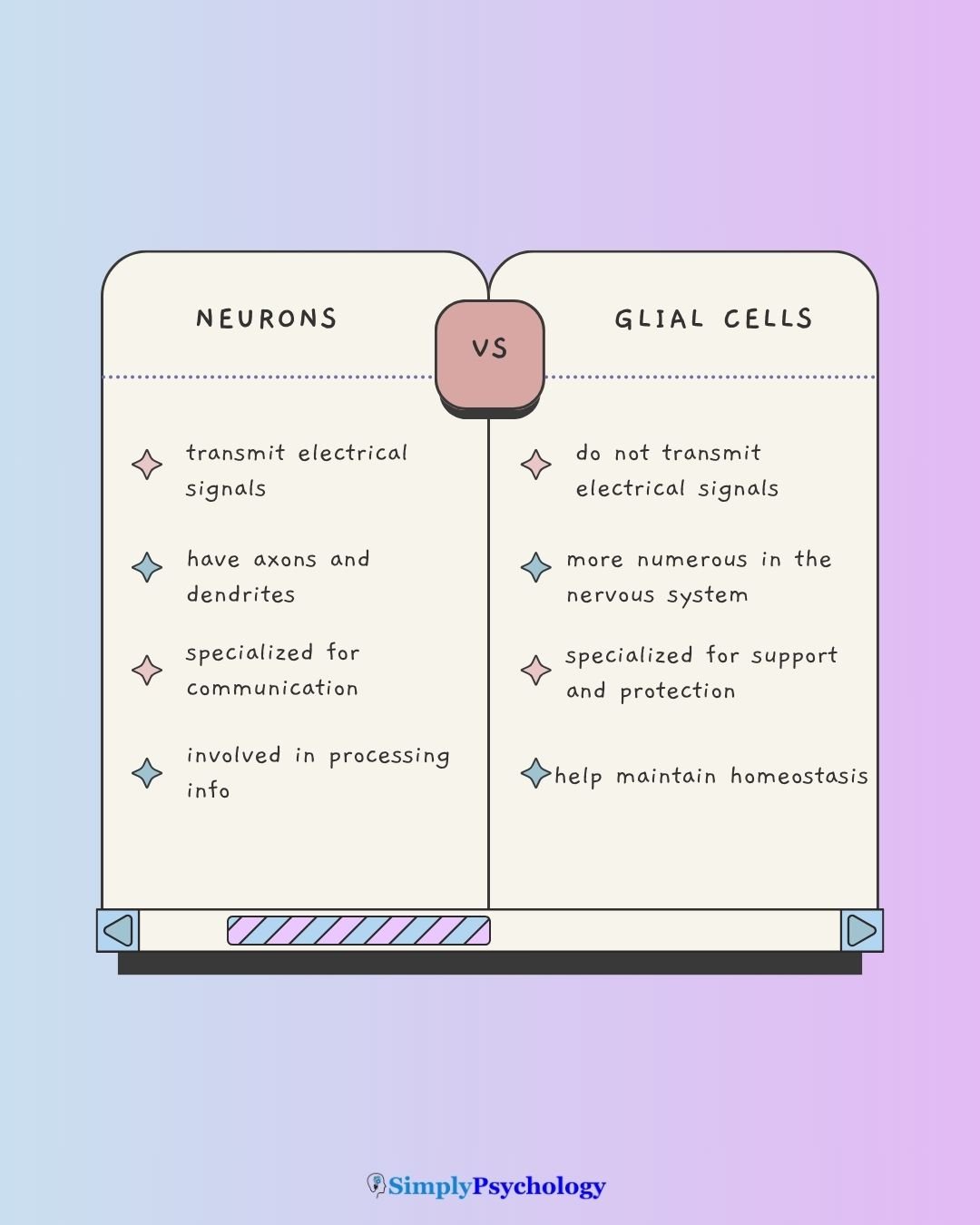
- Neurons transmit electrical signals and are responsible for thought, memory, and emotion.
- Glial cells do not transmit signals, but support, nourish, and protect neurons in many vital ways.
- Neurons have axons and dendrites for communication, while glial cells typically lack these structures.
- Glial cells include types like astrocytes, oligodendrocytes, and microglia (the brain’s resident immune cells), each with specialized roles such as forming myelin or defending against infection.
- Neurons and glial cells work together, and problems in either can contribute to neurological and psychological disorders like MS or schizophrenia.
What Are Neurons and Glial Cells?
Neurons and glial cells are the two primary types of cells that make up the nervous system.
The nervous system itself is the body’s intricate communication network. It is a complex system of channels that carry electrochemical signals throughout the body.
Neurons, also known as nerve cells, are considered the fundamental building blocks and interconnected information processors of this system.
They are essential for all nervous system tasks, including thought, memory, and emotion, as they react to stimuli, conduct impulses, and emit chemical regulators.
They come in several distinct types. Glial cells, or neuroglia, serve as vital support cells for neurons. They perform crucial functions such as providing physical scaffolding, nourishment, insulation for neuronal communication, removing waste products, and protecting neurons from harmful substances.
Both neurons and glial cells are essential components of the nervous system, but they fulfil different, complementary roles.
Consider their relative numbers. Glial cells are often said to outnumber neurons ten to one, but this figure is outdated. Careful whole-brain counts found roughly one glial cell for every neuron (Azevedo et al., 2009).
A review of over 150 years of cell-counting methods found no solid evidence for the old ten-to-one figure (von Bartheld et al., 2016). The exact ratio still varies by brain region.
Key Differences Between Neurons and Glial Cells
While both are indispensable, they serve distinct and complementary roles within this complex system.
Here are the key differences between them:
Neurons:
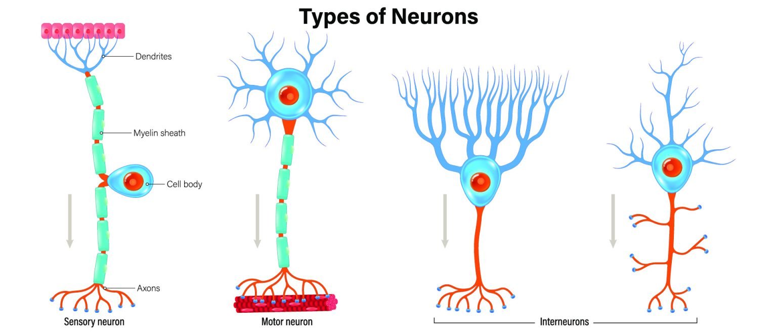
- Structure: Neurons possess specialized components for communication, including branching dendrites that act as input sites to receive signals. A single, long axon serves as the output, transmitting signals away from the cell body, typically ending in terminal buttons where messages are released.
- Function: They are the interconnected information processors essential for all nervous system tasks, reacting to stimuli, conducting impulses, and emitting chemical regulators necessary for thought, memory, and emotion.
- Signal Transmission: Neurons communicate through electrochemical events. An electrical impulse, called an action potential, travels rapidly down the axon. Upon reaching the terminal, this electrical message converts into a chemical message (neurotransmitters) released into the synaptic cleft to bind with adjacent neurons.

Glial Cells:
- Structure: Unlike neurons, glial cells generally lack axons and dendrites. They are primarily support cells rather than signaling units.
- Function: Glial cells perform crucial supportive functions for neurons. This includes providing physical scaffolding for the nervous system, offering nourishment, insulating neurons by forming the myelin sheath (which increases signal speed), removing waste products, and protecting neurons from harmful substances. They also help neurons align for efficient communication and mediate immune responses.
- Signal Transmission: Glial cells do not directly transmit electrochemical signals or communicate via neurotransmitters between neurons. Their role is to facilitate and maintain the environment for neuronal communication.

This distinction highlights how their specialized structures enable their unique yet complementary roles in the nervous system.
How do neurons and glial cells work together?
Neurons, the brain’s information processors, fundamentally rely on glial cells for optimal function.
Glia provide physical support, nourish neurons, and remove waste products, creating the ideal environment for communication within the nervous system.
How Glial Support Shapes Neural Communication
Their interaction is crucial for cognition and behavior. For example, glial cells form the myelin sheath, insulating axons and vastly increasing the speed of neural signal transmission, which is vital for efficient thought and action.
This close neuron-glia communication extends to supporting brain development and neuroplasticity, allowing the nervous system to adapt and form new connections.
Consider astrocytes as one example. These star-shaped glial cells physically wrap around synapses and can sense nearby neural activity.
They then respond by releasing their own signalling molecules, helping shape the strength of that synaptic signal (Araque et al., 1999).
This positions astrocytes as an active third partner in synaptic communication, not just a passive bystander. The effect is subtle but real.
Rather than being an incidental accompaniment to learning, this astrocyte-synapse signalling is proposed as one of the mechanisms through which everyday experience reshapes neural circuits over time.
Neuron-Glia Dysfunction in Disorders
Dysfunctions in this partnership have significant implications for understanding disorders.
In Multiple Sclerosis (MS), the widespread loss of the myelin sheath, known as demyelination, directly impedes rapid information transfer by neurons, leading to severe symptoms.
In Alzheimer’s disease, microglia are implicated too. Recent research found that microglia can abnormally prune, or eliminate, synapses very early in the disease, before hallmark plaques even appear (Hong et al., 2016).
This repurposes the same pruning process microglia normally use to refine healthy brain circuits during development.
For Schizophrenia, glial dysfunction is implicated as well. Diffusion tensor imaging, an MRI technique that maps the health of the brain’s white-matter connections, has found reduced myelination in people with schizophrenia (Scheel et al., 2013).
Researchers now propose that oligodendrocyte and myelin abnormalities directly contribute to schizophrenia and mood disorders, rather than simply resulting from them (Schmitt et al., 2019).
This link is still an active area of research. The condition is also marked by reduced overall brain volume and less gray matter in the frontal lobes.
Their combined, complementary roles are essential for all nervous system functions.
Types of neurons
Below are the types of neurons:
- Sensory (Afferent) Neurons: These neurons transmit information from the body’s periphery, such as sensory input, towards the central nervous system (CNS) and the brain.
- Motor (Efferent) Neurons: These communicate information in the opposite direction, from the brain and nervous system to muscles and glands, allowing for responses.
- Spindle Cells (Von Economo Neurons): A distinctive large neuron found almost exclusively in humans and great apes, thought to support fast, intuitive social judgements about others (Nimchinsky et al., 1999).
Types of glial cells
Below are some of the types of glial cells:
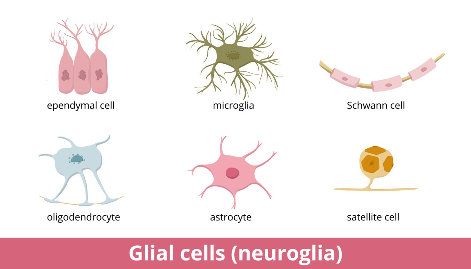
- Astrocytes: Maintain the chemical environment, support neurons structurally, and help form the blood-brain barrier.
- Oligodendrocytes: Produce the myelin sheath that insulates axons in the central nervous system (CNS).
- Radial Glia: Guide newly formed neurons to their correct position during brain development, and can themselves give rise to new neurons.
- Schwann Cells: Create the myelin sheath around axons in the peripheral nervous system (PNS).
- Microglia: Act as immune cells in the CNS, clearing debris and responding to injury or infection.
- Ependymal Cells: Line the brain’s ventricles and help circulate cerebrospinal fluid (CSF).
| Feature | Neurons | Glial Cells |
| Primary Function | Transmit electrical signals | Support, nourish, and protect |
| Physical Feature | Have axons and dendrites | Generally lack axons/dendrites |
| Electrical Activity | High (Action Potentials) | Low (Non-excitable) |
| Cell Division | Rarely divide in adults | Can divide (capable of mitosis) |
References
Azevedo, F. A. C., Carvalho, L. R. B., Grinberg, L. T., Farfel, J. M., Ferretti, R. E. L., Leite, R. E. P., Filho, W. J., Lent, R., & Herculano-Houzel, S. (2009). Equal numbers of neuronal and nonneuronal cells make the human brain an isometrically scaled-up primate brain. Journal of Comparative Neurology, 513(5), 532–541. https://doi.org/10.1002/cne.21974
Hong, S., Beja-Glasser, V. F., Nfonoyim, B. M., Frouin, A., Li, S., Ramakrishnan, S., Merry, K. M., Shi, Q., Rosenthal, A., Barres, B. A., Lemere, C. A., Selkoe, D. J., & Stevens, B. (2016). Complement and microglia mediate early synapse loss in Alzheimer mouse models. Science, 352(6286), 712–716. https://doi.org/10.1126/science.aad8373
Nimchinsky, E. A., Gilissen, E., Allman, J. M., Perl, D. P., Erwin, J. M., & Hof, P. R. (1999). A neuronal morphologic type unique to humans and great apes. Proceedings of the National Academy of Sciences, 96(9), 5268–5273. https://doi.org/10.1073/pnas.96.9.5268
Scheel, M., Prokscha, T., Bayerl, M., Gallinat, J., & Montag, C. (2013). Myelination deficits in schizophrenia: Evidence from diffusion tensor imaging. Brain Structure and Function, 218(1), 151–156. https://doi.org/10.1007/s00429-012-0389-2
Schmitt, A., Simons, M., Cantuti-Castelvetri, L., & Falkai, P. (2019). A new role for oligodendrocytes and myelination in schizophrenia and affective disorders? European Archives of Psychiatry and Clinical Neuroscience, 269(4), 371–372. https://doi.org/10.1007/s00406-019-01019-8
von Bartheld, C. S., Bahney, J., & Herculano-Houzel, S. (2016). The search for true numbers of neurons and glial cells in the human brain: A review of 150 years of cell counting. Journal of Comparative Neurology, 524(18), 3865–3895. https://doi.org/10.1002/cne.24040

