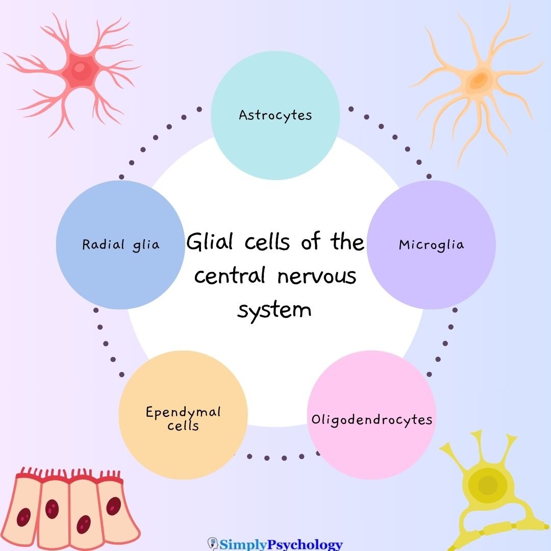
Glial cells, also called glial cells or neuroglia, are cells which are non-neuronal and are located within the central nervous system and the peripheral nervous system that provides physical and metabolic support to neurons, including neuronal insulation and communication, and nutrient and waste transport.
Glial cells are a general term for many types of glial cells, for example, microglial, astrocytes, and Schwann cells, each having their own functions within the body.
Each type of glial cell performs specific jobs that keep the brain functioning.
Primarily, glial cells provide support and protection to the neurons (nerve cells), maintain homeostasis, clean up debris, and form myelin. They essentially work to care for the neurons and the environment they are in.

What do glial cells do?
Glial cells perform many behind-the-scenes jobs that are essential to brain and nerve health:
- Support neurons physically and metabolically
- Insulate axons with myelin to speed up signals
- Maintain homeostasis in the brain’s internal environment
- Clear debris and dead cells
- Regulate neurotransmitter levels
- Assist in immune defense within the brain
- Guide brain development and support plasticity
Together, these tasks help neurons fire, recover, and stay healthy throughout life.
Think of Glial Cells Like a Support Crew
Astrocytes = Janitors + Supply Managers
They clean up debris, mop up excess chemicals, regulate nutrients, and even store energy—keeping neurons in top shape.Microglia = Immune Patrols
They’re the brain’s defense squad, constantly scanning for threats, cleaning up damage, and helping with repairs.Oligodendrocytes / Schwann Cells = Electricians
These cells insulate neuronal wiring (axons) with myelin, ensuring fast, efficient signal transmission.
Glial cells vs neurons
Glial cells differ from neurons in terms of structure. Neurons will have an axon and dendrites to transfer electrical signals between other nerve cells.
Glial cells, however, do not have axons or dendrites.
This means that glial cells do not participate directly in synaptic interactions and electrical signaling, although they are supportive in helping the neurons maintain these functions.
Also, although glial cells have complex extensions from their cell bodies since they do not have axons or dendrites, this makes them typically smaller than neurons.
Astrocytes, which are the largest type of glial cell, have a diameter of 40-50 microns.
Glial in the central nervous system (CNS)

Microglial
Microglia are immune cells that monitor for injury and disease, clear dead cells and toxins, and aid in brain development and plasticity.
Microglial dysfunction has been linked to chronic pain, fibromyalgia (Ohgidani et al., 2017), and autism (Tetreault et al., 2012).
Astrocytes
Astrocytes maintain the neuronal environment by regulating neurotransmitters, recycling glutamate, cleaning debris, removing excess potassium, forming the blood-brain barrier, and storing blood sugar for neurons.
Astrocyte dysfunction is linked to neurodegenerative diseases like ALS, Huntington’s, and Parkinson’s (Phatnani & Maniatis, 2015; Liddelow et al., 2017).
Oligodendrocytes
Oligodendrocytes form myelin sheaths around central nervous system axons and provide nutrients.
Oligodendrocyte issues are associated with multiple sclerosis, leukodystrophies, and mental health conditions like schizophrenia (Scheel et al., 2013; Schmitt et al., 2019) and bipolar disorder (Konradi et al., 2012).
Ependymal cells
Ependymal cells line brain ventricles and the spinal cord’s central canal, circulating and producing cerebrospinal fluid.
Their dysfunction is associated with hydrocephalus (Ji et al., 2022), multiple sclerosis (Hatrock et al., 2020), and may contribute to neurodegenerative diseases (Nelles & Hazrati, 2022).
Radial glial
Radial glia generate neurons, astrocytes, and oligodendrocytes, guide brain cell development, and contribute to brain plasticity. They are of interest in research on repairing brain damage.
A Second Look at Glial Cells
For years, glial cells were dismissed as “nerve glue” with little purpose. Today, we know they’re active participants in brain health—regulating mood, inflammation, development, and even memory. They’re not just support; they’re essential.

Glial in the peripheral nervous system (PNS)
Schwann cells
Schwann cells provide myelin insulation for peripheral nervous system axons and aid in nerve regeneration.
Abnormalities can lead to conditions like Guillain-Barre syndrome and Charcot-Marie-Tooth disease. Their dysfunction may trigger harmful inflammation in neuropathies (Ydens et al., 2013).

Satellite cells
Satellite cells surround nerve cell bodies in ganglia, regulating the neuronal environment and providing protection.
Their dysfunction may affect sensory processes and organ communication. Satellite cell issues are linked to muscle disorders (Servian-Morilla et al., 2021; Ganassi et al., 2022).
FAQs
What do glial cells do?
Glial cells are non-neuronal cells that provide support and protection for neurons in the central nervous system.
They regulate neurotransmitters, isolate neurons, destroy pathogens, guide neuron migration during development, promote synaptic plasticity, and remove dead neurons.
Glial cells are crucial for the proper functioning of the nervous system.
Do glial cells produce myelin?
Yes, certain types of glial cells called oligodendrocytes and Schwann cells produce the myelin sheath around neuronal axons in the central and peripheral nervous systems, respectively.
Myelin acts as an insulating layer that increases the speed of neural signaling by preventing leakage of electrical impulses out of the axon.
Myelin production by glial cells is crucial for proper neuronal function and communication.
Why are glial cells important for neurons and brain function?
Glial cells are crucial because they help maintain the microenvironment neurons require to function properly.
They provide nutrients and energy to neurons, regulate neurotransmitter levels, insulate axons, and protect neurons from damage and infection.
Can glial cell dysfunction impact mental health?
Yes, emerging research implicates glial cell abnormalities in conditions like depression, anxiety, schizophrenia, and bipolar disorder.
Dysfunctional astrocytes and microglia likely contribute to inflammation that damages neurons.
Could targeting glial cells lead to new treatments for neurodegenerative diseases?
Potentially. Research on manipulating reactive astrocytes and microglia to reduce inflammation in diseases like Alzheimer’s looks promising.
Enhancing oligodendrocytes may also help repair myelin damage. More studies are needed.

References
Ganassi, M., & Zammit, P. S. (2022). Involvement of muscle satellite cell dysfunction in neuromuscular disorders: Expanding the portfolio of satellite cell-opathies. European Journal of Translational Myology, 32(1).
Hatrock, D., Caporicci-Dinucci, N., & Stratton, J. A. (2020). Ependymal cells and multiple sclerosis: proposing a relationship. Neural regeneration research, 15(2), 263.
Jäkel, S., & Dimou, L. (2017). Glial cells and their function in the adult brain: a journey through the history of their ablation. Frontiers in cellular neuroscience, 11, 24.
Ji, W., Tang, Z., Chen, Y., Wang, C., Tan, C., Liao, J., … & Xiao, G. (2022). Ependymal cilia: Physiology and role in hydrocephalus. Frontiers in Molecular Neuroscience, 15, 927479.
Konradi, C., Sillivan, S. E., & Clay, H. B. (2012). Mitochondria, oligodendrocytes and inflammation in bipolar disorder: evidence from transcriptome studies points to intriguing parallels with multiple sclerosis. Neurobiology of disease, 45(1), 37-47.
Liddelow, S. A., Guttenplan, K. A., Clarke, L. E., Bennett, F. C., Bohlen, C. J., Schirmer, L., … & Barres, B. A. (2017). Neurotoxic reactive astrocytes are induced by activated microglia. Nature, 541(7638), 481-487.
Nelles, D. G., & Hazrati, L. N. (2023). The pathological potential of ependymal cells in mild traumatic brain injury. Frontiers in Cellular Neuroscience, 17, 1216420.
Ohgidani, M., Kato, T. A., Hosoi, M., Tsuda, M., Hayakawa, K., Hayaki, C., … & Kanba, S. (2017). Fibromyalgia and microglial TNF-α: translational research using human blood induced microglia-like cells. Scientific reports, 7(1), 11882.
Phatnani, H., & Maniatis, T. (2015). Astrocytes in neurodegenerative disease. Cold Spring Harbor perspectives in biology, 7(6), a020628.
Purves, D. A. GJ., Fitzpatrick, D., et al. (2001). Neuroscience 2nd edition. Neuroglial cells.
Scheel, M., Prokscha, T., Bayerl, M., Gallinat, J., & Montag, C. (2013). Myelination deficits in schizophrenia: evidence from diffusion tensor imaging. Brain Structure and Function, 218, 151-156.
Servián-Morilla, E., Cabrera-Serrano, M., Johnson, K., Pandey, A., Ito, A., Rivas, E., … & Paradas, C. (2020). POGLUT1 biallelic mutations cause myopathy with reduced satellite cells, α-dystroglycan hypoglycosylation and a distinctive radiological pattern. Acta neuropathologica, 139, 565-582.
Schmitt, A., Simons, M., Cantuti-Castelvetri, L., & Falkai, P. (2019). A new role for oligodendrocytes and myelination in schizophrenia and affective disorders?. European Archives of Psychiatry and Clinical Neuroscience, 269, 371-372.
Tetreault, N. A., Hakeem, A. Y., Jiang, S., Williams, B. A., Allman, E., Wold, B. J., & Allman, J. M. (2012). Microglia in the cerebral cortex in autism. Journal of Autism and Developmental Disorders, 42(12), 2569-2584.
Ydens, E., Lornet, G., Smits, V., Goethals, S., Timmerman, V., & Janssens, S. (2013). The neuroinflammatory role of Schwann cells in disease. Neurobiology of disease, 55, 95-103.

