Schwann cells are a special type of cell found in the peripheral nervous system (PNS), the network of nerves outside the brain and spinal cord.
They play a key role in helping nerves work efficiently by wrapping around nerve fibers and forming a protective layer called the myelin sheath.
This insulation allows electrical signals to travel quickly and smoothly along the nerves, which is essential for everything from moving your muscles to sensing temperature.

Key Takeaways
- Schwann cells are glial cells in the peripheral nervous system that insulate, support, and repair nerve fibers.
- They come in two types: myelinating (which wrap axons in myelin) and non-myelinating (which support small nerve fibers).
- These cells are vital for nerve signal speed, protection, and regeneration after injury.
- Damage to Schwann cells can cause or contribute to several neurological disorders.
- Their unique functions make them an exciting target for regenerative medicine and nerve repair research.
Did you know? Schwann cells are named after Theodor Schwann, a German physiologist who discovered these types of cells in the 19th century.
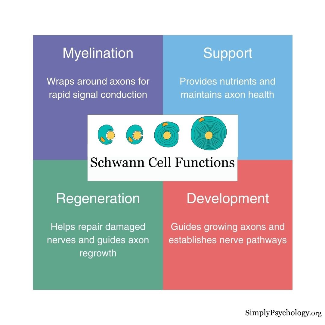
Schwann Cell Functions
Schwann cells have several important functions:
- Myelination: They create the myelin sheath, a fatty layer that insulates axons (the long parts of nerve cells) to speed up signal transmission.
- Support and protection: They supply nutrients, remove cellular waste, and shield nerve fibers from damage.
- Repair and regeneration: After injury, Schwann cells help clean up debris and guide new nerve growth by forming pathways and releasing growth factors.
- Development and communication: They help nerves develop correctly in the embryo and maintain healthy communication with neurons throughout life.
Schwann cells are also considered to be a type of glial cell.
Glia cells are non-neuronal cells that do not provide electrical impulses like neurons but function to maintain homeostasis, providing support and protection for neurons.
So, although Schwann cells do not conduct electrical activity themselves, they still help ensure the normal conduction of electrical signals.
Myelinating vs. Non-Myelinating Schwann Cells
There are two main types of Schwann cells:
- Myelinating Schwann cells wrap around one axon segment, creating a myelin sheath that increases the speed of electrical signals.
- Non-myelinating Schwann cells support small nerve fibers without forming myelin. They help organize and protect groups of unmyelinated axons.
Both types are essential for keeping peripheral nerves healthy and functioning.
Where Schwann Cells Come From
Schwann cells originate from the neural crest. This is a group of cells that migrates away from the developing spinal cord early in embryonic life.
These cells travel alongside growing nerve fibers and mature in stages, first into Schwann cell precursors, then into immature Schwann cells (Jessen, Mirsky, & Lloyd, 2015).
Each cell then picks one fate.
A process called radial sorting separates large-diameter axons, which get their own myelinating cell, from small-diameter axons, which are grouped together and served by a non-myelinating cell.
The deciding signal is a growth factor called neuregulin-1, found on the axon’s surface.
Its level tells the Schwann cell whether that axon needs a myelin sheath (Salzer, 2015).
This developmental flexibility does not disappear with age.
It is the same plasticity that lets a mature Schwann cell revert to a repair-like state after nerve injury (Jessen et al., 2015).
How Schwann Cells Form Myelin
The process of myelination begins during fetal development. Schwann cells wrap themselves around a single axon in a spiral, forming multiple layers.
The inner layers become compacted to form the insulating myelin sheath, while the outermost layer remains as the cell body (called the neurilemma), which supports and maintains the sheath.
This insulation lets electrical signals jump between gaps in the myelin, called nodes of Ranvier, rather than travelling continuously. This leaping is called saltatory conduction.
It lets a myelinated fiber conduct a nerve impulse up to 50 to 100 times faster than a bare, unmyelinated fiber, at a fraction of the metabolic cost.
Schwann Cells vs. Other Glial Cells
Schwann cells are the principal glial cells of the peripheral nervous system (PNS), primarily responsible for forming myelin sheaths around axons to facilitate rapid nerve impulse conduction.
In contrast, the central nervous system (CNS) contains several distinct types of glial cells with specialized functions:
- Oligodendrocytes: These cells myelinate axons in the CNS. Unlike Schwann cells, which myelinate a single axon segment, a single oligodendrocyte can extend its processes to myelinate multiple axons simultaneously.
- Astrocytes: Star-shaped cells that maintain the blood-brain barrier, regulate ion concentrations, and provide metabolic support to neurons. They also play a role in repairing the CNS after injury.
- Microglia: These act as the immune cells of the CNS, constantly surveying the environment for pathogens or damaged neurons and clearing debris through phagocytosis.
- Ependymal Cells: They line the ventricles of the brain and the central canal of the spinal cord, playing a crucial role in the production and circulation of cerebrospinal fluid.
While Schwann cells and oligodendrocytes share the function of myelination, their anatomical locations and capacities differ significantly.
Moreover, the CNS glial cells exhibit a broader range of functions beyond myelination, highlighting the specialized roles of glial cells across the nervous system.

How Do Schwann Cells Respond to Damaged Axons?
When a peripheral nerve is injured, Schwann cells take action:
- They clean up damaged myelin and axon fragments.
- They release chemical signals that attract immune cells to help with cleanup.
- They produce growth-promoting substances like neurotrophins.
- They form a tunnel-like structure to guide regrowing axons back to their target.
Without Schwann cells, nerve regeneration in the PNS would be slow or impossible.
What Causes Schwann Cell Damage?
Schwann cell damage is associated with demyelinating diseases of the PNS.
Damage to Schwann cells can occur due to various factors, including genetic mutations, autoimmune responses, infections, and trauma.
When Schwann cells are damaged, the myelin sheath that insulates and supports axons can be destroyed, leading to impaired nerve conduction and potential neurodegeneration.
Signs of damaged Schwann cells include:
- Weakened reflexes
- Muscle weakness
- Sensory loss
- Slower nerve conduction
- Paralysis
What Diseases Involve Schwann Cells?
Damage to Schwann cells or their myelin can lead to serious nerve disorders:
- Guillain-Barré Syndrome (GBS): An autoimmune disease that attacks Schwann cells, causing weakness and paralysis.
- Charcot-Marie-Tooth Disease (CMT): A genetic disorder affecting Schwann cell structure, leading to muscle weakness and loss of sensation.
- Diabetic Neuropathy: High blood sugar damages Schwann cells over time, leading to numbness, pain, and mobility issues.
- Schwannomas: Usually benign tumors that grow from Schwann cells, sometimes affecting nearby nerves.
Schwann cells from the PNS have been implanted into the central nervous system (CNS) to treat multiple sclerosis-type damage and spinal cord injuries in animal studies. These implanted cells have shown promise in promoting axonal regeneration and myelination, leading to improved nerve impulse conduction (Kohama et al., 2001).
That laboratory promise has now moved into early human trials. Surgeons transplant a patient’s own Schwann cells, grown from a small nerve biopsy, into the injury site.
One trial targeted recent spinal injuries and found no added neurological damage after a year (Anderson et al., 2017). A second, in long-standing injuries, paired the transplant with intensive rehabilitation and found it safe, with modest neurological improvement in one participant (Gant et al., 2022).
Both are small safety trials, not proof the treatment works. But they show the idea has reached human testing.
Critical Evaluation
The link between a Schwann cell’s structure and its insulating function is well supported. Culture studies, molecular signalling work, and nerve-conduction physiology all point to the same conclusion (Salzer, 2015).
The cell’s regenerative role is the strongest and most distinctive claim, but almost all of the causal evidence for it comes from animal studies rather than humans.
The c-Jun Discovery
A landmark study identified the switch that converts an ordinary Schwann cell into a repair cell after nerve injury (Arthur-Farraj et al., 2012).
- Aim: To test whether the transcription factor c-Jun, switched on rapidly in Schwann cells after injury, is required for nerve repair or merely a marker of it.
- Method: Researchers bred mice with the c-Jun gene deleted only in Schwann cells, cut the sciatic nerve, and compared their molecular injury response, myelin clearance, and functional recovery with normal mice.
- Results: Without c-Jun, the injured nerve formed a dysfunctional repair cell. Functional recovery failed, and neurons that would otherwise have survived and regrown instead died.
- Conclusion: A single glial transcription factor is essential for nerve repair. c-Jun drives Schwann cells to transform into repair cells, and without it the peripheral nervous system loses its ability to regenerate.
The study’s main limitation is that it establishes this cause-and-effect relationship in mice. Extending the finding to human nerve repair remains a plausible but unproven inference.
Contemporary Research
Research since 2015 has built on the c-Jun discovery. It confirms the repair cell is a genuinely distinct, actively generated cell type.
Jessen and Mirsky (2016) reviewed how injury converts myelinating and non-myelinating Schwann cells into this repair-specialised phenotype.
The verdict: true reprogramming, not passive decay.
Repair cells combine de-differentiation with active new gene expression. The programme down-regulates myelin genes while up-regulating trophic factors, myelin-clearing autophagy, and the Bands of Büngner that guide regrowing axons, all under c-Jun’s control.
Whole-genome analyses then confirmed this at the molecular level.
Generating the repair cell involves coordinated changes across the coding and non-coding transcriptome and the DNA methylome. This is evidence of a genuine reprogramming event, not simple dedifferentiation (Arthur-Farraj et al., 2017).
One finding tempers the picture. The repair phenotype is not durable.
Over the long timescales that regeneration takes in a large animal or a human, repair Schwann cells lose their pro-regenerative character.
This is one reason many human nerve injuries recover poorly despite the PNS’s regenerative machinery (Jessen & Mirsky, 2019).
References
Anderson, K. D., Guest, J. D., Dietrich, W. D., Bartlett Bunge, M., Curiel, R., Dididze, M., … Levi, A. D. (2017). Safety of autologous human Schwann cell transplantation in subacute thoracic spinal cord injury. Journal of Neurotrauma, 34(21), 2950–2963. https://doi.org/10.1089/neu.2016.4895
Arthur-Farraj, P. J., Latouche, M., Wilton, D. K., Quintes, S., Chabrol, E., Banerjee, A., … Jessen, K. R. (2012). c-Jun reprograms Schwann cells of injured nerves to generate a repair cell essential for regeneration. Neuron, 75(4), 633–647. https://doi.org/10.1016/j.neuron.2012.06.021
Arthur-Farraj, P. J., Morgan, C. C., Adamowicz, M., Gomez-Sanchez, J. A., Fazal, S. V., Beucher, A., … Aitman, T. J. (2017). Changes in the coding and non-coding transcriptome and DNA methylome that define the Schwann cell repair phenotype after nerve injury. Cell Reports, 20(11), 2719–2734. https://doi.org/10.1016/j.celrep.2017.08.064
Britannica, T. Editors of Encyclopaedia (2020, July 15). Schwann cell. Encyclopedia Britannica. https://www.britannica.com/science/Schwann-cell
Fallon, M., & Tadi, P. (2019). Histology, Schwann Cells.
Gant, K. L., Guest, J. D., Palermo, A. E., Vedantam, A., Jimsheleishvili, G., Bunge, M. B., … Levi, A. D. (2022). Phase 1 safety trial of autologous human Schwann cell transplantation in chronic spinal cord injury. Journal of Neurotrauma, 39(3–4), 285–299. https://doi.org/10.1089/neu.2020.7590
Jessen, K. R., & Mirsky, R. (2016). The repair Schwann cell and its function in regenerating nerves. The Journal of Physiology, 594(13), 3521–3531. https://doi.org/10.1113/JP270874
Jessen, K. R., & Mirsky, R. (2019). The success and failure of the Schwann cell response to nerve injury. Frontiers in Cellular Neuroscience, 13, 33. https://doi.org/10.3389/fncel.2019.00033
Jessen, K. R., Mirsky, R., & Lloyd, A. C. (2015). Schwann cells: Development and role in nerve repair. Cold Spring Harbor Perspectives in Biology, 7(7), a020487. https://doi.org/10.1101/cshperspect.a020487
Kohama, I., Lankford, K. L., Preiningerova, J., White, F. A., Vollmer, T. L., & Kocsis, J. D. (2001). Transplantation of cryopreserved adult human Schwann cells enhances axonal conduction in demyelinated spinal cord. Journal of Neuroscience, 21 (3), 944-950.
Oudega, M., & Xu, X. M. (2006). Schwann cell transplantation for repair of the adult spinal cord. Journal of neurotrauma, 23 (3-4), 453-467.
Salzer, J. L. (2015). Schwann cell myelination. Cold Spring Harbor Perspectives in Biology, 7(8), a020529. https://doi.org/10.1101/cshperspect.a020529
Sinha Dutta, S. (2020, February 4). What are Schwann Cells? News Medical Life Sciences. https://www.news-medical.net/health/What-are-Schwann-Cells.aspx#2

