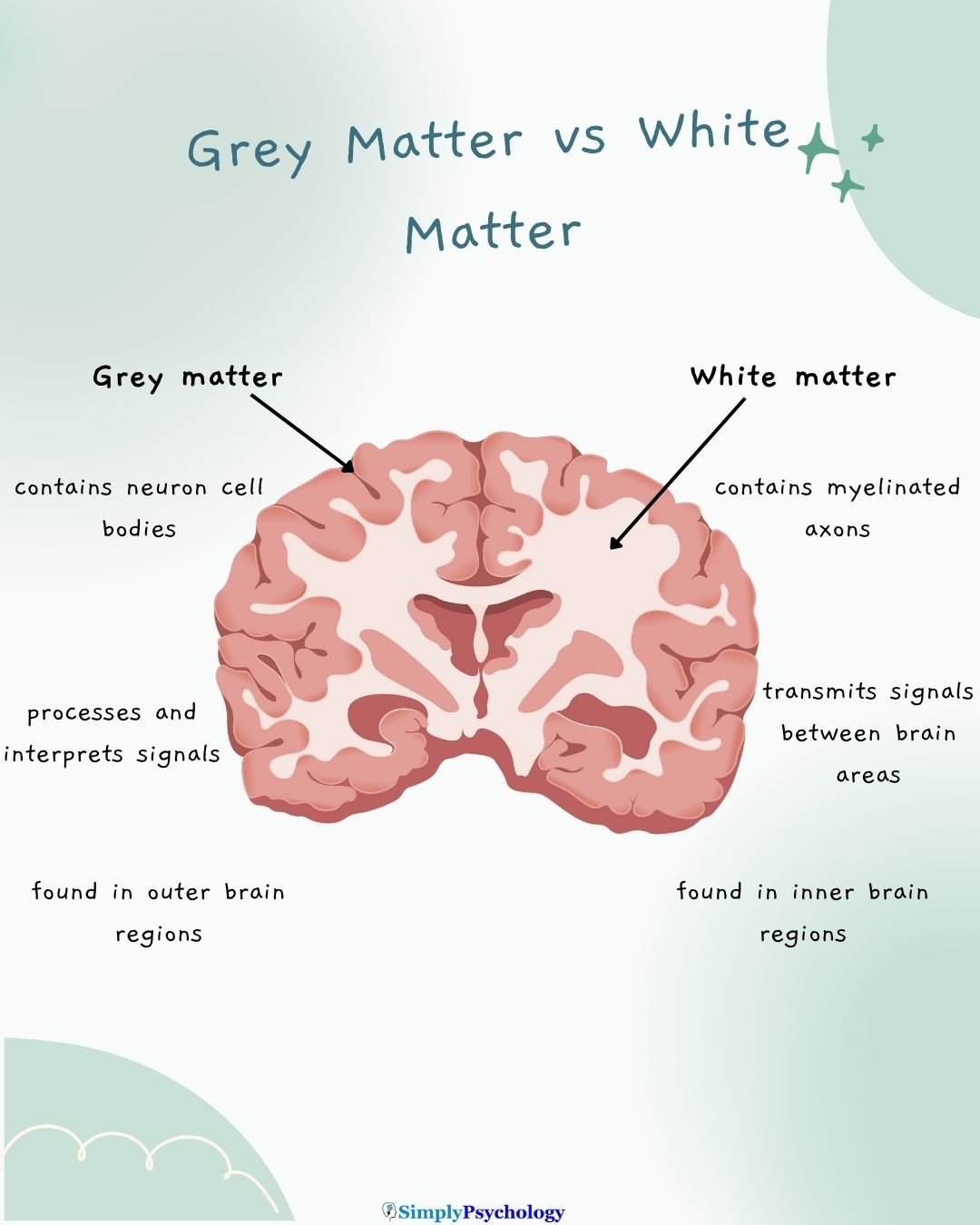
The central nervous system (CNS) is composed of white matter and grey matter. Grey matter, which makes up about half of the brain, consists primarily of neuronal cell bodies, dendrites, and unmyelinated axons.
Located primarily in the outer layers of the brain, grey matter is responsible for processing information, controlling muscle movement, and regulating sensory perception.

Where is grey matter found?
Its greyish-pink appearance comes from the high concentration of neuronal cell bodies it contains.
Grey matter is found in several key areas of the central nervous system:
- Brain:
- Forms the outermost layer of the cerebral cortex
- Present in both the cerebrum and cerebellum
- Found in deeper brain structures called nuclei
- Spinal Cord:
- Located in the center, surrounded by white matter
- Shaped like a butterfly when viewed in cross-section
This distribution allows grey matter to effectively process and integrate information throughout the central nervous system, facilitating various cognitive functions, sensory processing, and motor control.
What Grey Matter Consists Of
Grey matter is a complex tissue composed of various cellular structures:
- Neuronal Cell Bodies (Soma): These are the main components of grey matter, housing the nucleus of neurons. The cerebrum contains an estimated 10 to 50 billion neurons.
- Dendrites: These branching extensions of neurons receive signals from other neurons.
- Unmyelinated Axons: Unlike white matter, grey matter contains axons that are not covered by the fatty myelin sheath.
- Glial Cells: There are about ten times as many glial cells as neurons in grey matter. These include:
- Astrocytes: Support neurons and help regulate the chemical environment.
- Oligodendrocytes: Produce myelin in the central nervous system.
- Capillaries: Tiny blood vessels that supply oxygen and nutrients to the neural tissue.

Grey matter is particularly abundant in the cerebral cortex, which forms the outer layer of the cerebrum.
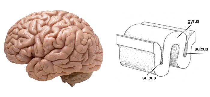
This cortex is characterized by gyri (ridges) and sulci (grooves), which increase the surface area of the brain, allowing for more neurons and enhanced processing capabilities.
Interestingly, the cerebellum, despite making up only 10% of the brain’s volume, contains more neuronal cell bodies than the rest of the brain combined.
This high density of neurons contributes to the cerebellum’s crucial role in motor control, balance, and coordination.
In the spinal cord, grey matter is arranged in a butterfly-shaped pattern when viewed in cross-section. It’s divided into three main regions:
- Anterior grey column: Important for motor movements.
- Posterior grey column: Receives sensory signals.
- Lateral grey column: Regulates the autonomic nervous system.
This unique composition and distribution of grey matter throughout the central nervous system enable it to perform its vital functions in information processing, sensory perception, and motor control.

Function
Grey matter serves to process information in the brain. The structures within the grey matter process signals from the sensory organs or from other areas of the grey matter.
This tissue directs sensory stimuli to the neurons in the central nervous system where synapses induce a response to the stimuli.
These signals reach the grey matter through the myelinated axons that make up the bulk of the white matter.
The grey matter that surrounds the cerebrum, also given the name cerebral cortex, is involved in several functions such as being involved in personality, intelligence, motor function, planning, organization, language processing, and processing sensory information.
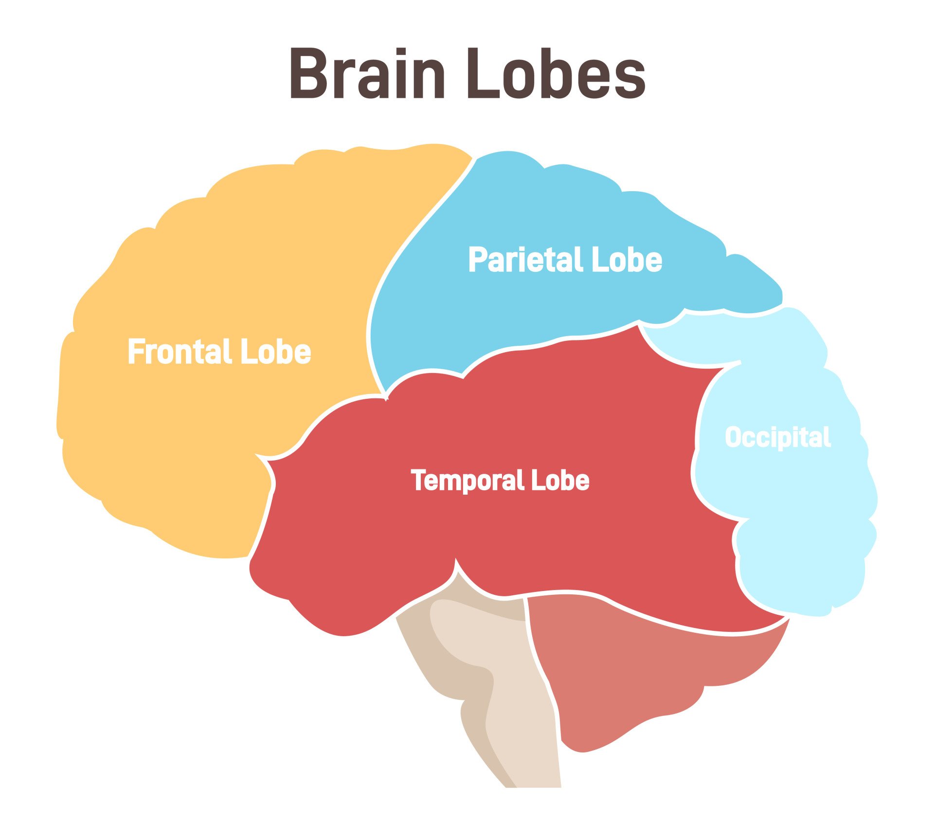
The cerebral cortex is divided into four lobes based on the gyri and sulci, which help mark these lobes:
- Frontal lobes: voluntary behavior such as decision-making, problem-solving, and thinking. It’s essential for cognition, intelligence, attention, and voluntary motor control.
- Parietal lobes: associated functions in perception and the integration of somatosensory information, visuospatial processing, spatial attention, spatial mapping, and number representation.
- Temporal lobes: essential for functions such as the comprehension of language, perception through hearing, vision, and smell, recognition, learning, and memory.
- Occipital lobe: functions related to vision such as encoding color, orientation, and motion.

Within these lobes are also specific sensory and motor areas. These can include:
- Primary Motor Cortex: Controls voluntary movements
- Somatosensory Cortex: Processes tactile information (touch, pressure, temperature, pain)
- Visual Cortex: Processes visual information
The grey matter in the cerebellum is related to motor control, balance, coordination, and automatic movements.
The three sections of grey matter in the spinal cord also serve their functions:
- The anterior grey column is important for all motor movements as it connects to the brain through a pathway called the pyramidal tract, which originates in the cerebral cortex.
- The posterior grey column plays an important function in receiving sensory signals, allowing for the constant interaction between the environment and the body.
- The lateral grey column, found in the middle of the grey matter of the spinal cord, is important for regulating the autonomic nervous system through its role in activating the sympathetic nervous system.
This means it helps to stimulate the body’s involuntary responses to stressful situations, such as accelerating heart rate and sending extra blood to the muscles.
White Matter vs. Grey Matter
Below are some of the key differences between white matter and grey matter:
| White Matter | Grey Matter |
| White in color | Grey in colour |
| Located deep in the brain; outer portion of the spinal cord | Located in outer layer of brain (cortex); central portion of spinal cord |
| Composed mainly of myelinated axons and oligodendrocytes | Composed mainly of neuron cell bodies, dendrites, and unmyelinated axons |
| Primary function is to transmit signals between different brain regions | Primary function is to process and analyze information |
| Fast signal transmission | Slower signal transmission |
| Shows structural changes with learning and experience | Undergoes pruning and reorganization during development |
| Appears bright on T1-weighted MRI images | Appears dark on T1-weighted MRI images |

Grey Matter Disorders
Because grey matter spans much of the brain and spinal cord—including the cerebral cortex, cerebellum, hippocampus, and basal ganglia—it is vulnerable to various forms of damage and disease.
Neurodegenerative Conditions
In Alzheimer’s disease, toxic plaques and tangles disrupt grey matter—especially in areas like the hippocampus, which plays a key role in memory.
As neurons die, cognitive abilities decline. Over time, this can lead to confusion, memory loss, and changes in behavior.
Parkinson’s disease also involves grey matter loss, particularly in the basal ganglia. This affects motor control and may result in tremors, stiffness, and impaired coordination.
Damage From Trauma or Oxygen Loss
Head injuries, strokes, or oxygen deprivation (hypoxia) can kill grey matter cells.
Because neurons in the cerebral cortex and cerebellum rely on constant oxygen and glucose, any interruption can cause permanent damage.
This might affect movement, balance, language, or emotional regulation—depending on which brain regions are involved.
Region-Specific Impacts
- Frontal lobes: Linked to personality changes, attention issues, and depression.
- Parietal lobes: May impair writing and sensory processing.
- Temporal lobes: Can affect speech comprehension, memory, and mood.
- Occipital lobes: Damage may cause visual problems or hallucinations.
- Cerebellum: Often leads to coordination and balance difficulties.
Research insights
In 2022, a brain imaging study found that people with major depression, bipolar disorder, or schizophrenia have less grey matter in the left hippocampus (a memory-related region) than healthy individuals.
In 2024, scientists reported that people who later developed dementia had a thinner cortical grey matter layer (the brain’s outer cortex) up to a decade before symptoms, suggesting this thinning is an early Alzheimer’s warning sign.
A 2021 study showed that older adults who stayed active with daily tasks (like household chores) had greater grey matter volume in their brains, highlighting that an active lifestyle can benefit brain structure.
In 2024, researchers used artificial intelligence to analyze MRI scans and predict with roughly 80% accuracy which people with mild memory problems will progress to Alzheimer’s disease.
How to Strengthen Grey Matter
The brain has remarkable potential to adapt, even after injury. Children tend to recover more effectively from grey matter damage than adults, largely because their neural networks are still developing and more plastic.
While there is no cure for neurodegenerative conditions like Alzheimer’s or Parkinson’s, research suggests several lifestyle choices can promote grey matter health and may reduce age-related decline.
Evidence-Based Ways to Support Grey Matter
- Aerobic exercise improves blood flow to the brain and has been linked to increased grey matter volume, especially in the hippocampus—a region essential for memory.
- Mindfulness and meditation enhance emotional regulation and stimulate growth in areas like the prefrontal cortex.
- Quality sleep allows for brain repair and toxin clearance, protecting grey and white matter alike.
- Nutrient-rich diets, particularly those high in omega-3s, antioxidants, and B vitamins, may support neuronal function and reduce inflammation.
- Limiting alcohol and psychoactive substances helps preserve brain tissue integrity.
- Mentally engaging activities—such as puzzles, strategy games, or learning a new language—can strengthen cognitive reserve and delay age-related changes.
- Fine motor hobbies like painting or knitting activate sensory and motor circuits, promoting coordination and cortical engagement.
- Wearing protective gear, such as helmets during cycling or sports, helps prevent traumatic injuries to vulnerable grey matter areas.
Emerging Therapies: Neuroplasticity in Action
Research into electroconvulsive therapy (ECT) shows promising results. One study (Camilleri et al., 2020) found that ECT increased grey matter volume in the medial temporal lobe, an area involved in emotion and memory.
Although further research is needed, ECT may offer future therapeutic options for restoring grey matter loss in severe cases.
References
Brosch, K., Stein, F., Schmitt, S., Pfarr, J., Ringwald, K. G., Meller, T., Steinsträter, O., Waltemate, L., Lemke, H., Meinert, S., Winter, A., Breuer, F., Thiel, K., Grotegerd, D., Hahn, T., Jansen, A., Dannlowski, U., Krug, A., Nenadić, I., . . . Kircher, T. (2022). Reduced hippocampal gray matter volume is a common feature of patients with major depression, bipolar disorder, and schizophrenia spectrum disorders. Molecular Psychiatry, 27(10), 4234-4243. https://doi.org/10.1038/s41380-022-01687-4
Camilleri, J. A., Hoffstaedter, F., Zavorotny, M., Zöllner, R., Wolf, R. C., Thomann, P., Redlich, R., Opel, N., Dannlowski, U., Grozinger, M., Demirakca, T., Sartorius, A., Eickhoff, S. B. & Nickl-Jockschat, T. (2020). Electroconvulsive therapy modulates grey matter increase in a hub of an affect processing network. NeuroImage: Clinical, 25, 102114.
Koblinsky, N.D., Meusel, LA.C., Greenwood, C.E. et al. Household physical activity is positively associated with gray matter volume in older adults. BMC Geriatr 21, 104 (2021). https://doi.org/10.1186/s12877-021-02054-8
Mercadante, A. A., & Tadi, P. (2020). Neuroanatomy, Gray Matter.
Satizabal, C. L., Beiser, A. S., Fletcher, E., Seshadri, S., & DeCarli, C. (2024). A novel neuroimaging signature for ADRD risk stratification in the community. Alzheimer’s & Dementia, 20(3), 1881-1893.

