The parietal lobe, located in the upper middle part of the cerebral cortex, integrates sensory information from across the body into a single, coherent picture. It also plays a central role in spatial orientation and the processing of touch.

The parietal lobe is crucial for tasks such as attention, spatial reasoning, and distinguishing between self and others, thereby influencing perception and interpersonal interactions.
What does the parietal lobe do?
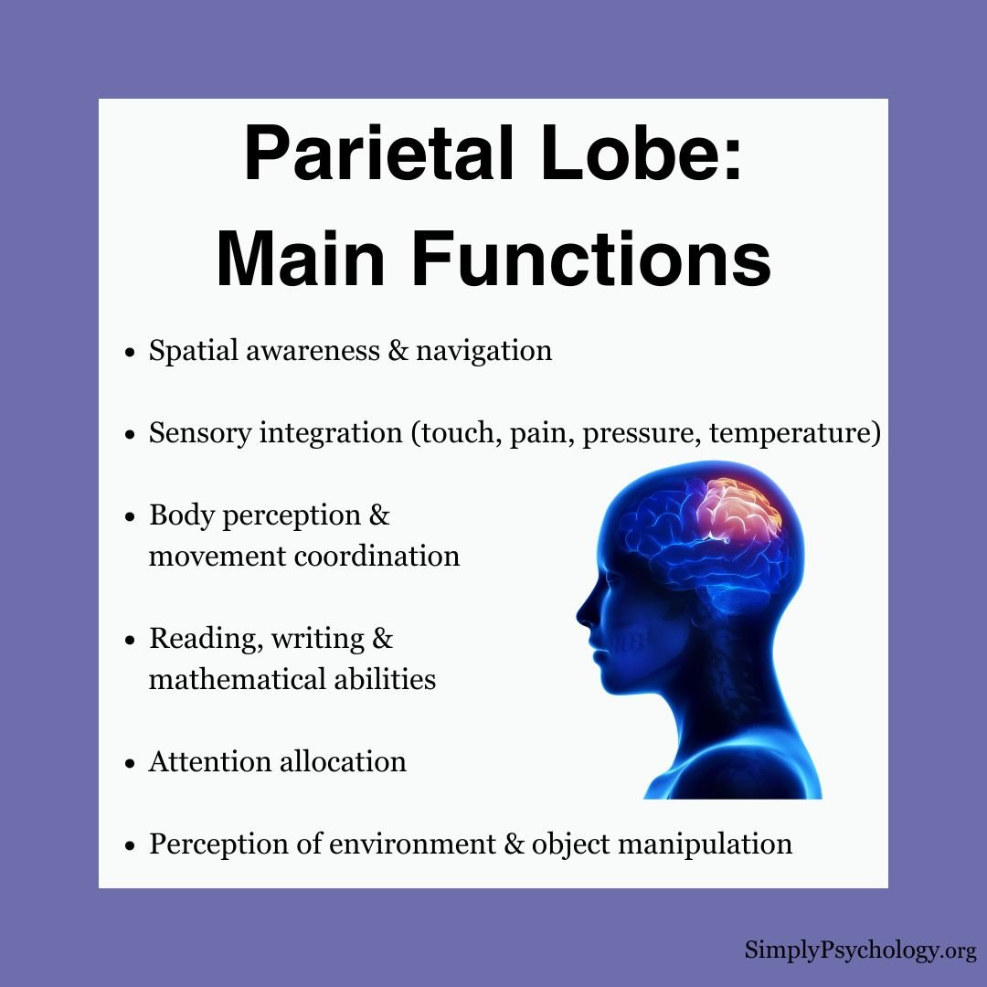
The parietal lobe plays a crucial role in integrating sensory information from various modalities and is associated with several key functions:
Spatial Awareness and Navigation
- The parietal lobe helps us perceive and navigate our environment by creating spatial maps and representations of the world around us.
Sensory Integration and Processing
- The parietal lobe combines information from different senses—like touch, temperature, pain, and pressure—into a single, unified experience. This helps you recognize what you’re feeling and where it’s happening.
- It also supports multisensory integration, allowing your brain to blend touch, vision, and movement—for example, helping you reach for your phone in the dark using feel alone.
Body Perception and Movement
- It contributes to the perception of the body and coordinates movement, turning what the eyes see into a plan for reaching or grasping.
- The left side of the parietal lobe is believed to keep track of the location of moving body parts.
- This visual-to-movement link runs through the dorsal (“where/how”) visual stream, distinct from the ventral stream that identifies what an object is (Goodale & Milner, 1992).
Language and Mathematics
- The parietal lobe is involved in reading, writing, and number representation (mathematics).
How Your Parietal Lobe Works Behind the Scenes
- Folding laundry without watching your hands? Your parietal lobe helps you track finger movements and feel fabric textures to match socks or smooth out a shirt.
- Navigating a dark room? It lets you gauge space and position using memory, touch, and subtle feedback from your muscles.
- Reaching for a glass without knocking over the salt shaker? That’s spatial awareness and coordination in action—skills your parietal lobe fine-tunes constantly.
Where is the parietal lobe located?
The brain’s parietal lobe is situated between the frontal and occipital lobes and above the temporal lobes. The parietal lobes take up premises in the brain’s right and left hemispheres.
It makes up roughly 19% of the cortex. Like the rest of the cortex, it is built from neurons that relay signals to one another, supported by glial cells.

Substructures of the Parietal Lobe
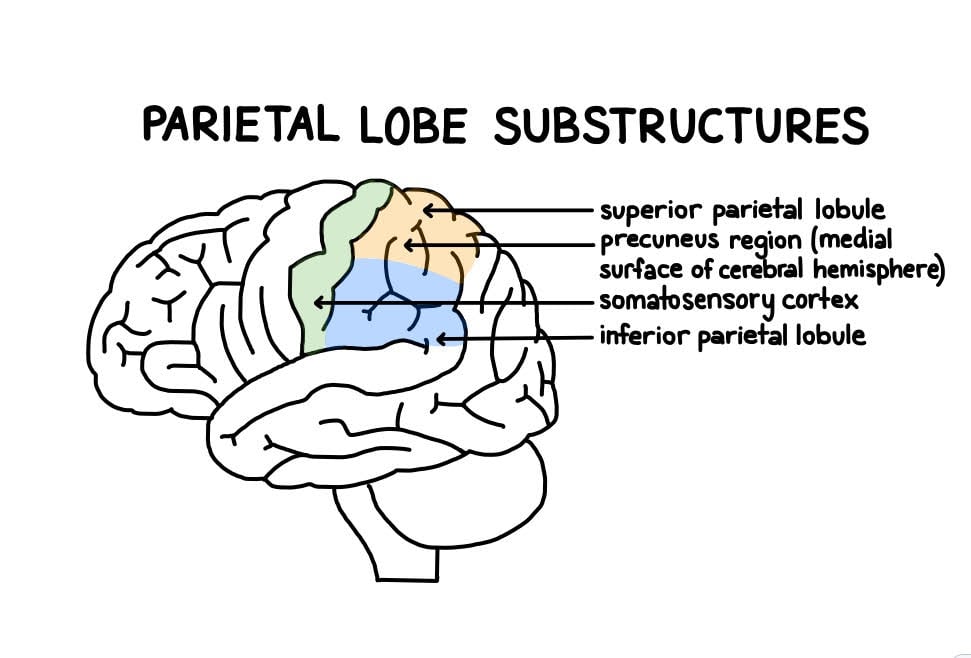
The parietal lobe is structurally divided into the somatosensory cortex, inferior parietal lobe, superior parietal lobe, intraparietal sulcus, and precuneus. The precuneus is a hub on the inner surface linked to memory and self-awareness.
Somatosensory Cortex
The somatosensory cortex’s main function is to receive and process sensory information from the entire body, such as touch, temperature, and pain.
It creates a body map in the brain. This lets it localize sensations, judge different degrees of pressure, and recognize the shape and texture of objects through touch.
Damage to this area could result in difficulties in perceiving touch and recognizing objects by touch.
How Does Your Brain Know What You’re Holding?
Imagine reaching into your bag or pocket to find your keys without looking. You can feel the shape, texture, and size—and somehow, you just know when you’ve got them. That’s your parietal lobe at work.
This brain region processes touch, pressure, and spatial awareness, allowing you to recognize objects by feel alone. It creates a “map” of your body and surroundings, helping you interact with the world—without needing to rely on sight.
Inferior Parietal Lobe
The inferior parietal lobe sits below the intraparietal sulcus. It contains two key regions: the supramarginal gyrus, involved in phonological processing and skilled hand movements, and the angular gyrus, a hub for reading, writing and arithmetic.
The two sides do different jobs.
On the left, this region is woven into the language network and supports reading, writing and calculation. On the right, it is more important for spatial attention and visuomotor processing, and has been linked to recognising emotion in faces.
The pattern flips for damage too. Left-side damage is linked to impaired speech repetition and difficulty with maths and calculation (acalculia). Right-side damage more often disrupts spatial attention and the integration of separate parts into a coherent whole.
Superior Parietal Lobe
The superior parietal lobe is concerned with spatial orientation and sensorimotor integration, and it receives a lot of visual and sensory signals from the hands.
Damage to the superior parietal lobe is most closely linked to optic ataxia. The person can see an object clearly, yet the hand fumbles or misses when reaching for it. The visual-to-motor link that guides the reach has broken down.
Related parietal damage can also disrupt touch-based object recognition. Right-hemisphere damage, in particular, can produce hemispatial neglect, a reduced awareness of one side of space.
Intraparietal Sulcus
The intraparietal sulcus is the deep groove that separates the superior from the inferior parietal lobule. It is one of the most closely studied strips of cortex in the field.
Its banks translate raw sensory coordinates into the frames needed for eye movements, reaching, grasping, and spatial attention. The same circuitry is also central to number processing. It houses a “mental number line” that represents quantity regardless of whether numbers appear as digits, words, or dots (Dehaene et al., 2003).
This is why arithmetic can feel spatial, and why damage here can selectively impair calculation, a deficit known as acalculia.
Precuneus
The precuneus sits on the medial surface of the parietal lobe, tucked between the somatosensory cortex and the occipital lobe. Its hard-to-reach location long delayed its study. It is now seen as one of the busiest hubs in the cortex (Cavanna & Trimble, 2006).
Functional neuroimaging links the precuneus to visuospatial imagery, episodic memory retrieval, and taking a first-person perspective on one’s own body and actions. It is also a default-mode network hub. That network runs when the mind rests rather than focuses on a task.
This area is also thought to play a role in self-awareness and consciousness.
What Happens When the Parietal Lobe is Damaged?
Damage to the parietal lobes, which can result from conditions like stroke, vascular disease, tumors, traumatic brain injury, or infections, can lead to a variety of symptoms:
- Hemispatial neglect: inability to locate/recognize objects, events, and body parts
- Visual attention deficits and difficulty discriminating sensory information
- Disorientation, lack of coordination, and impaired spatial awareness
- Impaired reading, writing, and drawing abilities without speech deficits
- Mathematical difficulties (acalculia) and language disorders (aphasia) from left-side damage
- Gerstmann’s Syndrome: a tetrad from damage near the left angular gyrus (Gerstmann, 1940) — finger agnosia (trouble naming one’s own fingers), left/right confusion, and difficulty with maths, reading and writing.
- Right-side damage: perceptual deficits integrating parts into wholes and spatial skill issues
- Right-side damage: self-care difficulties, impaired construction tasks, and contralateral neglect
The clearest demonstration of what neglect actually disrupts comes from a classic study of patients with right-hemisphere damage.
- Aim: to test whether neglect is only a problem of seeing the left side of a scene, or whether it also affects the brain’s internal, imagined map of space (Bisiach & Luzzatti, 1978).
- Method: patients with left neglect were asked to picture a familiar square from memory and describe every building they could “see,” first from one end and then from the opposite end.
- Results: from each imagined viewpoint, patients described only the buildings on the right, always omitting whichever buildings had shifted to the left of their mental image.
- Conclusion: neglect is not just a visual problem. It also distorts the brain’s internal spatial map, so the same content is reported or ignored depending purely on which side of the imagined scene it falls on.
Research has provided further insights into parietal lobe damage:
- Fridriksson et al. (2010) found that damage to the left inferior parietal lobes can impair speech repetition.
- Freund (2003) showed that anterior parietal damage can weaken motor control, while posterior damage affects virtually all aspects of somatosensory function.
- Zhou et al. (2007) found that gray and white matter volume reductions in parietal regions are common in schizophrenia and may account for some symptoms.
- Functional neuroimaging suggests the precuneus is involved in visuospatial imagery, episodic memory retrieval, first-person perspective, self-awareness and consciousness (Cavanna & Trimble, 2006).
Overall, parietal lobe damage can significantly affect perception, sensory integration, spatial awareness, language, and cognitive ability, with distinct deficits following left- versus right-side lesions.
Ongoing research continues to elucidate the complex functions of this brain region.
Key Takeaways
- Location: Sits behind the frontal lobe and above the temporal lobe, forming roughly a fifth of the cerebral cortex.
- Core Role: Integrates touch, body position and vision into a working map of the body and the space around it.
- Substructures: The somatosensory cortex, superior and inferior parietal lobules, intraparietal sulcus and precuneus each handle a different job, from touch to number to memory.
- Left vs Right: Left-side damage tends to disrupt language and calculation; right-side damage more often causes neglect of the left side of space.
- Hemispatial Neglect: Right-side damage especially can leave a person unaware of one side of their body or surroundings, despite intact eyesight.
- Network View: Modern research treats these abilities as the work of connected brain networks, not a single “spatial centre” in one lobe.
Critical Evaluation
The parietal lobe’s role in touch, space and attention is well supported, but treating it as a single “spatial organ” oversimplifies what the evidence actually shows. Three points are worth weighing before drawing firm conclusions:
- Not One Function: different jobs — touch, movement, number, language — are handled by different subregions, not by the lobe as a whole.
- A Network, Not a Lobe: modern evidence places these functions in large-scale brain networks, with the parietal lobe as one connected node rather than a stand-alone control centre.
- Imprecise Lesions: much of the evidence comes from strokes that damage grey matter and neighbouring fibre tracts together, making it hard to pin a function to one exact spot.
Not One Function
Somatosensory processing, reaching and grasping, spatial attention, number and language each depend on a different piece of the parietal lobe. The postcentral gyrus reads out touch and joint position on an orderly body map.
Behind it, the superior parietal lobule guides reaching. The intraparietal sulcus represents number and steers eye movements, while the left angular and supramarginal gyri support reading, writing and calculation.
These are separate systems sharing one lobe, not one function wearing different hats. A lesion in the somatosensory strip disrupts touch. A lesion just centimetres away, in the angular gyrus, can leave touch intact yet derail arithmetic and writing.
Treating the whole lobe as “the spatial brain” therefore risks a category error. The honest summary is that several distinct functions are carried by several distinct subregions, wired into different large-scale circuits (Corbetta & Shulman, 2002; Dehaene et al., 2003).
A Network, Not a Lobe
Spatial attention, reaching and number processing do not live in the parietal lobe alone. They depend on large-scale frontoparietal networks (Corbetta & Shulman, 2002).
Hemispatial neglect makes the point clearly. It is no longer read as damage to one “neglect centre.” It reflects a breakdown in how attention networks interact with each other. Two patients with similar parietal damage can show quite different patterns of neglect, depending on which network connections survive.
This network view does not weaken the parietal lobe’s importance. It explains it. The lobe is a critical hub inside several circuits, and losing that hub disrupts everything wired through it, even when the damage itself is small.
That is why imaging now maps neglect onto disrupted networks, not onto a single point of damage.
Imprecise Lesions
Vallar and Perani (1986) mapped neglect in 110 stroke patients and found it most reliably tied to the inferior parietal lobule. Yet a meaningful number of patients with damage in that same region showed no neglect at all. Some patients even developed neglect after damage elsewhere entirely.
Natural strokes rarely respect tidy anatomical borders. A single lesion usually destroys grey matter and the white-matter fibre tracts running beneath it at the same time.
Its size and shape also vary enormously from one patient to the next. Working backward from such damage to a “normal” function is therefore an indirect, error-prone method, however consistent the overall pattern looks across a large sample.
A single case can mislead. Some of the most influential evidence rests on just one or two patients. The original demonstration that neglect can distort even an imagined scene shows this. A single striking case is powerful, yet it is a fragile base for general claims.
Contemporary Research
Recent work asks less whether the parietal lobe supports space and attention, and more how its parts are wired into networks that can fail after injury.
- Aim: to find out why visual neglect looks so different from one patient to the next, and whether each symptom traces to a different piece of damage, in grey matter or in the connecting white-matter tracts (Toba et al., 2017).
- Method: 25 patients with recent right-hemisphere stroke were tested on several separate neglect tasks (line bisection, drawing, cancellation, and reading), then had their lesions mapped with structural MRI and diffusion tractography.
- Results: damage to the angular and supramarginal gyri predicted poor performance across every task, but damage to specific white-matter tracts predicted specific symptoms: one tract for line bisection and copying, a different tract for cancellation.
- Conclusion: neglect is not one deficit from one lesion. It is the disruption of overlapping frontoparietal networks, and disconnection matters as much as destruction.
Other recent work fits the same picture from different angles. Resting-state imaging shows disrupted connectivity between attention and sensorimotor networks after neglect (Baldassarre et al., 2014).
Imaging of healthy brains has traced a similar network origin for the mild leftward attention bias everyone shows, called pseudoneglect (Chen et al., 2019). Brain stimulation confirms the intraparietal sulcus takes part in more than one network at once (Capotosto et al., 2023).
The signal is consistent. Work on number processing, meanwhile, continues to test what exactly its cells encode (Bulthé et al., 2015). Together, this evidence points the same way: the parietal lobe works as a set of connected hubs, not a single control room. The picture keeps sharpening.
References
Baldassarre, A., Ramsey, L., Hacker, C. L., Callejas, A., Astafiev, S. V., Metcalf, N. V., Snyder, A. Z., & Corbetta, M. (2014). Large-scale changes in network interactions as a physiological signature of spatial neglect. Brain, 137(12), 3267-3283.
Bisiach, E., & Luzzatti, C. (1978). Unilateral neglect of representational space. Cortex, 14(1), 129-133.
Bulthé, J., De Smedt, B., & Op de Beeck, H. P. (2015). Visual number beats abstract numerical magnitude: Format-dependent representation of Arabic digits and dot patterns in human parietal cortex. Journal of Cognitive Neuroscience, 27(7), 1376-1387.
Capotosto, P., Sulpizio, V., Galati, G., & Baldassarre, A. (2023). Visuo-spatial attention and semantic memory competition in the parietal cortex. Scientific Reports, 13, 6218.
Cavanna, A. E., & Trimble, M. R. (2006). The precuneus: a review of its functional anatomy and behavioural correlates. Brain, 129(3), 564-583.
Ferro, A., Bonivento, C., Delvecchio, G., Bellani, M., Perlini, C., Dusi, N., Marinelli, V., Ruggeri, M., Altamura, C., Crespo-Facorro. & Brambilla, P. (2017). Longitudinal investigation of the parietal lobe anatomy in bipolar disorder and its association with general functioning. Psychiatry Research: Neuroimaging, 267, 22-31.
Freund, H. J. (2003). Somatosensory and motor disturbances in patients with parietal lobe lesions. Advances in Neurology, 93, 179-193.
Chen, J., Lee, A. C. H., O’Neil, E. B., Abdul-Nabi, M., & Niemeier, M. (2019). Mapping the anatomy of perceptual pseudoneglect: A multivariate approach. NeuroImage, 207, 116402.
Corbetta, M., & Shulman, G. L. (2002). Control of goal-directed and stimulus-driven attention in the brain. Nature Reviews Neuroscience, 3(3), 201-215.
Dehaene, S., Piazza, M., Pinel, P., & Cohen, L. (2003). Three parietal circuits for number processing. Cognitive Neuropsychology, 20(3-6), 487-506.
Fridriksson, J., Kjartansson, O., Morgan, P. S., Hjaltason, H., Magnusdottir, S., Bonilha, L., & Rorden, C. (2010). Impaired speech repetition and left parietal lobe damage. Journal of Neuroscience, 30(33), 11057-11061.
Gerstmann, J. (1940). Syndrome of finger agnosia, disorientation for right and left, agraphia and acalculia. Archives of Neurology and Psychiatry, 44(2), 398-408.
Goodale, M. A., & Milner, A. D. (1992). Separate visual pathways for perception and action. Trends in Neurosciences, 15(1), 20-25.
Lou, H. C., Luber, B., Crupain, M., Keenan, J. P., Nowak, M., Kjaer, T. W., Sackein, A. A. & Lisanby, S. H. (2004). Parietal cortex and representation of the mental self. Proceedings of the National Academy of Sciences, 101 (17), 6827-6832.
Milner, A. D. (1998). Streams and consciousness: visual awareness and the brain. Trends in Cognitive Sciences, 2 (1), 25-30.
Radua, J., Phillips, M. L., Russell, T., Lawrence, N., Marshall, N., Kalidindi, S., El-Hage, W., McDonald, C., Giampietro, V., Brammer, David, A. S. & Surguladze, S. A. (2010). Neural response to specific components of fearful faces in healthy and schizophrenic adults. Neuroimage, 49 (1), 939-946.
Toba, M. N., Migliaccio, R., Batrancourt, B., Bourlon, C., Duret, C., Pradat-Diehl, P., Dubois, B., & Bartolomeo, P. (2017). Common brain networks for distinct deficits in visual neglect: A combined structural and tractography MRI approach. Neuropsychologia, 115, 167-178.
Torrey, E. F. (2007). Schizophrenia and the inferior parietal lobule. Schizophrenia Research, 97 (1-3), 215-225.
Vallar, G., & Perani, D. (1986). The anatomy of unilateral neglect after right-hemisphere stroke lesions: A clinical/CT-scan correlation study in man. Neuropsychologia, 24(5), 609-622.
Vance, A., Silk, T. J., Casey, M., Rinehart, N. J., Bradshaw, J. L., Bellgrove, M. A., & Cunnington, R. (2007). Right parietal dysfunction in children with attention deficit hyperactivity disorder, combined type: a functional MRI study. Molecular Psychiatry, 12 (9), 826-832.
Weiskrantz, L. (1977). Trying to bridge some neuropsychological gaps between monkey and man. British Journal of Psychology, 68 (4), 431-445.
Zhou, S. Y., Suzuki, M., Takahashi, T., Hagino, H., Kawasaki, Y., Matsui, M., Seto, H. & Kurachi, M. (2007). Parietal lobe volume deficits in schizophrenia spectrum disorders. Schizophrenia Research, 89 (1-3), 35-48.

