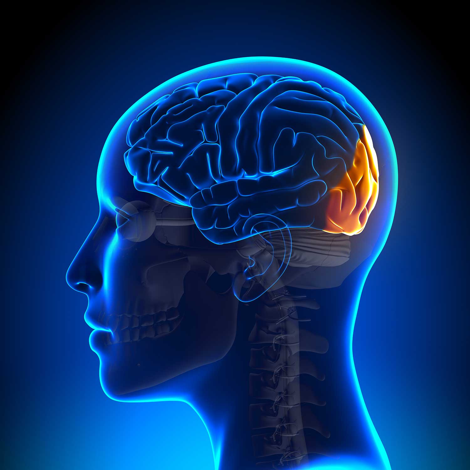
The occipital lobe is the brain’s visual processing center. Located at the back of the head, it helps us make sense of what we see, from colors and shapes to motion and depth.
Though it’s the smallest of the four major brain lobes, it’s crucial for everyday activities like reading, recognizing faces, and navigating the world.
Functions of the Occipital Lobe
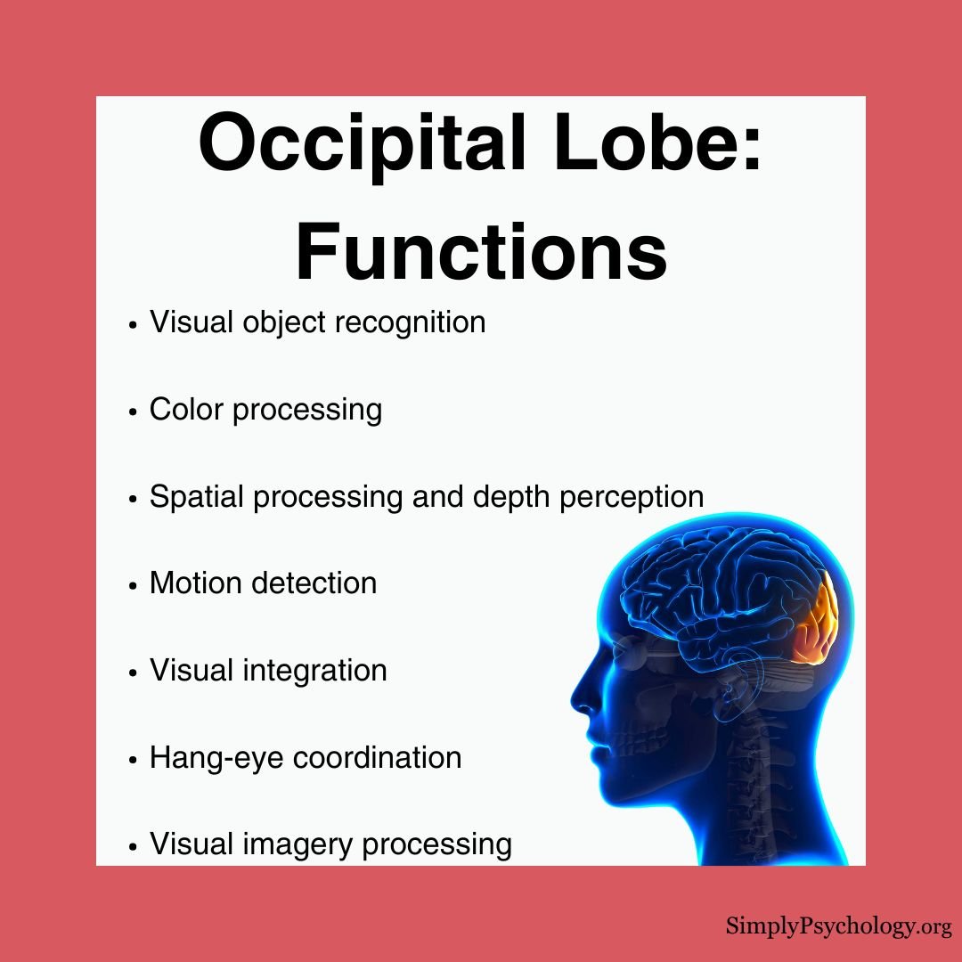
The occipital lobe handles a wide range of visual processing tasks. Here’s what it does:
1. Visual Recognition
- Identifies faces, objects, and written words
- Distinguishes fine details like texture, shape, and pattern
- Helps mentally rotate or imagine objects from different angles
2. Spatial and Motion Processing
- Maps the 3D world and calculates distance
- Supports depth perception and hand-eye coordination
- Tracks the movement and direction of objects
3. Color Perception
- Translates light wavelengths into color
- Helps us recognize color in different lighting
- Detects subtle color differences for tasks like matching shades
4. Integration With Other Brain Regions
- Works with the temporal lobe to identify and remember what we see
- Connects with motor areas to guide eye movements
- Coordinates with the parietal lobe for spatial awareness and mental imagery
Visual Phenomena Involving the Occipital Lobe
This lobe plays a surprising role in unique visual experiences:
- Dream imagery: It helps generate images we “see” in dreams.
- Synaesthesia: Some people experience colors when reading letters or numbers.
- Blindsight: People with damage to the primary visual cortex may still point to a light or detect movement while insisting they see nothing.
- Visual hallucinations: Damage or stimulation can produce vivid visual experiences.
Location
The occipital lobe is located at the very back of the brain beneath the occipital bone and behind the parietal and temporal lobes.
It is the smallest lobe of the brain, accounting for around 12% of the total surface area of the brain’s cortex.
There is one occipital lobe in each hemisphere. A deep groove, the cerebral fissure, separates the two hemispheres.
The lobe’s borders are set by grooves (sulci), not a hard wall. Its main landmarks make it easy to find.
- Parieto-occipital sulcus: On the inner surface, this deep groove separates the occipital lobe from the parietal lobe in front.
- Preoccipital notch: On the outer surface, no deep groove marks the border. Convention draws it from the parieto-occipital sulcus to this small indentation.
- Occipital pole: The rearmost tip of the hemisphere, where the center of vision (the fovea) is represented.
- Calcarine sulcus: This deep horizontal groove holds the primary visual cortex. The cuneus above it maps the lower visual field, and the lingual gyrus below maps the upper.
Each occipital lobe processes the opposite half of the visual field, not the opposite eye. The left visual field, seen by both eyes, is routed to the right occipital lobe. Partial crossing of the optic nerves makes this possible.

Structure
The occipital lobes can be divided into several functional areas. Some have clear anatomical markers.
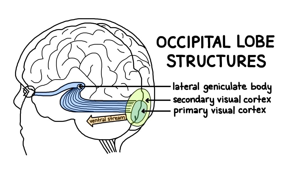
The primary visual cortex (V1) sits in the calcarine sulcus. A visible white stripe within it, the stria of Gennari, gives it the name “striate cortex”.
Neurons in the occipital cortex keep an orderly map of the visual field, called retinotopy. Neighboring points on the retina project to neighboring points on the cortex.
The map is distorted. The central few degrees of vision, the fovea, claim a hugely enlarged share of cortex near the occipital pole, while the periphery is squeezed into a small strip. This is called cortical magnification.
Magnification is why we see fine detail only where we look, and why reading needs constant eye movements to bring words onto the fovea. Functional MRI first measured these maps in living people in the mid-1990s (Sereno et al., 1995).
The motor cortex also plays a role in the muscles of the eyes, which the occipital lobes rely on heavily.
Primary Visual Cortex (V1 or Brodmann Area 17)
- Detects motion, light, color, and location
- First stop for visual information from the eyes
V1 is the first cortical stage of vision. It holds the brain’s most detailed map of the visual field, and visual information must pass through it to reach conscious awareness.
The classic evidence comes from David Hubel and Torsten Wiesel, who shared the 1981 Nobel Prize for it (Hubel & Wiesel, 1959, 1962, 1968).
- Aim: To find out how single neurons in the primary visual cortex respond to light, and how the cortex is organized.
- Method: They recorded single cells in anesthetized cats (1959, 1962) and later monkeys (1968) while bars, edges and spots were projected onto a screen.
- Results: Cells barely responded to diffuse light but fired to edges of a particular orientation. Cells sharing an orientation were stacked in columns, alongside separate columns for each eye.
- Conclusion: V1 contains feature detectors that recode the image as oriented edges, arranged in a columnar map of the visual field.
Hubel and Wiesel described a hierarchy of cells. Simple cells respond to an edge in one position. Complex cells respond to that edge anywhere in a wider area. Hypercomplex cells also need the edge to be a set length.
The evidence is exceptionally strong. It was replicated across two species and links structure directly to function. But it is reductionist: edge detectors alone cannot explain how a whole face or scene is recognized. The recordings also came from anesthetized animals viewing simple stimuli.
Secondary Visual Cortex (V2 to V5, Areas 18 & 19)
- V2: Much of area 18. It refines input from V1 and processes contours, including illusory and figure-ground borders.
- V3 and V3A: Handle dynamic form, combining motion with shape.
- V4: Analyzes color and complex form, keeping a surface’s color stable under changing light.
- V5 (MT): Sits at the border with the temporal lobe. It tracks the direction and speed of moving objects.
These areas work in parallel, each analyzing one attribute of the scene. That is why damage to one can remove color or motion while leaving the rest of vision intact.
Ventral Stream (“What” Pathway)
- Sends information to the temporal lobe
- Helps us understand what we’re seeing (e.g., recognizing a face or a cup)
Ungerleider and Mishkin (1982) found that monkeys with damage to the inferotemporal cortex struggled to tell objects apart. This stream supports recognition of objects, faces and words.
Dorsal Stream (“Where” Pathway)
- Sends information to the parietal lobe
- Tells us where things are in space and how they’re moving
Monkeys with damage to the posterior parietal cortex instead struggled with spatial tasks (Ungerleider & Mishkin, 1982). Milner and Goodale later argued that this stream is better described as vision for action than as “where”.
Patient D.F. had ventral-stream damage. She could not report or match the tilt of a slot, yet she rotated her hand correctly to post a card through it (Goodale & Milner, 1992; Milner, 1998).
Patients with dorsal damage show the mirror pattern. They can describe an object but cannot shape the hand to grasp it.
Lateral Geniculate Nucleus and Lingual Gyrus
- Lateral geniculate nucleus (LGN): A relay in the thalamus, outside the occipital lobe. It keeps the two eyes’ inputs separate and forwards them to V1.
- Optic radiations: Fibers that fan from the LGN back to V1. Those carrying the upper visual field loop forward through the temporal lobe first.
- Lingual gyrus: Occipital cortex below the calcarine sulcus. It maps the upper half of the opposite visual field.
The main route for conscious vision, the geniculostriate pathway, runs from the retina through the optic chiasm and LGN to V1. An older route through the superior colliculus, a midbrain structure that helps orient the eyes, bypasses V1. It is the leading explanation for blindsight.
What Happens When the Occipital Lobe Is Damaged?
Damage to the occipital lobe can affect visual perception in several ways:
Common Symptoms
- Partial or complete blindness
- Visual hallucinations
- Difficulty reading, writing, or recognizing objects
- Poor depth perception or motion tracking
- Inability to distinguish colors or shapes
How Damage Occurs
Injuries can result from trauma, stroke, tumors, or infections. Even small areas of damage can create “blind spots” in a person’s field of view.
In some cases, the brain adapts by rerouting information, but widespread damage may cause permanent vision loss.
Syndromes After Occipital Damage
The side of the lesion decides which side of space is affected. The stage of processing destroyed decides which aspect of vision is lost. Several named syndromes show this:
- Cortical blindness: Extensive damage to V1 in both hemispheres abolishes conscious vision. The eyes work, and the pupils still react to light.
- Hemianopia: Damage to one occipital lobe blinds the opposite half of the visual field in both eyes. The very center of vision is often spared.
- Anton’s syndrome: A person made blind by damage to both lobes denies being blind and describes sights they cannot see (Anton, 1899).
- Visual agnosia: Basic sight survives but recognition fails. In prosopagnosia, the loss is specific to faces.
- Achromatopsia: Damage around V4 removes color vision but leaves form and motion intact (Zeki, 1990).
- Akinetopsia: Damage to V5 removes motion perception. Patient L.M. saw coffee freeze as it poured (Zihl et al., 1983).
Blindsight: Vision Without Awareness
Blindsight shows that some vision can survive without awareness. It is the mirror image of Anton’s syndrome, where a person cannot see but does not know it.
The classic demonstration came from Weiskrantz and colleagues (1974).
- Aim: To test whether a patient with a blind region caused by occipital damage kept any visual ability there.
- Method: Patient D.B. had part of his right occipital lobe removed, leaving the left half of his visual field blind. He was asked to guess the location, movement and orientation of stimuli shown there.
- Results: D.B. performed far above chance. He could point to stimuli, detect movement and tell line orientations apart, while insisting he saw nothing.
- Conclusion: Some visual ability survives loss of V1 without awareness. V1 seems necessary for visual experience but not for all visually guided behavior.
Blindsight is most often explained by the older pathway, through the superior colliculus, that bypasses V1. Skeptics have questioned it.
The evidence rests on a few intensively studied patients, and residual vision might reflect spared islands of V1 or scattered light. Replication in further patients and in monkeys, plus careful controls for light scatter, has strengthened the case.
Associated Conditions
Riddoch Syndrome
A rare condition linked to the occipital lobes is Riddoch syndrome, also called the Riddoch phenomenon. Riddoch (1917) noticed that some patients with occipital injuries could perceive moving objects in a blind part of their visual field, but not stationary ones.
Autism
Studies using MRI have revealed significant occipital cortex enlargement in autistic individuals compared to control groups.
Functional imaging tells a similar story. A meta-analysis of such studies (Samson et al., 2012) compared autistic and non-autistic people. During visual tasks, autistic people showed more activity in occipital, temporal and parietal regions.
This fits reports that autistic people often perform better on visual search and discrimination tasks. The findings are correlational, though. They cannot show that occipital differences cause the enhanced perception.
Depression
A study of 25 adults over 55 with late-life depression used single photon emission computed tomography (SPECT), a scan that maps blood flow. It found reduced blood flow in the parieto-occipital lobes compared with healthy controls (Ishizaki et al., 2008).
Treatment shifted the pattern. After a mean of 13.7 weeks of drug treatment, blood flow rose in the right parieto-occipital regions and parts of the left prefrontal cortex. The study was correlational. The parieto-occipital region also includes parietal cortex, so it cannot show that visual processing drives mood.
Epilepsy
Occipital lobe epilepsy presents unique challenges for treatment. While surgery can be successful, precise identification of the affected area through brain imaging remains difficult.
The strongest evidence on surgery is a systematic review and meta-analysis (a study pooling the results of many studies) of 27 case series covering 584 patients (Harward et al., 2018). Overall, 65% were seizure-free afterward.
Seizure freedom was more likely in patients under 18, with a focal lesion, or with an abnormal preoperative MRI. There was a cost to vision. Of the 175 patients whose vision was assessed, 57% showed some visual decline.
A study of 21 children aged 6 to 14 with idiopathic occipital lobe epilepsy (no identified cause) found no significant difference from matched healthy controls in basic neuropsychological functions.
Even so, the children scored lower on attention, memory and intellectual functioning. The deficits were subtle (Gülgönen et al., 2000).
Schizophrenia
Brain imaging studies have revealed reduced gray matter volume in the visual association cortex of male patients with chronic schizophrenia (Onitsuka et al., 2007).
The finding is consistent with the visual processing difficulties seen in schizophrenia. It is correlational, so it cannot show cause.
The connection between occipital lobe abnormalities and visual hallucinations in schizophrenia continues to be an active area of research.
Critical Evaluation of Occipital Lobe Research
The occipital lobe’s role in vision is among the best-supported findings in biopsychology. Even so, the evidence has limits, and each is worth weighing.
Strengths: Converging Evidence
Several independent methods point the same way. Single-neuron recordings show V1 cells acting as edge detectors (Hubel & Wiesel, 1959, 1962, 1968). Damage to the calcarine sulcus produces clean field cuts. Selective loss of color (Zeki, 1990) and of motion (Zihl et al., 1983) shows separate specialist areas.
Modern scanning adds a fourth line of evidence: high-resolution mapping of the living human brain (Benson et al., 2018). The methods converge.
Because cellular, lesion and imaging evidence agree independently, the account makes precise clinical predictions. A lesion in the calcarine cortex, for example, predicts a specific blind area on the opposite side of the visual field. That is rare in biopsychology.
The occipital lobe is therefore a model case of anchoring a sensory function to a defined patch of cortex. Few brain regions have such secure ground beneath them.
Limitations of the Evidence
Much of the strongest evidence comes from naturally occurring damage, such as strokes and war wounds. These lesions rarely respect anatomical borders. They harm gray matter and the underlying optic radiations together, and they vary widely between patients.
Inferring normal function from imprecise damage is therefore indirect. It is one reason the exact boundaries of human V4 and V5 were disputed for years.
Some influential claims also rest on single cases: patient D.B. for blindsight (Weiskrantz et al., 1974) and patient L.M. for akinetopsia (Zihl et al., 1983). The cleanest cellular evidence comes from cats and monkeys, so applying it to humans was long an inference rather than a demonstration.
The psychiatric findings are weaker still.
Occipital differences in schizophrenia (Onitsuka et al., 2007) and autism (Samson et al., 2012) come from correlational imaging studies. They cannot show that occipital differences cause a condition rather than accompany it.
Is the Occipital Lobe a Single Vision Center?
Calling the occipital lobe “the visual cortex” hides how different its parts are. V1’s edge detectors, V4’s color analyzers and V5’s motion detectors are separate systems that happen to share a lobe. That is why a lesion in V4 removes color while a lesion in V5 removes motion.
Much of vision is completed outside the lobe. Recognizing a face or guiding a reach relies on temporal and parietal cortex (Ungerleider & Mishkin, 1982; Goodale & Milner, 1992).
The better description is an entry stage of a distributed system. Treating the lobe as a self-contained box where vision happens is a reductionist error. Vision is a team effort. A network account fits the evidence far better than any single “vision center” idea, because it explains selective losses.
Serial or Recurrent Processing?
Researchers also disagree about how processing runs inside the visual system.
Felleman and Van Essen (1991) mapped the connections among more than thirty areas of macaque visual cortex and arranged them into a strict hierarchy. Each stage takes its main input from the stage below.
Lamme and Roelfsema (2000) disagreed. They argued that a one-way cascade cannot explain figure-ground segregation, which needs recurrent processing. A fast feedforward sweep gives only rudimentary, largely unconscious detection. Later feedback from higher areas to V1 is what lets a scene be consciously split into figure and ground.
The two views do not clash on anatomy, because feedback connections belong to the hierarchy Felleman and Van Essen catalogued.
They clash on logic. Is vision completed in one pass on the way up, or does a loop let later stages keep revising earlier ones?
Contemporary Research
Recent work has moved from inferring the lobe’s organization to measuring it directly in living people. The anchor is a large ultrahigh-field mapping study from the Human Connectome Project.
- Aim: To map the retinotopic organization of human visual cortex in a large sample. The study also produced population receptive field (pRF) estimates: the region of visual field that drives each patch of cortex.
- Method: Benson et al. (2018) scanned 181 healthy young adults with 7-Tesla fMRI at 1.6-mm resolution as they viewed rotating wedges, expanding rings and moving bars.
- Results: Orderly retinotopic maps appeared across occipital, temporal, parietal and even frontal cortex. Receptive fields grew larger up the visual hierarchy and away from the fovea. Maps were reliable within individuals but varied between them.
- Conclusion: The study confirmed, non-invasively and at population scale, the map and magnification architecture first inferred from wounded soldiers and animal recordings.
It sits near the top of the evidence hierarchy for descriptive human neuroanatomy: a large sample, high resolution, open data and demonstrated reliability. Its limit is that fMRI measures blood-flow signals from populations of neurons. It cannot resolve individual orientation columns, and the sample was healthy young adults.
Columns and Receptive Fields in Living Brains
Columns are now within reach. In a rare person born without an optic chiasm, sub-millimeter imaging showed left- and right-hemifield maps in V1 interleaved into columns (Olman et al., 2018).
The columns matched the scale of ocular-dominance columns, the first in vivo human evidence of this layout.
Dumoulin and Wandell (2008) showed that human pRF sizes in V1 to V3 agree well with receptive fields recorded in monkeys at matching eccentricities. That links the whole-brain and cellular evidence.
Surgery Outcomes
For epilepsy surgery, the best synthesis is the Harward et al. (2018) meta-analysis described above: about two-thirds of patients became seizure-free, at some cost to vision.
Weighted by design quality, the mapping and meta-analytic evidence is strongest. The disorder-specific findings discussed earlier remain the weakest link.
Key Takeaways
- Definition: The occipital lobe is the rearmost lobe of the cerebral cortex and the brain’s main visual processing center.
- Location: It sits at the back of the head, behind the parietal and temporal lobes, and is the smallest of the four lobes.
- Visual Map: The primary visual cortex (V1) holds an orderly map of the visual field. The center of vision gets far more space than the periphery.
- Two Streams: Processed signals leave along a ventral “what” stream to the temporal lobe and a dorsal “where/how” stream to the parietal lobe.
- Damage: Depending on the area hit, damage causes blind spots, cortical blindness, or loss of color or motion perception.
- Evidence: The core findings are well supported. Links to conditions such as schizophrenia and autism are correlational and tentative.
Anton, G. (1899). Über die Selbstwahrnehmung der Herderkrankungen des Gehirns durch den Kranken bei Rindenblindheit und Rindentaubheit. Archiv für Psychiatrie und Nervenkrankheiten, 32(1), 86–127.
Benson, N. C., Jamison, K. W., Arcaro, M. J., Vu, A. T., Glasser, M. F., Coalson, T. S., Van Essen, D. C., Yacoub, E., Ugurbil, K., Winawer, J., & Kay, K. (2018). The Human Connectome Project 7 Tesla retinotopy dataset: Description and population receptive field analysis. Journal of Vision, 18(13), 23. https://doi.org/10.1167/18.13.23
Binder, D. K., Von Lehe, M., Kral, T., Bien, C. G., Urbach, H., Schramm, J., & Clusmann, H. (2008). Surgical treatment of occipital lobe epilepsy. Journal of Neurosurgery, 109 (1), 57-69.
Dumoulin, S. O., & Wandell, B. A. (2008). Population receptive field estimates in human visual cortex. NeuroImage, 39(2), 647–660. https://doi.org/10.1016/j.neuroimage.2007.09.034
Felleman, D. J., & Van Essen, D. C. (1991). Distributed hierarchical processing in the primate cerebral cortex. Cerebral Cortex, 1(1), 1–47. https://doi.org/10.1093/cercor/1.1.1
Goodale, M. A., & Milner, A. D. (1992). Separate visual pathways for perception and action. Trends in Neurosciences, 15(1), 20–25. https://doi.org/10.1016/0166-2236(92)90344-8
Gülgönen, S., Demirbilek, V., Korkmaz, B., Dervent, A., & Townes, B. D. (2000). Neuropsychological functions in idiopathic occipital lobe epilepsy. Epilepsia, 41 (4), 405-411.
Harward, S. C., Chen, W. C., Rolston, J. D., Haglund, M. M., & Englot, D. J. (2018). Seizure outcomes in occipital lobe and posterior quadrant epilepsy surgery: a systematic review and meta-analysis. Neurosurgery, 82 (3), 350-358.
Hubel, D. H., & Wiesel, T. N. (1959). Receptive fields of single neurones in the cat’s striate cortex. The Journal of Physiology, 148(3), 574–591. https://doi.org/10.1113/jphysiol.1959.sp006308
Hubel, D. H., & Wiesel, T. N. (1962). Receptive fields, binocular interaction and functional architecture in the cat’s visual cortex. The Journal of Physiology, 160(1), 106–154. https://doi.org/10.1113/jphysiol.1962.sp006837
Hubel, D. H., & Wiesel, T. N. (1968). Receptive fields and functional architecture of monkey striate cortex. The Journal of Physiology, 195(1), 215–243. https://doi.org/10.1113/jphysiol.1968.sp008455
Ishizaki, J., Yamamoto, H., Takahashi, T., Takeda, M., Yano, M., & Mimura, M. (2008). Changes in regional cerebral blood flow following antidepressant treatment in late‐life depression. International Journal of Geriatric Psychiatry, 23 (8), 805-811.
Jessell, T. M. (1991). Principles of neural science (pp. 173-193). J. H. Schwartz, & E. R. Kandel (Eds.). New York: Elsevier.
Lamme, V. A. F., & Roelfsema, P. R. (2000). The distinct modes of vision offered by feedforward and recurrent processing. Trends in Neurosciences, 23(11), 571–579. https://doi.org/10.1016/S0166-2236(00)01657-X
McCarley, R. W., Wible, C. G., Frumin, M., Hirayasu, Y., Levitt, J. J., Fischer, I. A., & Shenton, M. E. (1999). MRI anatomy of schizophrenia. Biological Psychiatry, 45 (9), 1099-1119.
Milner, A. D. (1998). Streams and consciousness: Visual awareness and the brain. Trends in Cognitive Sciences, 2(1), 25–30. https://doi.org/10.1016/S1364-6613(97)01116-9
Olman, C. A., Bao, P., Engel, S. A., Grant, A. N., Purington, C., Qiu, C., Schallmo, M.-P., & Tjan, B. S. (2018). Hemifield columns co-opt ocular dominance column structure in human achiasma. NeuroImage, 164, 59–66. https://doi.org/10.1016/j.neuroimage.2016.12.063
Onitsuka, T., McCarley, R. W., Kuroki, N., Dickey, C. C., Kubicki, M., Demeo, S. S., Frumin, M., Kikinis, R., Jolesz, F. A. & Shenton, M. E. (2007). Occipital lobe gray matter volume in male patients with chronic schizophrenia: A quantitative MRI study. Schizophrenia Research, 92 (1-3), 197-206.
Park, K. C., Yoon, S. S., & Rhee, H. Y. (2011). Executive dysfunction associated with stroke in the posterior cerebral artery territory. Journal of Clinical Neuroscience, 18 (2), 203-208.
Piven, J., Arndt, S., Bailey, J., & Andreasen, N. (1996). Regional brain enlargement in autism: a magnetic resonance imaging study. Journal of the American Academy of Child & Adolescent Psychiatry, 35 (4), 530-536.
Riddoch, G. (1917). Dissociation of visual perceptions due to occipital injuries, with especial reference to appreciation of movement. Brain, 40(1), 15–57. https://doi.org/10.1093/brain/40.1.15
Samson, F., Mottron, L., Soulières, I., & Zeffiro, T. A. (2012). Enhanced visual functioning in autism: An ALE meta‐analysis. Human Brain Mapping, 33 (7), 1553-1581.
Sereno, M. I., Dale, A. M., Reppas, J. B., Kwong, K. K., Belliveau, J. W., Brady, T. J., Rosen, B. R., & Tootell, R. B. (1995). Borders of multiple visual areas in humans revealed by functional magnetic resonance imaging. Science, 268(5212), 889–893. https://doi.org/10.1126/science.7754376
Sperling, J. M., Prvulovic, D., Linden, D. E., Singer, W., & Stirn, A. (2006). Neuronal correlates of colour-graphemic synaesthesia: AfMRI study. Cortex, 42 (2), 295-303.
Tate, D. F., Bigler, E. D., McMahon, W., & Lainhart, J. (2007). The relative contributions of brain, cerebrospinal fluid-filled structures and non-neural tissue volumes to occipital-frontal head circumference in subjects with autism. Neuropediatrics, 38(01), 18-24.
Ungerleider, L. G., & Mishkin, M. (1982). Two cortical visual systems. In D. J. Ingle, M. A. Goodale, & R. J. W. Mansfield (Eds.), Analysis of visual behavior (pp. 549–586). MIT Press.
Weiskrantz, L., Warrington, E. K., Sanders, M. D., & Marshall, J. (1974). Visual capacity in the hemianopic field following a restricted occipital ablation. Brain, 97(1), 709–728. https://doi.org/10.1093/brain/97.1.709
Zeki, S. (1990). A century of cerebral achromatopsia. Brain, 113(6), 1721–1777. https://doi.org/10.1093/brain/113.6.1721
Zihl, J., von Cramon, D., & Mai, N. (1983). Selective disturbance of movement vision after bilateral brain damage. Brain, 106(2), 313–340. https://doi.org/10.1093/brain/106.2.313

