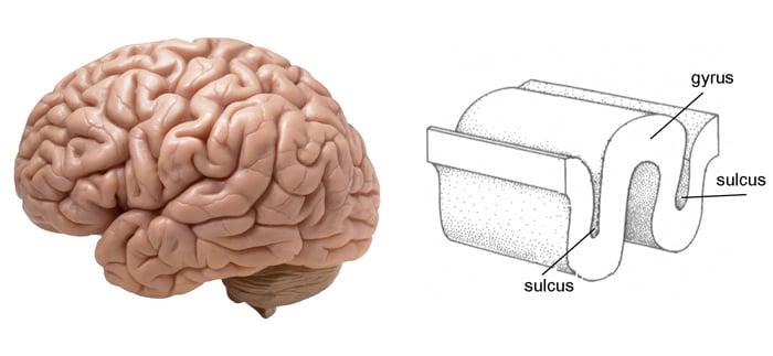
The brain’s outer surface isn’t smooth—it’s full of folds and grooves that give it a wrinkled appearance. These folds aren’t random. They’re made up of gyri (ridges) and sulci (grooves), and they play a crucial role in how our brains are organized and function.
Understanding these structures helps us map out different brain regions and link them to specific functions like movement, memory, and language.

What Are Gyri and Sulci?
- Gyri (singular: gyrus) are the raised ridges or folds on the surface of the brain.
- Sulci (singular: sulcus) are the grooves or furrows that separate the gyri.
This pattern of folds increases the brain’s surface area, packing in more neurons without increasing the brain’s size. The more surface area, the greater the brain’s ability to process complex information.
Why Is the Brain Folded?
The folding of the brain allows a large cortex to fit into the limited space of the skull. This is vital for higher-level functions like reasoning, memory, and language.
The layout of these folds isn’t exactly the same in every person, but key gyri and sulci appear consistently across human brains. These consistent features help scientists navigate and study the brain.
Gyri
Gyri are made up of grey matter—clusters of nerve cell bodies and dendrites that handle thinking, sensing, and processing.
By increasing surface area, gyri allow more neural connections and higher cognitive capacity. Each gyrus is linked to a particular function.

Types of Gyri
1. Cingulate gyrus
The cingulate gyrus is a component of the limbic system, consisting of a curved fold covering the corpus callosum (a bundle of nerve fibers connecting the right and left cerebral hemispheres).
The anterior portion has a significant role in processing emotions, enabling emotional vocalization, and facilitating bonding between caregiver and child.
The posterior portion handles spatial memory and coordinates movement, orientation, and navigation through its connections with the parietal and temporal lobes.
2. Precentral gyrus
The precentral gyrus is located in the posterior position of the frontal lobe and contains the primary motor cortex.
This structure creates and organizes the homunculus (‘little man’) map of the body. It controls motor movements on the body’s opposite side to where it is located within the brain.
3. Superior temporal gyrus
The superior temporal gyrus houses the auditory cortex, which processes sounds through precisely mapped sound frequencies.
Within this gyrus lies Wernicke’s area, which is crucial for language comprehension and the ability to produce spoken words.
Disorders Related to Gyri
When gyri don’t form properly, it can lead to brain malformations:
- Schizophrenia has been linked to structural abnormalities in the superior temporal gyrus, especially in patients who experience auditory hallucinations (Barta et al., 1990).
- Lissencephaly: A condition where the brain appears smooth due to a lack of gyri.
- Pachygyria: Abnormally large gyri.
- Polymicrogyria: Excessively small and numerous gyri with shallow sulci, often leading to developmental delays, seizures, and speech or motor difficulties.
Sulci
Sulci are the grooves that divide the brain’s ridges. The deeper grooves are called fissures.
These structures:
- Increase the surface area of the brain.
- Divide the brain into lobes and functional areas.
Some sulci appear early in fetal development (primary sulci), while others emerge later in life (secondary and tertiary sulci), often shaped by experience and brain growth.

Types of Sulci
1. Longitudinal fissure
The longitudinal fissure is a deep furrow that serves as the primary division between the left and right hemispheres of the brain.
Within this fissure lies the corpus callosum, which connects the hemispheres and enables the transfer of visual, auditory, and somatosensory information between them.
2. Central sulcus
The central sulcus, also known as the sulcus of Rolando, creates an important separation between the parietal and frontal lobes.
It defines the boundary between the primary motor cortex and primary somatosensory cortex.
Interestingly, its size relates to handedness, being larger in the left hemisphere for right-handed individuals and vice versa.
3. Parieto-occipital sulcus
The parieto-occipital sulcus forms a deep groove that creates the division between the parietal and occipital lobes.
Unlike primary sulci, this structure forms after birth as a secondary sulcus.
4. Lateral sulcus
The lateral sulcus, also known as the Sylvian sulcus, creates a deep groove that separates the parietal and temporal lobes, with the insular cortex nestled deep within it.
Disorders Related to Sulci
Changes in sulci can reflect or contribute to neurological issues:
- Perisylvian Syndrome: A rare condition involving the lateral sulcus, often resulting in speech and language impairments.
- Abnormal Central Sulcus Development: Can affect motor control and movement planning.
These structural variations can have long-term impacts on brain function and behavior.
Table: Major Gyri and Sulci of the Brain
| Structure | Type | Location | Function |
|---|---|---|---|
| Precentral Gyrus | Gyrus | Frontal lobe, anterior to central sulcus | Primary motor cortex – controls voluntary movement |
| Postcentral Gyrus | Gyrus | Parietal lobe, posterior to central sulcus | Primary somatosensory cortex – processes touch and proprioception |
| Cingulate Gyrus | Gyrus | Medial surface above corpus callosum | Emotion processing, bonding, spatial memory |
| Superior Temporal Gyrus | Gyrus | Temporal lobe, beneath lateral sulcus | Auditory processing, language comprehension (Wernicke’s area) |
| Central Sulcus | Sulcus | Between frontal and parietal lobes | Separates motor and sensory cortices |
| Lateral Sulcus (Sylvian) | Sulcus | Between frontal/parietal and temporal lobes | Houses auditory cortex and insula; involved in language |
| Longitudinal Fissure | Sulcus | Midline, between hemispheres | Divides brain into left and right hemispheres |
| Parieto-occipital Sulcus | Sulcus | Between parietal and occipital lobes | Separates visual processing from spatial/motor integration areas |
Brain Folding and Development
The brain starts forming folds during prenatal development. These folds reflect both genetics and environmental influences.
- Primary sulci appear early and are mostly hardwired.
- Secondary and tertiary sulci develop later and may be influenced by experience.
- Regions involved in complex functions like language tend to have more intricate folds.
Evolutionary Perspective
Cortical folding is a key marker of advanced brain function. Animals with more folds—such as humans, dolphins, and elephants—show greater cognitive abilities.
The gyrification index, which measures the degree of folding, is much higher in humans than in animals with smooth brains (like rodents). This folding allows for:
- More advanced problem-solving, language, and social skills
- More cortical surface area
- Denser neural networks
Summary
Gyri and sulci are more than just wrinkles on the brain—they’re the foundation for how the brain is organized, how it grows, and how it functions. From emotion and memory to movement and speech, these folds help make us who we are.
References
Banker, L., & Tadi, P. (2020). Neuroanatomy, Precentral Gyrus. StatPearls [Internet].
Barta, P. E., Pearlson, G. D., Powers, R. E., Richards, S. S., & Tune, L. E. (1990). Auditory hallucinations and smaller superior temporal gyral volume in schizophrenia. The American Journal of Psychiatry.
Haines, D. E., & Mihailoff, G. A. (2017). Fundamental Neuroscience for Basic and Clinical Applications E-Book . Elsevier Health Sciences.
Spreafico, R., & Tassi, L. (2012). Cortical malformations. Handbook of clinical neurology, 108, 535-557.

