Your body’s connection to voluntary movement
The somatic nervous system (SNS) is a division of the peripheral nervous system. It connects your brain and spinal cord (the central nervous system) to the rest of your body. This link runs both ways.
The autonomic nervous system handles automatic functions like heart rate and digestion. The SNS is different. It controls actions you do on purpose, such as picking up a glass or tying your shoes.
The SNS also carries sensory information (like touch, pain, and temperature) from the body to the brain. This helps you interpret and react to the world around you.

Key Takeaways
- The somatic nervous system (SNS) controls voluntary movements like walking, talking, and reaching for objects.
- It processes sensory input—such as touch, temperature, and pain—and sends that information to the brain.
- The SNS uses sensory and motor neurons to communicate between the brain and the body.
- Reflex arcs allow the body to react quickly to danger without needing the brain’s involvement.
- Damage to the SNS can cause muscle weakness, numbness, and nerve pain, but healthy habits may reduce your risk.
Somatic Nervous System Function
The somatic nervous system has two primary jobs:
1. Processing Sensory Input
Sensory receptors in your skin, eyes, ears, nose, and tongue detect changes in your environment, such as heat, texture, sound, or light. This information travels through sensory (afferent) neurons to your brain, where it’s interpreted.
Some receptors, called proprioceptors, sit inside your muscles. They report on muscle length and tension, which is how you sense where your arm is with your eyes closed.
Each sensory neuron’s signal first reaches a cluster of cell bodies called a dorsal root ganglion, just outside the spinal cord. From there it travels into the spinal cord and up to the brain.
Example: When you touch a hot stove, sensory neurons send a message to your brain letting you know it’s painful.
2. Controlling Voluntary Movement
Once your brain processes this information, it sends signals through motor (efferent) neurons to your skeletal muscles. These signals tell your body to move.
A single motor neuron and every muscle fibre it controls form a motor unit, the smallest building block of a voluntary contraction. Small motor units control fine movements, like those of the fingers, while large motor units produce powerful, coarse movements, like those of the thigh.
Moving a joint usually needs two muscles working together. Bending your elbow, for example, requires your biceps to contract while your triceps relaxes. This automatic pairing is called reciprocal innervation.
Example: After seeing a ball fly toward you, your brain sends a signal to your arm muscles to catch it.
How the Somatic Nervous System Works
Below is a step-by-step guide to how the somatic nervous system works:
- Detecting the Environment
Sensory receptors in places like your skin and eyes notice changes—like heat or light. - Creating a Signal
Sensory neurons generate electrical impulses that travel through your spinal nerves to the spinal cord. - Brain Processing
The brain receives and interprets the signals and decides how to respond. - Sending a Response
Motor neurons carry the brain’s instructions back down the spinal cord to your muscles. - Muscle Activation
The targeted skeletal muscles contract, completing the voluntary actio
Reflex Arcs
Not all movements need the brain’s involvement. Sometimes the body reacts so quickly that the brain isn’t part of the loop. This is called a reflex arc. It is a fixed circuit, mostly inside the spinal cord, that produces a fast response without the brain’s involvement.
A reflex arc lets your body respond to danger immediately, without waiting for the brain. Sensory neurons send signals straight to the spinal cord, which triggers a motor response.
Example: You accidentally touch something hot and pull your hand away before you even feel the pain.
Monosynaptic and Polysynaptic Reflexes
The simplest reflexes are monosynaptic: the sensory neuron connects directly to the motor neuron, with a single synapse between them. The knee-jerk reflex works this way, taking as little as 30 milliseconds.
More complex reflexes are polysynaptic. They route through one or more interneurons rather than a single synapse. Pulling your hand back from a hot surface is a withdrawal reflex. Pain signals excite the muscles that pull your hand back, while relaxing the muscles that would resist it.
At the same time, the opposite leg often stiffens. This crossed-extensor reflex helps you keep your balance while the injured leg lifts clear.
Common reflexes include:
- Blinking: when something approaches your eye
- Knee-jerk reflex: tested with a reflex hammer at the doctor’s office
- Withdrawal reflex: pulling away from something painful or hot
- Plantar reflex: curling of the toes when the sole of the foot is stroked

Components of the Somatic Nervous System
- Sensory (Afferent) Neurons: Carry signals from sensory receptors to the brain and spinal cord.
- Motor (Efferent) Neurons: Deliver signals from the brain and spinal cord to muscles.
- Ganglia: Clusters of neuron cell bodies located outside the brain and spinal cord.
- Glial Cells: Support cells that help neurons function effectively.
- Nuclei: Collections of neuron cell bodies in the central nervous system with a shared function.

Somatic vs. Autonomic nervous system
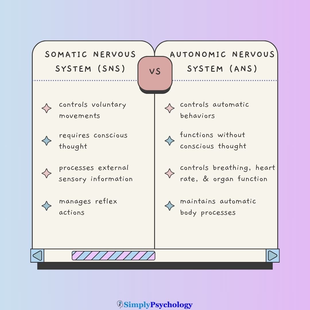
The SNS and the autonomic nervous system (ANS) are both part of the peripheral nervous system (PNS), but they have different functions:
| Somatic Nervous System (SNS) | Autonomic Nervous System (ANS) |
|---|---|
| Controls voluntary movements | Controls automatic behaviors |
| Requires conscious thought | Functions without conscious thought |
| Controls skeletal muscle movement | Controls breathing and heart rate |
| Processes external sensory information | Regulates internal organ function |
| Manages reflex actions | Maintains automatic body processes |
Working Together:
Both systems collaborate to maintain homeostasis (“homo-” meaning same and “-stasis” meaning state of equilibrium). The SNS handles external responses to environmental changes, while the ANS manages internal bodily functions.
For example, when experiencing cold (detected by SNS), the body can both move to a warmer place (SNS response) and adjust internal temperature regulation (ANS response).
What Happens If the Somatic Nervous System Is Damaged?
Since the SNS is essential for sensing the environment and moving your body, damage can lead to:
- Muscle weakness or loss of control
- Numbness or loss of sensation
- Sharp or burning nerve pain
Common causes of SNS damage include:
- Diabetes (leading to peripheral neuropathy, damage to the nerves that causes numbness, tingling, or pain)
- Injuries or trauma affecting nerves
- Autoimmune diseases (e.g., multiple sclerosis)
In motor neuron disease, the neurons that control muscles gradually die, causing weakness and muscle wasting.
In multiple sclerosis, the protective covering of nerves (myelin sheath) breaks down, disrupting communication between brain and body.

Can SNS Damage Be Prevented?
Some nerve disorders, particularly inherited ones, cannot currently be prevented. For the most common acquired cause, diabetic neuropathy, you can substantially cut your risk through:
- Keeping blood glucose well controlled, especially if you have diabetes
- Eating a healthy, balanced diet
- Avoiding alcohol and tobacco
- Exercising regularly
- Treating vitamin deficiencies
These habits support nerve health and overall wellness.
Real-World Applications
This system does more than explain how you move. Doctors, coaches, and engineers all put its wiring to work in real, practical ways.
Clinical and Neurological Testing
Reflex testing is one of the oldest tools in a neurological exam. Tapping the knee tests whether a spinal segment’s reflex arc is working.
An absent reflex points to damage in that arc. An unusually strong reflex, or toes that curl upward when the sole of the foot is stroked, is called the Babinski sign. It points to a loss of the brain’s control over spinal reflexes, such as after a stroke.
Reflexes aren’t the only diagnostic tool. Each spinal nerve also serves a specific strip of skin, called a dermatome. Because dermatome maps are consistent from person to person, doctors can use a pattern of numbness to pinpoint a suspected disc problem or nerve-root compression without needing a scan.
Doctors also use nerve conduction studies and EMG tests to measure how fast signals travel along somatic nerves. This shows which part is damaged. That distinction matters for treatment.
Sport and Motor Learning
The stretch reflex matters for sport, not just the clinic. It reacts within tens of milliseconds, far faster than a conscious correction, which typically takes 150 to 200 milliseconds.
This is why so much rapid balance correction in trail running, gymnastics, or skiing happens at the level of the spinal cord rather than the brain. Plyometric training, which loads a muscle through a rapid stretch before it shortens, is designed to take advantage of this reflex.
Coaches also use motor unit recruitment to explain training differences. Maximal-strength training recruits more motor units, while skill and power training refines their timing.
A second, smaller set of neurons, called gamma motor neurons, continuously adjusts how sensitive the muscle spindles are. This keeps the stretch reflex responsive throughout a movement, which is one reason a trained athlete’s movements can look smooth rather than jerky.
Rehabilitation and Neurotechnology
After a stroke or spinal injury, therapists often see spasticity. This is an overactive stretch reflex. It follows damage to the brain’s control over spinal circuits and is treated with stretching, positioning, or nerve blocks.
The same architecture is now the design target for bidirectional prosthetic hands. These devices read a person’s muscle signals.
They control a robotic grip and also electrically stimulate the person’s sensory nerves, creating a real sense of touch that conventional prosthetics cannot offer. The full study appears below, under Contemporary Research.
This feedback loop matters because it lets a wearer judge how hard they’re gripping something, helping prevent crushing a fragile object or dropping it. Engineers see accurate sensory feedback as central to making a prosthetic limb feel like a natural part of the body, not just an external tool.
Critical Evaluation
Sherrington’s classic experiments explain simple reflexes well. Researchers still debate how far that framework stretches to skilled, voluntary movement.
Sherrington’s Discovery of the Reflex Arc and the Synapse (1906)
Charles Scott Sherrington first described the reflex arc in 1906. His study remains foundational.
Aim: Sherrington wanted to find out how simple reflexes are produced. He also wondered whether studying reflexes could reveal how neurons communicate with each other. At the time, no one knew how a signal crossed the gap between separate nerve cells.
Method: Sherrington worked with anaesthetised cats and dogs. Their spinal cords had been surgically separated from the brain, isolating the reflex from voluntary control. He applied controlled pinches and taps to the skin, then measured the reflexes’ timing and strength precisely.
Results: Weak stimuli could add up. Several together could trigger a reflex one alone could not. One reflex also inhibited its opposite muscle.
Conclusion: Sherrington concluded that neurons meet at a specialised junction, which he named the synapse. This junction explained the summation and inhibition effects he had observed. He argued the nervous system works through integration, combining excitatory and inhibitory signals rather than firing isolated reflexes.
Evaluation: Sherrington inferred the synapse’s existence purely from behavioural timing. Decades later, Eccles (1964) confirmed it directly by recording electrical signals inside neurons.
The main limitation is that the work relied on anaesthetised, spinally injured animals. That raises a real question. How fully do these findings apply to intact human movement?
Contemporary Research
Two different kinds of recent evidence build on Sherrington’s model. One asks how often the somatic system’s nerves fail. The other asks whether their signals can be engineered around.
A 2020 systematic review and meta-analysis pooled 33 studies covering more than 150,000 people with diabetes. It found that peripheral neuropathy, damage to the somatic system’s sensory and motor fibres, affected 38.5% of them overall (Sun et al., 2020).
Rates were higher in lower- and middle-income countries, and the studies used different diagnostic criteria, so the exact figure is a useful estimate rather than a fixed number.
A newer, smaller-scale study asks the opposite question. Can a damaged somatic pathway be replaced? George et al. (2019) built a bidirectional prosthetic hand that reads a person’s muscle signals to control a robotic grip.
It also electrically stimulates the person’s own sensory nerves, creating a real sense of touch. The results were striking. When the stimulation copied how a real nerve naturally encodes touch, participants identified objects faster and judged their size, hardness, and fragility more accurately than a standard, feedback-free prosthesis.
Together, the two studies show that the somatic architecture Sherrington first mapped can now be measured at scale, and partly rebuilt by hand.
Strengths and Limitations
Sherrington’s model remains hugely influential, but it has real limits.
- Explains reflexes well: the monosynaptic and polysynaptic circuits above accurately predict how simple reflexes behave, and Eccles (1964) directly confirmed the synapse Sherrington inferred.
- Says less about voluntary movement: Miles and Evarts (1979) argued that skilled actions, like speaking or playing an instrument, are planned in advance rather than purely reactive. Reflex-chaining alone cannot explain them.
- Built on animal evidence: the core findings come from spinally injured cats and dogs. Human testing and George et al.’s (2019) work above have since narrowed the gap to humans.
- Voluntary and involuntary blur together: the gamma motor system unconsciously fine-tunes muscle sensitivity during “voluntary” movement (Merton, 1972). Conscious control is less complete than it feels.
References
Akinrodoye, M. A., & Lui, F. (2022). Neuroanatomy, somatic nervous system. In StatPearls. StatPearls Publishing.
Cuevas, J. (2015). The Somatic Nervous System. Reference Module in Biomedical Sciences. Elsevier.
Eccles, J. C. (1964). The Physiology of Synapses. Springer-Verlag.
George, J. A., Kluger, D. T., Davis, T. S., Wendelken, S. M., Okorokova, E. V., He, Q., Duncan, C. C., Hutchinson, D. T., Thumser, Z. C., Beckler, D. T., Marasco, P. D., Bensmaia, S. J., & Clark, G. A. (2019). Biomimetic sensory feedback through peripheral nerve stimulation improves dexterous use of a bionic hand. Science Robotics, 4(32), eaax2352. https://doi.org/10.1126/scirobotics.aax2352
Merton, P. A. (1972). How we control the contraction of our muscles. Scientific American, 226(5), 30–37. https://doi.org/10.1038/scientificamerican0572-30
Miles, F. A., & Evarts, E. V. (1979). Concepts of motor organization. Annual Review of Psychology, 30, 327–362. https://doi.org/10.1146/annurev.ps.30.020179.001551
Rea, P. (2014). Introduction to the nervous system. In P. Rea (Ed.), Clinical Anatomy of the Cranial Nerves (pp. 1–7). Academic Press.
Sherrington, C. S. (1906). The Integrative Action of the Nervous System. Charles Scribner’s Sons.
Sun, J., Wang, Y., Zhang, X., Zhu, S., & He, H. (2020). Prevalence of peripheral neuropathy in patients with diabetes: A systematic review and meta-analysis. Primary Care Diabetes, 14(5), 435–444. https://doi.org/10.1016/j.pcd.2019.12.005



