The central nervous system (CNS) is the brain and spinal cord, which process information and send out commands. The peripheral nervous system (PNS) is every nerve outside them, relaying signals between the CNS and the rest of the body.
Key Takeaways
- CNS: The brain and spinal cord. It receives, interprets and stores information, then issues commands.
- PNS: Every nerve outside the CNS: 12 pairs of cranial nerves and 31 pairs of spinal nerves.
- PNS Divisions: The somatic system controls voluntary movement. The autonomic system runs involuntary functions such as heartbeat and digestion.
- Injury Response: Peripheral nerves can sometimes regrow after damage. CNS neurons generally cannot, so spinal cord injury often causes lasting paralysis.
- Reflexes: The spinal cord can trigger fast, protective reflexes without waiting for the brain.
- Simplification: The CNS/PNS split is a useful map, not a rigid wall. The gut’s enteric system, for example, can coordinate digestion on its own.

CNS vs PNS
Below are some of the main differences between the CNS and the PNS:
Location
The Central Nervous System (CNS) consists solely of the brain and spinal cord.
Bone protects both components. The brain sits within the skull, and the spinal cord within the spinal column.
In contrast, the Peripheral Nervous System (PNS) encompasses all nerves located outside of the brain and spinal cord.
It forms a vast network that extends throughout the entire body, connecting the CNS to muscles, organs, and sensory receptors.
Structure
The CNS contains two kinds of tissue. Gray matter, found in the brain and at the core of the spinal cord, is made mainly of nerve cell bodies and synapses. White matter is the tracts of axons that carry signals up and down.
The spinal cord is the body’s main communication cable.
Nearly every sensory signal heading to the brain, and every voluntary motor command heading away from it, passes through its white-matter tracts.
The PNS is structured from thick bundles of axons, functioning as “long neural wires” or a two-way “superhighway” for signals.
Single neurons straddle the boundary between the two systems:
- Sensory Neurons: The cell body sits just outside the spinal cord, in the dorsal root ganglion. One axon branch runs to a receptor, the other into the cord’s gray matter.
- Motor Neurons: The cell body sits inside the cord’s gray matter. The axon leaves through the ventral root to reach a muscle.
- Interneurons: These relay neurons connect other neurons and stay almost entirely within the CNS.
Function
The CNS is responsible for receiving, processing, interpreting, and storing incoming sensory information.
It is primarily involved in complex cognitive functions like sensing, perception, thinking, and emotions, and then dispatches commands for appropriate bodily responses.
The spinal cord acts as the primary means for transmitting messages between the brain and the rest of the body. Think of it as the body’s main cable.
The PNS serves as the body’s communication network, relaying sensory information from the body’s periphery to the CNS and carrying motor commands from the CNS to muscles and glands.
Its somatic subdivision handles voluntary movements and sensory input from the body’s surface, while its autonomic subdivision manages involuntary internal functions such as heart rate and digestion.
The autonomic system itself has two divisions. The sympathetic division prepares the body for stress (the “fight-or-flight” response). The parasympathetic division calms the body and restores normal functions.
Response to Injury
Injuries to the CNS, particularly the spinal cord, often cause significant and sometimes permanent loss of function, such as paralysis. Lower segments are cut off from the brain.
CNS neurons have limited regenerative capacity. A severed spinal cord or brain tract generally cannot regrow.
The PNS, however, may show better capacity for regeneration or functional adaptation after injury, although recovery depends on the specific nerve and injury severity.
PNS axons are wrapped by Schwann cells, not the oligodendrocytes that wrap CNS axons, and Schwann cells support regrowth.
A severed peripheral nerve can sometimes regrow toward its target.
CNS vs PNS Comparison Table
| Feature | Central Nervous System (CNS) | Peripheral Nervous System (PNS) |
| Location | Brain and spinal cord; encased in bone. | All nerves outside brain and spinal cord; extends throughout body. |
| Structure | Gray matter (nerve cell bodies and synapses) and white matter (axon tracts), in the brain and spinal cord. | Made of thick bundles of axons called nerves; includes somatic and autonomic subdivisions. |
| Function | Processes, interprets, stores sensory info; sends commands; responsible for thought, emotion. | Relays sensory info to CNS; relays motor commands from CNS to body; controls voluntary and involuntary actions. |
| Injury Response | Damage often leads to permanent functional loss (e.g., paralysis). | May show better capacity for regeneration or functional adaptation. |
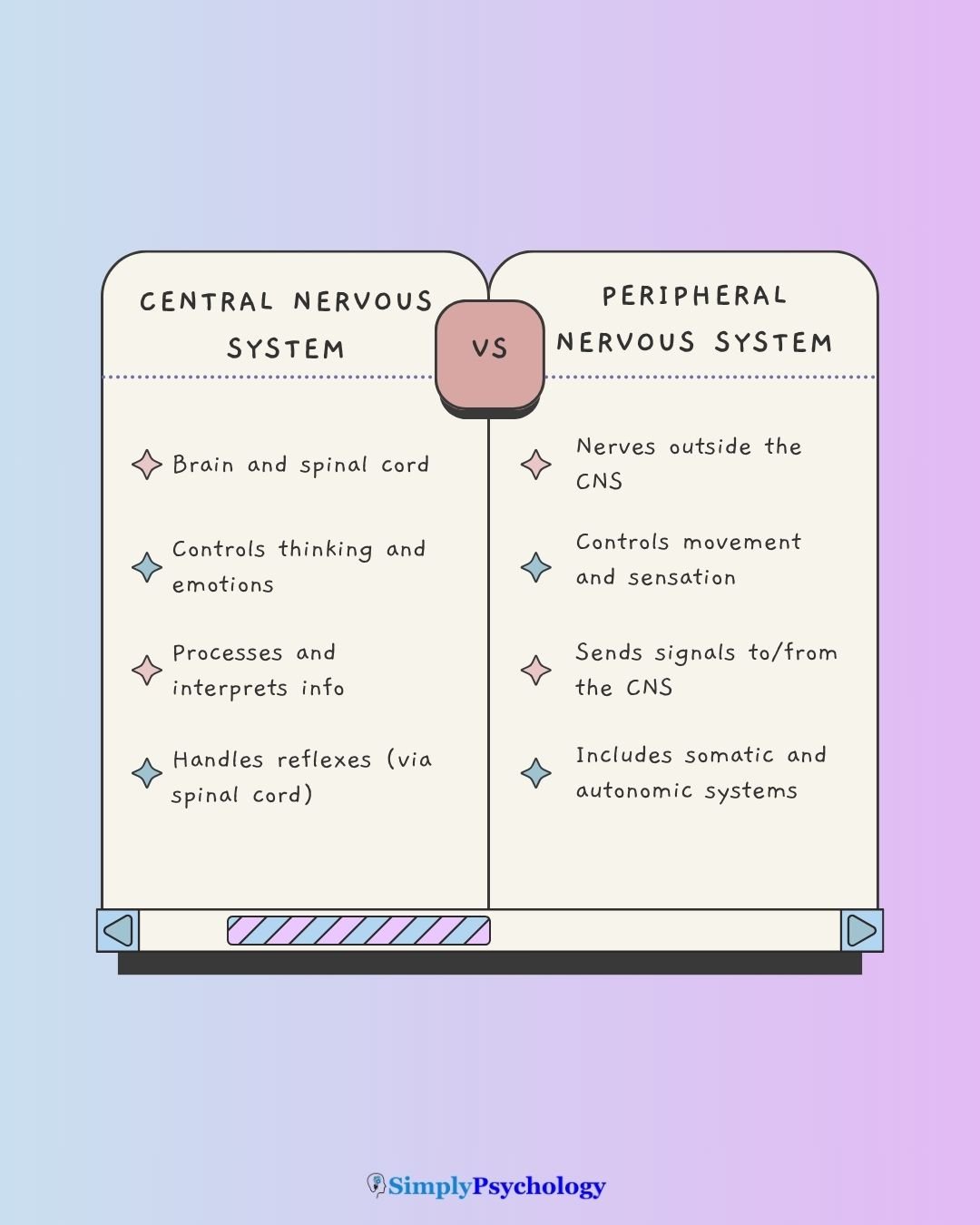
How do the CNS and PNS work together?
The Central Nervous System (CNS) and Peripheral Nervous System (PNS) continuously collaborate to enable all bodily functions and interactions with the world.
The Functional Flow
The relationship between the CNS and PNS is defined by a continuous feedback loop of information.
This flow follows three distinct phases:
- Sensory Input (PNS): Sensory information from our environment is detected by receptor cells and transmitted via afferent (sensory) neurons of the PNS to the CNS (brain and spinal cord).
- Integration (CNS): The brain and spinal cord act as the processing center. Interneurons within the CNS analyze the sensory input, compare it to stored memories or homeostatic set points, and formulate a specific response.
- Motor Output (PNS): Once the CNS processes and interprets this information, it dispatches commands via efferent (motor) neurons of the PNS to muscles and glands, dictating responses.
This constant electrochemical communication across nerves facilitates all our experiences and actions.
Example: Touching a Hot Surface
If you touch a hot stove, sensory receptors in your skin (PNS) immediately detect the heat. Afferent neurons swiftly transmit this “hot” signal up your arm to your spinal cord (CNS).
For a rapid reflex arc, the spinal cord can issue a motor command directly through efferent neurons to your arm muscles (PNS) to pull away, saving precious seconds without immediate brain processing.
Meanwhile, the “pain” signal continues to the brain for conscious perception and further response planning.

How Spinal Reflex Arcs Work
A reflex arc is a fast, automatic loop that the spinal cord completes on its own. A sensory neuron carries the signal in through the dorsal root. After one or more synapses, a motor neuron carries the command out through the ventral root to a muscle.
Reflex arcs come in two basic forms:
- Monosynaptic: One synapse links the sensory neuron directly to the motor neuron. The knee-jerk reflex is the classic example.
- Polysynaptic: At least two synapses, with an interneuron between them. Pulling a hand off a hot surface works this way, which allows more flexible, multi-muscle coordination.
In the knee-jerk reflex, a tap on the tendon briefly stretches the quadriceps at the front of the thigh. A stretch receptor called the muscle spindle detects this and fires a sensory neuron.
That neuron synapses directly onto a motor neuron. The quadriceps contracts and the lower leg kicks forward. At the same time, an inhibitory interneuron relaxes the opposing hamstring, a pairing called reciprocal inhibition.
Doctors exploit this predictability. Tapping a tendon and grading the response helps localize damage to a specific spinal segment or nerve.
An abnormal Babinski sign, where the big toe extends upward when the sole is stroked, points to damage in the descending pathways from the brain.
Liddell and Sherrington’s (1924) Stretch Reflex Study
Liddell and Sherrington (1924) gave the classic evidence for the single-synapse stretch reflex.
- Aim: To determine whether the tonic stretch reflex, a sustained contraction when a muscle is passively stretched, runs through one spinal synapse or several.
- Method: Working with decerebrate cats (forebrain disconnected from the spinal cord), they passively stretched limb muscles and recorded reflex tension. The very short delay before contraction showed how many synapses the signal crossed.
- Results: A sustained stretch produced a sustained contraction of the same muscle, and the short delay indicated a single spinal synapse. The reflex was paired with inhibition of the opposing muscle group.
- Conclusion: The stretch reflex is a monosynaptic arc with reciprocal inhibition of the antagonist. The spinal cord can therefore coordinate precise movement without the cerebral cortex.
The evidence is unusually direct, because the decerebrate preparation removed cortical influence. But the work used anesthetized, non-human animals, and most reflexes are polysynaptic and more modifiable by experience than this simple circuit.
Everyday Tasks
Both systems are crucial for daily life.
Voluntary actions like typing rely on CNS planning and somatic motor commands from the PNS. Involuntary functions such as heartbeat and digestion rely on CNS oversight and autonomic control from the PNS.
Picking up a glass shows both at once. Your somatic system moves the muscles. Meanwhile your autonomic system keeps your heart beating and your gut digesting, with no decision from you.
It’s a continuous feedback loop that allows us to sense, think, feel, and act.
Common disorders and their impact
Common disorders affecting the nervous system can stem from issues within either the Central Nervous System (CNS) or the Peripheral Nervous System (PNS), leading to distinct symptoms and treatment approaches.
CNS-Related Conditions
Conditions impacting the CNS include stroke, multiple sclerosis (MS), and traumatic brain injury (TBI).
TBI, for example, often results in significant and sometimes permanent functional loss. CNS neurons have limited regenerative capacity.
Schizophrenia, primarily affecting the brain, is characterized by major disturbances in thought, perception, emotion, and behavior, often leading to social and occupational impairment.
Mood disorders and anxiety disorders are also linked to brain chemistry imbalances, with psychotropic medications often used to restore neurotransmitter balance.
PNS-Related Conditions
Disorders of the PNS include neuropathy and nerve compression, which often cause symptoms such as “numbness and tingling”. Neuropathy can result from diabetes, nutritional deficiency or nerve compression.
These symptoms can feel very distressing. The outlook is often more hopeful than for a comparable brain or spinal cord injury. Peripheral nerves can sometimes regrow toward their targets, whereas a severed spinal cord tract generally cannot.
What Is the Central Nervous System (CNS)?
The Central Nervous System (CNS) is the body’s control center, consisting of the brain and spinal cord.
As the “headquarters” of the nervous system, the CNS primarily receives, processes, interprets, and stores incoming sensory information.
Brain structures interpret this information. Commands are then dispatched via the peripheral nervous system to direct bodily responses.
The CNS is responsible for sensing, perception, thinking, awareness, emotions, and planning.
The spinal cord also runs reflexes on its own. These fast, automatic reactions are completed within the cord, so the brain learns what happened only afterwards.

What Is the Peripheral Nervous System (PNS)?
The Peripheral Nervous System (PNS) comprises all nerves located outside the brain and spinal cord.
Its main role is to relay messages between the Central Nervous System (CNS) and the body’s muscles, organs, and senses.
The PNS consists of two sets of nerves:
- Cranial Nerves: Twelve pairs leave the brain through openings in the skull. Most serve the head and neck, but the vagus nerve (X) reaches the heart, lungs and gut.
- Spinal Nerves: Thirty-one pairs leave the spinal cord between the vertebrae: 8 cervical, 12 thoracic, 5 lumbar, 5 sacral and 1 coccygeal.
Every spinal nerve is “mixed”. It carries motor (efferent) fibers out to the muscles and sensory (afferent) fibers back from receptors. At the spinal cord, each nerve splits in two. The dorsal root carries only sensory fibers, and the ventral root carries only motor fibers.

The PNS has two key subdivisions:

1. The Somatic Nervous System (SNS)
The Somatic Nervous System (SNS) controls voluntary movements of the skeletal muscles. It also carries sensory information, such as touch, pain and temperature, from the body to the central nervous system. In everyday terms, it manages conscious actions and external sensations.
It works through two types of neurons, plus reflex arcs:
- Sensory (Afferent) Neurons: These carry signals toward the CNS from sensory receptors (skin, eyes, ears).
- Motor (Efferent) Neurons: These carry signals away from the CNS to the skeletal muscles, allowing for voluntary actions like walking or typing.
- Reflex Arcs: The SNS also handles involuntary muscle “jerks” (like pulling your hand off a hot stove) where the signal bypasses the brain and loops through the spinal cord for a faster response.
Most somatic motor neurons have their cell bodies inside the spinal cord. Their axons are wrapped in myelin. This insulating coat speeds conduction, allowing the fast, precise contractions that voluntary movement needs.
“Voluntary” describes this system’s typical mode of operation, not an absolute rule. Reflexes use the same sensory neurons, motor neurons and muscles as voluntary movement, yet they bypass conscious decision-making.
2. The Autonomic Nervous System (ANS)
The Autonomic Nervous System (ANS) governs the body’s internal organs and glands, largely outside conscious control. It runs heart rate, digestion, breathing and blood pressure. Its aim is homeostasis, the body’s stable internal balance.
It has two main divisions: the sympathetic system (fight-or-flight) and the parasympathetic system (rest-and-digest). They generally act in opposition to regulate organs, smooth muscles and glands.
Unlike somatic pathways, autonomic pathways are slower and largely unmyelinated. They use a two-neuron relay through a ganglion, a cluster of nerve cell bodies outside the CNS.
A third branch, the enteric nervous system, is a mesh of neurons in the gut wall. It can drive digestion even when its links to the rest of the ANS are cut.
The ANS acts through two channels. It stimulates organs directly through nerves, and it triggers hormone release from the endocrine glands, with the hypothalamus coordinating both. This is how a stressful thought becomes a bodily reaction.
Although called involuntary, autonomic activity can be partly trained. Biofeedback shows a person their own heart rate or breathing in real time, helping them shift toward parasympathetic dominance.
Sympathetic Division (“Fight or Flight“)
This system mobilizes the body’s resources during energy-expending activities or perceived threats.
- Physical Effects: Dilates pupils for better vision, increases heart rate and blood pressure, inhibits digestion, and triggers the release of adrenaline.
- Goal: Rapid survival response.
Parasympathetic Division (“Rest and Digest”)
This system dominates during quiet, relaxed periods to conserve energy and maintain long-term health.
- Physical Effects: Constricts pupils, slows the heart rate, stimulates digestion and salivation, and allows for waste elimination.
- Goal: Recovery and “housekeeping.”
Critical Evaluation of the CNS/PNS Model
The CNS/PNS division is one of the most widely taught frameworks in biopsychology. It rests on real anatomical differences with clear clinical payoff, but the tidy diagram oversimplifies the biology in three ways:
- Fuzzy Labels: “Voluntary” and “involuntary” describe typical modes, not fixed rules, and the brain constantly adjusts spinal reflexes.
- Peripheral Computation: The gut’s enteric system coordinates digestion with its own neurons, challenging a strict CNS-commands, PNS-obeys hierarchy.
- Structure vs Function: A functional map groups neurons by signal direction, which cuts across the CNS/PNS boundary.
Voluntary and Involuntary Are Not Fixed
The somatic system is defined by voluntary control, yet it also runs automatic reflex arcs. The autonomic system is defined by involuntary control, yet training can shift it.
The line blurs in both directions. Biofeedback and slow breathing, for example, move the balance toward parasympathetic dominance.
So the labels describe each system’s typical mode, not an absolute rule. Reflexes are not purely bottom-up either.
Descending pathways from the cortex and brainstem constantly adjust spinal reflex circuits to suit the current motor task (Grillner, 1975). The adult Babinski sign makes this visible at the bedside. When the brain’s normal suppression of a primitive infant reflex breaks down, the big toe extends upward (Van Gijn, 1978).
A purely bottom-up account of spinal reflexes is therefore only a first approximation.
The Gut Has Its Own Nervous System
A strictly top-down model says the CNS commands and the PNS simply obeys. The enteric nervous system challenges that.
This mesh of hundreds of millions of neurons lies in the gut wall. It can coordinate digestion even when its connections to the rest of the nervous system are cut (Gershon, 1998).
The enteric system is not wholly autonomous, because sympathetic and parasympathetic input modulates it.
Even so, it does real computation of its own. Some researchers therefore treat it as a semi-autonomous system in its own right, not just a branch of the ANS.
Review work now links enteric dysfunction to disorders once treated as purely central. Parkinson’s disease pathology, for example, is increasingly understood to appear in enteric neurons years before it appears in the brain (Rao & Gershon, 2016).
Structure Versus Function
The CNS/PNS map is structural. It asks where nervous tissue physically sits.
A functional map asks a different question: does a pathway carry information toward an integrating center (afferent) or away from one (efferent)? That distinction cuts across the CNS/PNS boundary (Swanson, 2000).
A sensory neuron shows the difference. Its cell body sits in a peripheral ganglion, part of the PNS, yet its central synapse lies inside the spinal cord, part of the CNS.
The structural map splits that neuron in two. The functional map treats the whole afferent pathway as one unit.
Both views are correct. The structural map suits locating a lesion or explaining where a drug acts. The functional view suits explaining how a signal is transformed as it travels.
Contemporary Research
Recent spinal cord injury research shows that the reflex circuit still matters. Restoring movement means working with its wiring, not overriding it.
Formento et al. (2018) combined computational modeling with direct testing in people with chronic spinal cord injury. The stimulation pattern mattered. Continuous epidural electrical stimulation, which restores stepping in animal models, blocked much of the natural proprioceptive signaling (muscle and joint position) in humans.
It also disrupted the reciprocal-inhibition networks that Liddell and Sherrington described. Burst, spatiotemporally patterned stimulation left room for natural signals to keep flowing. Voluntary control of the leg muscles became much more robust.
Wagner et al. (2018) found a converging result.
Stimulation timed with a person’s own attempted movement, combined with rehabilitation training, restored voluntary stepping in people with chronic, incomplete spinal cord injury. Some gains persisted after the stimulation was switched off.
Both studies are small, uncontrolled human case series, not randomized trials. They are best read as converging with a larger animal and computational evidence base, not as strong evidence alone.
Summary
The central and peripheral nervous systems work together to control everything from basic reflexes to complex thoughts.
The CNS, made up of the brain and spinal cord, is the body’s processing hub, handling thinking, emotions, and coordination.
The PNS connects the CNS to the rest of the body, relaying sensory input and carrying out responses through nerves.
To remember the difference, think of the CNS as the “control center” and the PNS as the “communication network.” The CNS interprets information, while the PNS delivers messages to and from it.
References
Formento, E., Minassian, K., Wagner, F., Mignardot, J. B., Le Goff-Mignardot, C. G., Rowald, A., Bloch, J., Micera, S., Capogrosso, M., & Courtine, G. (2018). Electrical spinal cord stimulation must preserve proprioception to enable locomotion in humans with spinal cord injury. Nature Neuroscience, 21(12), 1728–1741. https://doi.org/10.1038/s41593-018-0262-6
Gershon, M. D. (1998). The second brain: A groundbreaking new understanding of nervous disorders of the stomach and intestine. HarperCollins.
Grillner, S. (1975). Locomotion in vertebrates: Central mechanisms and reflex interaction. Physiological Reviews, 55(2), 247–304. https://doi.org/10.1152/physrev.1975.55.2.247
Liddell, E. G. T., & Sherrington, C. S. (1924). Reflexes in response to stretch (myotatic reflexes). Proceedings of the Royal Society of London. Series B, Containing Papers of a Biological Character, 96(675), 212–242. https://doi.org/10.1098/rspb.1924.0023
Rao, M., & Gershon, M. D. (2016). The bowel and beyond: The enteric nervous system in neurological disorders. Nature Reviews Gastroenterology & Hepatology, 13(9), 517–528. https://doi.org/10.1038/nrgastro.2016.107
Swanson, L. W. (2000). What is the brain? Trends in Neurosciences, 23(11), 519–527. https://doi.org/10.1016/S0166-2236(00)01639-8
Van Gijn, J. (1978). The Babinski sign and the pyramidal syndrome. Journal of Neurology, Neurosurgery & Psychiatry, 41(10), 865–873. https://doi.org/10.1136/jnnp.41.10.865
Wagner, F. B., Mignardot, J.-B., Le Goff-Mignardot, C. G., Demesmaeker, R., Komi, S., Capogrosso, M., Rowald, A., Seáñez, I., Caban, M., Pirondini, E., Vat, M., McCracken, L. A., Heimgartner, R., Fodor, I., Watrin, A., Seguin, P., Paoles, E., Van Den Keybus, K., Eberle, G., … Courtine, G. (2018). Targeted neurotechnology restores walking in humans with spinal cord injury. Nature, 563(7729), 65–71. https://doi.org/10.1038/s41586-018-0649-2

