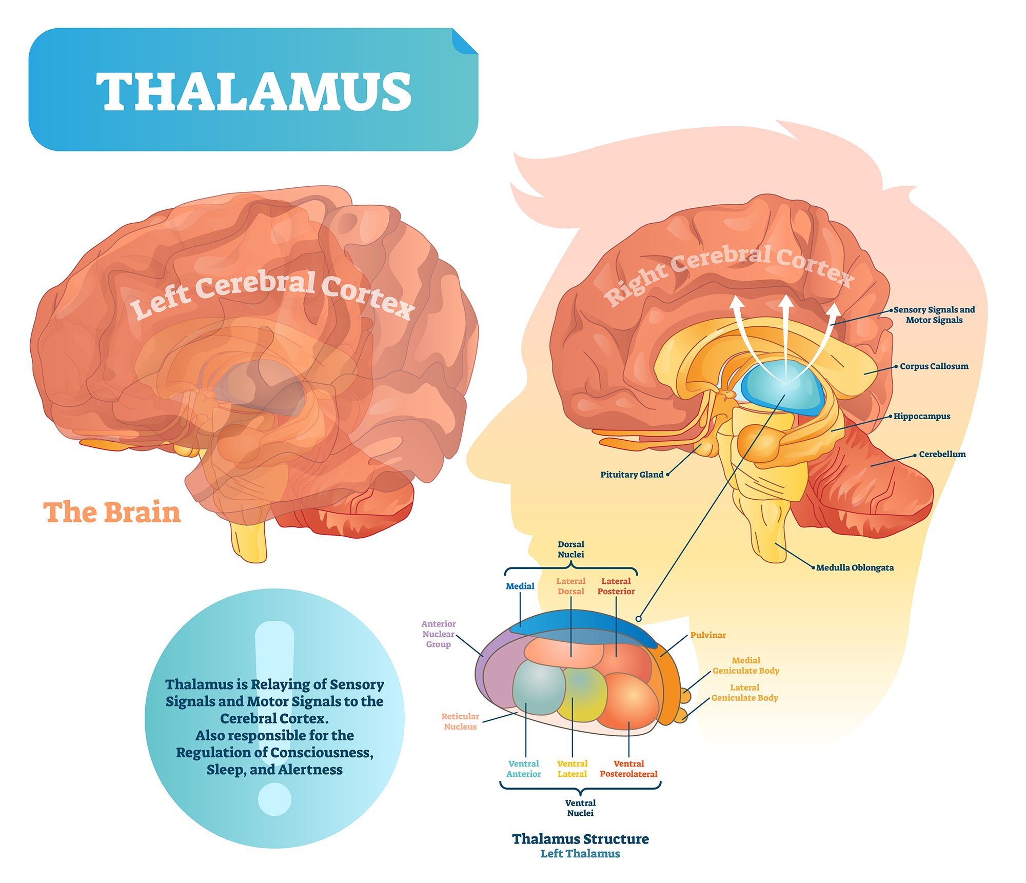
The thalamus is a structure of thebrain that processes and transmits sensory (except for smell) and motor information from the body to the cerebral cortex.
The thalamus is often described as the brain’s relay station as much of the information that reaches the cerebral cortex first stops in the thalamus before being sent to its destination.
Imagine the thalamus is a train station where passengers (or sensory and motor signals) come to catch their train to their intended location (within the brain).

Key Takeaways
- The thalamus is the brain’s central relay station, directing most sensory and motor signals to the cerebral cortex.
- It plays a vital role in sensation, movement, attention, sleep, and memory.
- Each part of the thalamus, called a nucleus, has a specialized job—processing different types of information.
- Damage to the thalamus can affect memory, coordination, alertness, and sensory perception.
- New research shows the thalamus is more adaptable than once believed, with early development and adult neuroplasticity shaping how it works.
What Does the Thalamus Do?
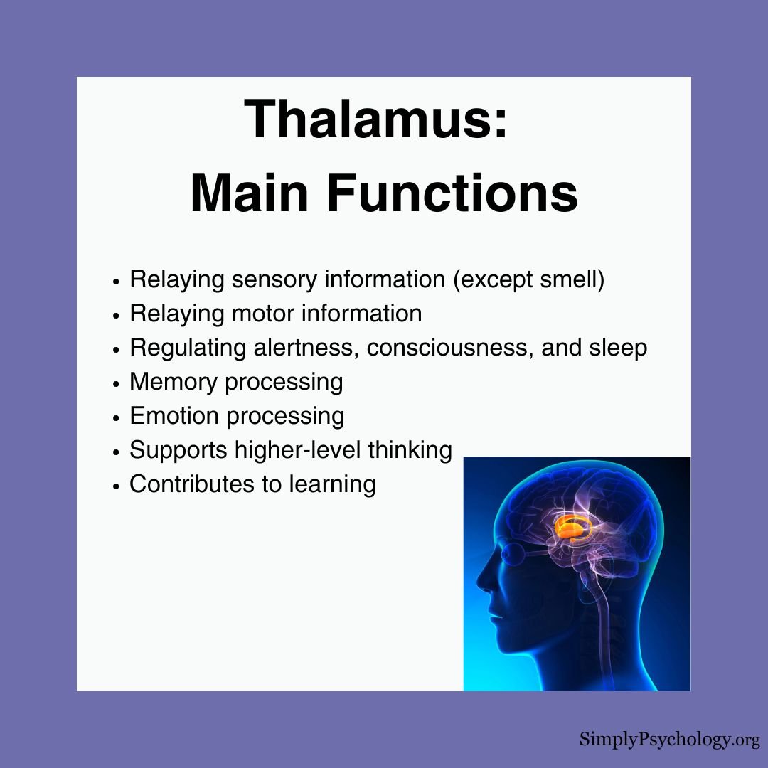
The thalamus plays a key role in multiple brain functions by helping different parts of the brain communicate effectively. Its main functions include:
Sensory Processing & Relay
- Processes all sensory information except smell
- Each type of sensory input has a dedicated nucleus within the thalamus
- Transmits processed information to specific areas of the cerebral cortex
- Connections are contralateral with sensory organs (opposite side) but ipsilateral with the cortex (same side)
Motor Signal Control
- Relays motor signals between the cerebellum, basal ganglia, and motor cortex
- Helps coordinate and refine movement
- Contributes to posture control and balance
Attention & Filtering
- Acts as a gatekeeper for sensory information
- Filters which signals reach higher brain regions
- Helps determine which stimuli deserve attention
- Particularly important in visual attention through the pulvinar nucleus
Consciousness & Arousal
- Regulates sleep and wakefulness
- Maintains alertness and awareness
- Contributes to overall consciousness levels
- Damage can lead to coma or sleep disorders
Memory & Cognition
- Connects with the hippocampus for episodic memory formation
- Works with the limbic system for emotional processing
- Contributes to learning and memory organization
- Supports higher-level thinking through connections with the prefrontal cortex

Anatomy
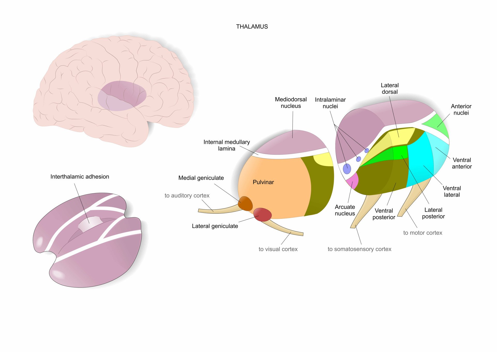
There are two thalami, one in each hemisphere of the brain. They are oval-shaped in appearance, almost looking like eggs, with two protuberances on the surface.
One of these is known as themedial geniculate bodies, which are important for auditory information processing.
The other is the lateral geniculate bodies, which are responsible for the processing of visual sensory inputs.
The thalamus is mostly comprised of gray matter but is surrounded by two white matter layers.
🧠 Did You Know? Your Thalamus Started Forming Before You Were Born
The thalamus starts developing while you’re still in the womb. It grows from a section of the early brain called the diencephalon.
A special signal called Sonic Hedgehog helps shape the thalamus and tells it how to organize itself.
Another part, the ZLI (zona limitans intrathalamica), works like a guide, dividing the thalamus into different zones that later control things like sight, touch, movement, and memory.
By the time you’re almost ready to be born, many of the thalamus’s connections to the rest of the brain are already set up—getting you ready to learn, feel, and explore the world.
Location
The thalamus is situated above the brainstem in the middle of the brain.
The thalamus is a part of an area called the diencephalon, which includes the hypothalamus, subthalamus, and epithalamus.
Thalamus vs. Hypothalamus
The thalamus and hypothalamus serve distinct functions in the brain.
The thalamus acts as the brain’s relay station, processing and directing sensory and motor signals to the correct areas of the cerebral cortex.
The hypothalamus, located below the thalamus, is the control center for many automatic functions including hormone production, temperature regulation, hunger, thirst, sleep cycles, and emotional responses.
Think of the thalamus as a switchboard operator directing signals, while the hypothalamus is more like the body’s thermostat and control center.
Nuclei within the thalamus
The thalamus is made up of a series of nuclei, all of which are responsible for the relay of different sensory signals.
The nuclei are both excitatory and inhibitory in nature and receive sensory or motor information from the body, presenting selected information via the nerve fibers to the cerebral cortex.


Below are some of the main groups of nuclei within the thalamus and what they are responsible for:
Anterior nucleus
The anterior nucleus is thought to be involved with memory due to its extensive connectivity to the hippocampus.
It is also connected to the mammillothalamic tract (from the mammillary nucleus of the mammillary bodies to the hypothalamus) and the cingulate gyrus (involved in processing emotions and behavior regulation).
As these areas are linked with the limbic system, they are involved in organizing memory and emotion.
Dorsomedial nucleus
The dorsomedial nucleus is thought to be involved in emotional behavior and memory.
This nucleus relays information from the amygdala and olfactory cortex, which then projects to the prefrontal cortex and the limbic system, in turn relaying them to the prefrontal association cortex.
Because of this, the dorsomedial nucleus has an important role in attention, organization, planning, and higher cognitive thinking.
Ventral posterolateral and ventral posteromedial nucleus
These both act as relay nuclei sending somatosensory information to the somatosensory cortex, a region that receives and processes sensory information about the body.
Further, the ventral posteromedial nucleus receives sensory information regarding the face from the trigeminal nerve.
Ventral anterior and ventrolateral nucleus
These two nuclei are the motor relay nuclei, which receive inputs from the cerebellum and the basal ganglia.
They are thought to be involved in motor functions, and both have pathways leading to the substantia nigra, premotor cortex, reticular formation, and the corpus striatum.
Lateral posterior nucleus
The lateral posterior nucleus is believed to be involved in integrating sensory input and associating it with cognitive functions.
Its other functions include being able to determine visual stimuli that stands out the most and visually guided behaviors.
Pulvinar nucleus nucleus
The pulvinar supports visual attention. It connects to the visual cortex, amygdala, and striatum (which helps with motivation and decision-making).
It plays a key role in helping us focus on important visual details and guides precise movements based on what we see.
Medial geniculate and lateral geniculate nucleus
These are the brain’s audio-visual switchboards:
- The medial geniculate nucleus handles auditory signals, passing them from the midbrain to the auditory cortex.
- The lateral geniculate nucleus receives visual input from the eyes and sends it to the visual cortex.
Reticular nucleus
Unlike other nuclei, the reticular nucleus doesn’t send signals to the cortex. Instead, it wraps around the thalamus and helps control what other thalamic nuclei do.
It filters information flowing through the thalamus, playing a key role in attention and sleep regulation.
🧠 Did You Know? The Thalamus Is More Than Just a Relay Station
Recent research has uncovered fascinating insights into the thalamus:
- Early Development: Scientists have found that the thalamus starts forming its different parts earlier in fetal development than previously thought. By 14 weeks after conception, distinct regions are already taking shape, guided by specific genes.
- Neuroplasticity: Scientists used to think only the cortex could change and adapt. But new research shows the thalamus also has neuroplasticity—which means it can rewire itself based on your experiences. This ability to adapt continues even in adulthood, helping the brain respond to learning, injuries, and new environments.
- Growing Connections: As we grow from babies to young adults, the thalamus forms and refines connections with other brain areas. These connections are crucial for developing attention, memory, and other important skills.
Damage
Since the thalamus is involved in relaying signals from many brain structures, damage to this area can impact many functions. Symptoms associated with thalamic damage include:
- Memory difficulties or amnesia
- Difficulties with attention
- Impaired movement and posture
- Loss of alertness and activation
- Impaired processing of sensory information
- Sleep difficulties and insomnia
- Language difficulties such as thalamic aphasia
- Sensory loss and movement disorders
- Thalamic stroke which can lead to chronic pain
People with schizophrenia were found to have significantly reduced thalamic volume compared to those without schizophrenia (Coscia et al., 2009).
This reduced size was thought to be associated with weakened neuropsychological functioning and specific difficulties with language, motor, and executive skills.
Lower and weaker functional connectivity in the thalamus was also found in autistic individuals (Tomasi & Volkow, 2019).
‘Non-typical’ thalamic connectivity in temporal and motor areas was also found in autistic individuals (Woodward et al., 2017).
References
Alhesain, M., Sankar, N., Smith, C., Kerwin, J., Laws, R., Lindsay, S., & Clowry, G. J. (2025). Development of the early fetal human thalamus: From a protomap to emergent thalamic nuclei. Frontiers in Neuroanatomy, 19, 1530236. https://doi.org/10.3389/fnana.2025.1530236
Coscia, D. M., Narr, K. L., Robinson, D. G., Hamilton, L. S., Sevy, S., Burdick, K. E., Gunduz-Bruce, H., McCormack, J., Bilder, R. M. & Szeszko, P. R. (2009). Volumetric and shape analysis of the thalamus in first‐episode schizophrenia. Human brain mapping, 30 (4), 1236-1245.
Mandal, A. (2021, February 15). What is the Thalamus? News Medical Life Sciences. https://www.news-medical.net/health/What-is-the-Thalamus.aspx#:~:text=The%20thalamus%20is%20a%20small,signals%20to%20the%20cerebral%20cortex
Park, S., Haak, K. V., Oldham, S., Cho, H., Byeon, K., Park, B., Thomson, P., Chen, H., Gao, W., Xu, T., Valk, S., Milham, M. P., Bernhardt, B., Di Martino, A., & Hong, S. (2024). A shifting role of thalamocortical connectivity in the emergence of cortical functional organization. Nature Neuroscience, 27(8), 1609-1619. https://doi.org/10.1038/s41593-024-01679-3
Sonoda, T., Stephany, C. É., Kelley, K., Kang, D., Wu, R., Uzgare, M. R., … & Chen, C. (2025). Experience influences the refinement of feature selectivity in the mouse primary visual thalamus. Neuron, 113(9), 1352-1362. 10.1016/j.neuron.2025.02.023
Tomasi, D., & Volkow, N. D. (2019). Reduced local and increased long-range functional connectivity of the thalamus in autism spectrum disorder. Cerebral Cortex, 29 (2), 573-585.
Torrico, T. J., & Munakomi, S. (2019). Neuroanatomy, Thalamus.
Woodward, N. D., Giraldo-Chica, M., Rogers, B., & Cascio, C. J. (2017). Thalamocortical dysconnectivity in autism spectrum disorder: an analysis of the autism brain imaging data exchange. Biological Psychiatry: Cognitive Neuroscience and Neuroimaging, 2 (1), 76-84.

