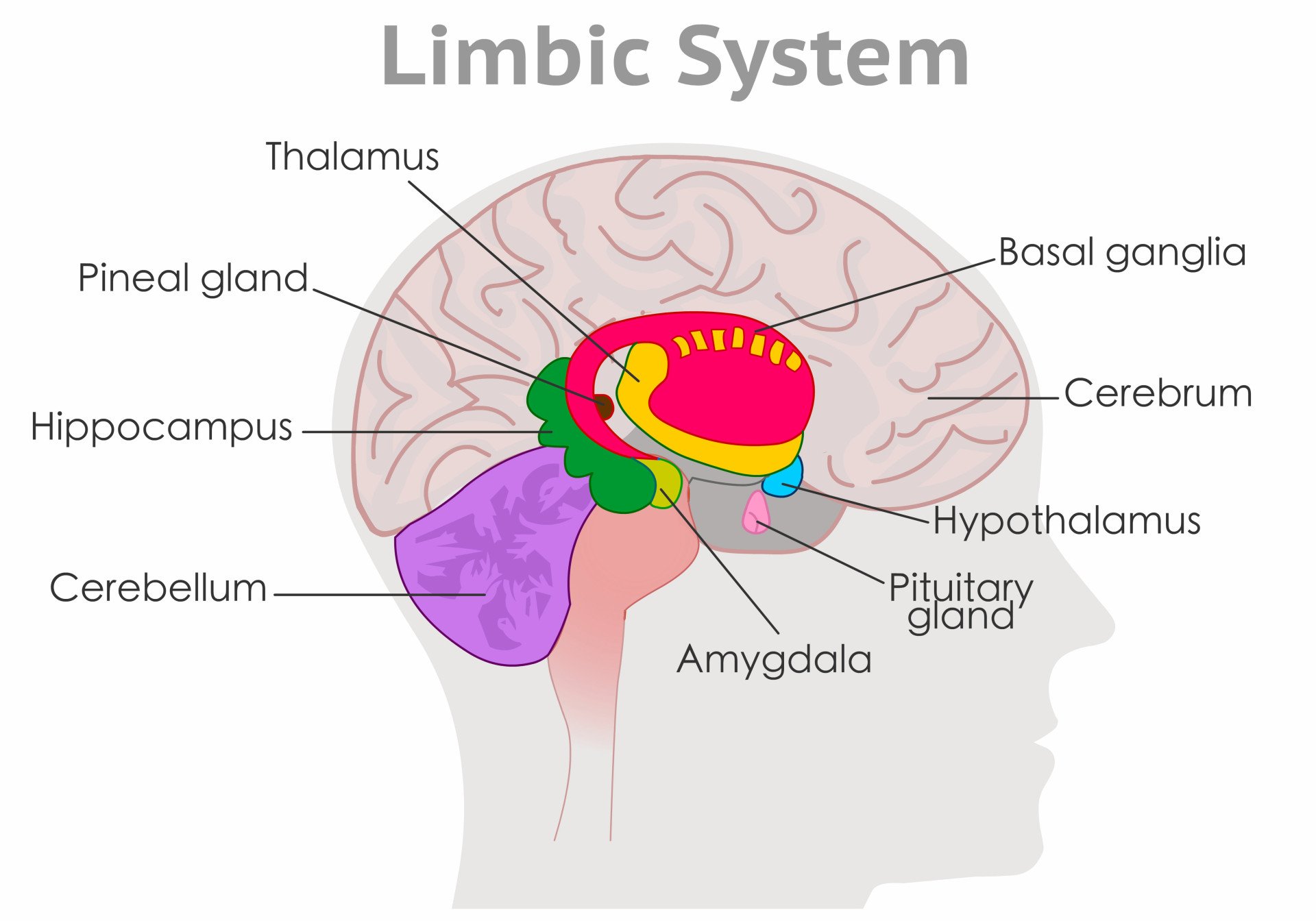
The brain is categorized into three primary developmental regions: the forebrain (prosencephalon), midbrain (mesencephalon), and hindbrain (rhombencephalon).
Forebrain: The largest division. The cerebrum handles high-level processing. The diencephalon at its core holds the thalamus (a sensory relay station) and the hypothalamus (which regulates temperature, hunger and thirst).
Midbrain: A central connector that manages visual and auditory reflexes. It contains structures like the substantia nigra, a dopamine-producing nucleus needed to start voluntary movement.
Hindbrain: The region responsible for vital life-support functions via the medulla and pons, and motor coordination via the cerebellum.
The forebrain, midbrain, and hindbrain are the three main brain divisions. The forebrain handles higher cognitive functions, the midbrain controls motor movement and sensory processing, and the hindbrain manages coordination, balance, and autonomic functions like breathing and heart rate.
The three divisions take shape early in development. In the third to fourth week, the neural tube swells at its head end into three bulges called primary vesicles. By about the fifth week, two of them divide again, giving five secondary vesicles.
Secondary vesicle
Main structures
Division
Telencephalon
Cerebral hemispheres (cerebrum), basal ganglia, limbic system
Forebrain
Diencephalon
Thalamus, hypothalamus, pineal gland
Forebrain
Mesencephalon
Tectum, tegmentum (including the reticular formation)
Midbrain
Metencephalon
Pons, cerebellum
Hindbrain
Myelencephalon
Medulla oblongata
Hindbrain
The midbrain is the exception: it never divides again. Every mature structure traces back to one of these five vesicles.
The forebrain constitutes the largest and most complex division of the mammalian central nervous system.
This region, technically termed the prosencephalon, facilitates sophisticated cognitive processes like reasoning, language, and conscious perception.
It sits at the very front of the skull. Because it houses the mechanisms for voluntary action, the forebrain defines much of what is considered human behavior.
The forebrain has two main parts:
Telencephalon: The cerebral hemispheres, including the cerebral cortex, the basal ganglia and much of the limbic system.
Diencephalon: The central core, containing the thalamus, hypothalamus and pineal gland.
Cerebral Cortex
The cerebral cortex is the thin outer layer of grey matter, about 1 cm deep, that covers the cerebral hemispheres. The hemispheres themselves, cortex plus the thicker white matter beneath it, make up roughly two-thirds of the brain’s total mass.
Each cerebral hemisphere can be subdivided into four lobes (frontal, parietal, temporal, and occipital lobes), each associated with different functions.
Frontal Lobe: It manages executive functions: high-level mental processes like planning, language, and decision-making.
Parietal Lobe: This region processes somatosensation: the perception of touch, temperature, and body position (spatial awareness).
Temporal Lobe: It facilitates audition: the formal sense of hearing and processing of sound. Wernicke’s area, usually in the left hemisphere, supports language comprehension. Damage to it leaves speech fluent but meaningless.
Occipital Lobe: This posterior region is dedicated to visual processing and interpretation. Brodmann Area 17, the primary visual cortex, determines the size, shape, and location of objects in the field of vision.
The frontal lobe’s role in personality is illustrated by Phineas Gage, a railway worker whose frontal lobes were pierced by an iron bar in 1848. He survived the accident. However, Gage underwent a lasting change in personality and social conduct (Harlow, 1868).
The right and left hemispheres are connected by a thick band of nerve fibers called the corpus callosum.
Surgeons sometimes sever this structure to control severe epilepsy. The result is split-brain. The two hemispheres can no longer share information and start acting independently of each other.
Roger Sperry pioneered this research in the 1960s (Sperry, 1961). His split-brain studies showed that each hemisphere can act on information the other is unaware of.
The primary motor cortex and somatosensory cortex are the best-mapped areas, and both show contralateral control. Each hemisphere serves the opposite side of the body.
Wilder Penfield mapped these strips by electrically stimulating the exposed cortex of conscious neurosurgical patients. The map is distorted. Neighboring body parts occupy neighboring cortex, and the space given to each reflects its sensitivity or dexterity, not its size. This is the homunculus (Penfield & Boldrey, 1937).
Most of the cortex is association cortex. Primary motor and sensory areas cover only about 25 percent of its surface.
The other 75 percent is where thinking, reasoning, planning and deciding probably occur. Humans have more of it than any other species.
Beneath the cortex sit several other structures, including the thalamus, hypothalamus, pituitary gland, basal ganglia, hippocampus, and amygdala. Some of these form the limbic system, which governs emotional behavior, motivation, and learning.
Limbic System
The forebrain also features important structures in the limbic system, which controls basic emotions and psychological drives.
The limbic system is made up of a number of different structures, but three of the most important are the hippocampus, the amygdala, and the hypothalamus.
A critical component of the limbic system is the amygdala, an almond-shaped set of neurons responsible for processing fear.
Heinrich Klüver and Paul Bucy studied this in the 1930s. They removed the amygdalae from rhesus monkeys and found the animals lost their normal fear response, becoming placid even around threats (Klüver & Bucy, 1939).
Humans show the same pattern, now called Klüver-Bucy syndrome, after damage to the amygdala (Hayman et al., 1998).
Another key limbic structure is the hippocampus. It stores long-term memories. Henry Gustave Molaison, known as H.M., had two-thirds of his hippocampi removed in 1953 to control severe epilepsy (Scoville & Milner, 1957).
As a result, H.M. suffered severe anterograde amnesia. He could no longer form new memories. He also struggled to recall events from just before the surgery, a pattern known as graded retrograde amnesia (Halber, 2018).
Basal Ganglia and Movement Control
Deep in the white matter of each hemisphere sit clusters of grey matter called the basal ganglia (“nerve knots”). They include the corpus striatum (“striped body”), made of the caudate nucleus and the lentiform nucleus (putamen plus globus pallidus).
Traditional lists add the amygdala and the substantia nigra. The substantia nigra is usually classed with the midbrain.
Closely linked to the thalamus and cortex, the basal ganglia help control muscle tone, posture, and the initiation and scaling of voluntary movement. They work through loops that run from the cortex through the striatum and thalamus and back to the cortex.
That central role in movement is why their dysfunction produces two families of movement disorders:
Parkinson’s disease: Loss of dopamine input from the substantia nigra causes slowness and rigidity (hypokinetic symptoms).
Huntington’s disease: Degeneration of the caudate and putamen causes involuntary movements (hyperkinetic symptoms).
Thalamus, Hypothalamus, and Pineal Gland
The diencephalon is the central core of the forebrain, wrapped almost entirely by the cerebral hemispheres. It contains the thalamus, the hypothalamus and the pineal gland. Only the hypothalamus and the anterior thalamus are usually counted as part of the limbic system.
The thalamus acts as a relay station, passing sensory information from the body to the rest of the brain (Mandal, 2019). With the single exception of smell, every sensory signal is routed through it on its way to the cortex. It also supports consciousness, alertness, and sleep.
Its specialized clusters of neurons (nuclei) include:
Ventrobasal complex: Takes in information from the body via the spinal cord.
Modern research shows the thalamus does more than relay signals passively: it actively gates and coordinates the ongoing conversation between cortical areas (Sherman, 2016).
Located between the two halves of the thalamus, the pineal gland is a pea-shaped endocrine gland. It secretes the hormone melatonin in response to darkness.
This helps time the sleep-wake cycle. It also keeps circadian rhythms, the body’s roughly 24-hour clock, in step with day and night. In effect, it turns light into a chemical signal.
The hypothalamus is located at the base of the brain near the pituitary gland, and it is involved with pleasure, food, body temperature, and sex (Seladi-Schulman, 2018). If the body temperature gets too high, the hypothalamus instructs the body to sweat.
If the body is cold, the hypothalamus will cause shivers. Irregular hypothalamic behavior can, in turn, lead to sweating and shivering when these reactions are not needed.
The hypothalamus works through two routes. It sends electrochemical signals to the autonomic nervous system, the network that runs involuntary body functions. It also controls the pituitary gland, the endocrine “master gland” that regulates the body’s other hormone-releasing glands.
The two are linked by blood vessels and neurons. Through this link, the hypothalamus launches the slow hormonal stress response known as the HPA axis.
Lesion studies tie specific hypothalamic tissue to motivation. Hetherington and Ranson (1940) destroyed the ventromedial hypothalamus in rats, which produced overeating (hyperphagia) and gross obesity. Lesions of the lateral hypothalamus have the opposite effect: the animals refuse to eat (aphagia).
Midbrain (Mesencephalon)
The second area of the brain is the midbrain, which lies on top of the brainstem.
The midbrain is involved in auditory and visual processing (Peters, 2017).
The midbrain is also called the mesencephalon. It sits in the middle of the brain, forming the topmost part of the brainstem and connecting the forebrain above to the hindbrain and spinal cord below.
The midbrain begins, like the forebrain and hindbrain, as one of three primary vesicles in the embryo. But it never splits further. That is why it stays small and simple in the adult brain, unlike its two neighbors, which each divide again as development continues.
Despite its small size, the midbrain plays a diverse and vital role in motor control, sensory processing, and the regulation of consciousness.
Midbrain Structure: Tectum and Tegmentum
A narrow, fluid-filled canal called the cerebral aqueduct runs through the core of the midbrain. It carries cerebrospinal fluid (CSF) between the third and fourth ventricles and divides the midbrain into two main regions.
1. The Tectum (Roof)
The dorsal (top) surface of the midbrain is called the tectum. It features two pairs of swellings known as the colliculi:
Superior Colliculus: Also known as the optic tectum, it receives direct visual input from the eyes and drives fast, reflexive eye and head movements toward a visual target.
Inferior Colliculus: This structure serves as a crucial relay station for auditory (hearing) information as it travels from the inner ear to the thalamus.
2. The Tegmentum (Floor)
Lying ventral to (underneath) the tectum is the tegmentum, which forms the intermediate level of the midbrain. It contains several specialized and highly colorful structures:
Substantia Nigra: Meaning “black substance,” this region contains dopamine-producing neurons whose projection to the striatum is essential for smoothly initiating voluntary movement. Degeneration of these neurons is the primary cause of Parkinson’s disease.
Red Nucleus: A structure with a distinctive pinkish hue that also contributes to motor control via the rubrospinal tract.
Periaqueductal Gray Matter: This area surrounds the cerebral aqueduct and is an important center for modulating and inhibiting somatic pain sensations.
Cranial Nerve Nuclei: The tegmentum houses the nuclei for the third (oculomotor) and fourth (trochlear) cranial nerves, which supply the muscles that control eye movement.
Reynolds (1969) showed how powerful the periaqueductal gray’s pain control is. He implanted electrodes in the midbrain gray matter of eight rats. In three of them, stimulation abolished responses to painful stimuli while movement stayed normal. Those rats then underwent abdominal surgery without any anesthetic.
The finding revealed a built-in pain-suppression system running from the periaqueductal gray down to the spinal cord, which the body’s own opioids help drive. It helps explain why pain can be dampened in an emergency.
Note: The midbrain also features the crus cerebri (cerebral peduncles), which carry large bundles of descending nerve fibers.
Key Functions of the Midbrain
Information Conduit: Major nerve tracts run up and down through the midbrain. The corticospinal tract, for example, descends through it from the cerebral cortex to the spinal cord to carry voluntary motor commands.
Arousal, Attention, and Sleep: The midbrain houses important parts of the reticular formation (or reticular activating system), a widespread network of neurons that filters background stimuli and produces general arousal. It regulates the sleep-wake cycle and alerts the cerebral cortex to novel or challenging tasks.
Coma Risk: Severe damage to the midbrain’s reticular formation can result in a prolonged sleep-like state or coma.
Reflex Integration: The midbrain is a correlation center for sensory and motor reflexes. It manages the orienting reflex, an automatic response to a novel sight or sound, such as turning your head suddenly to a loud noise.
Neurochemical Regulation: Four concentrated groups of neurons in and around the midbrain use specific neurotransmitters to control the activation levels of the rest of the brain.
Chemical Sources: The ventral tegmental area and substantia nigra release dopamine, the raphe nuclei release serotonin, and the locus coeruleus (in the pons, just below the midbrain) releases noradrenaline.
Reward and Aggression: Stimulating the midbrain’s ventral tegmental area can elicit predatory aggressive behaviors or activate the brain’s reward and reinforcement systems.
Hindbrain (Rhombencephalon)
The hindbrain is located at the back of the head and looks like an extension of the spinal cord. It contains the medulla oblongata, pons, and cerebellum. The brainstem itself is just the medulla, pons, and midbrain; the cerebellum is a separate structure alongside it.
The hindbrain mostly coordinates autonomic functions that are essential to survival.
Parts of the Hindbrain: Medulla, Pons, and Cerebellum
The hindbrain consists of three primary structures, which collectively govern vital bodily functions and movement:
1. Medulla Oblongata
Often simply called the medulla, this fibrous structure is roughly 2 cm long. It acts as an enlarged, elaborated extension of the spinal cord.
This is also where the major descending motor tracts cross from one side of the brain to the other. This crossover is called decussation. As a result, each half of the brain controls the opposite side of the body.
The medulla is responsible for controlling vital, automatic reflexes such as breathing, heart rate, blood pressure, swallowing, vomiting, coughing, and sneezing. It never switches off.
Because it regulates these critical, life-sustaining functions, damage to the medulla is frequently fatal. Large doses of certain drugs, such as opiates, are life-threatening for the same reason: they suppress medullary activity.
The medulla also houses the nuclei for several cranial nerves, which control sensations and muscle movements in the head.
2. Pons
Located anterior and ventral to the medulla, the pons derives its name from the Latin word for “bridge”.
True to its name, it serves as a massive switchboard and connection point within the brain.
The pons connects the two halves of the cerebellum. It also lets nerve fibers cross between the two sides of the brain, integrating movement on the left and right sides of the body.
It relays motor commands from the cerebral cortex down to the cerebellum. Several cranial nerves have nuclei (clusters of neurons) here, including the large trigeminal nerve, which carries facial sensation and controls chewing. It helps regulate sleep, arousal and the rhythm of breathing.
3. Cerebellum
The cerebellum, or “little brain,” is a highly convoluted structure located at the back of the hindbrain.
Though it accounts for only about 11% of the brain’s total weight, it contains more neurons than the rest of the brain combined. Its primary functions include:
Motor Control and Balance: The cerebellum coordinates voluntary (skeletal) muscle activity, posture, and fine movements. It receives input from the spinal cord about the body’s position and compares it with intended movement goals from the cerebral cortex to calculate the precise muscle contractions needed.
Procedural Memory: It stores motor programs and procedural memories, acting as an “automatic pilot” that allows us to perform complex, well-rehearsed tasks (like walking or picking up a glass) without conscious thought.
Cognitive and Sensory Timing: Beyond movement, the cerebellum is involved in shifting attention between auditory and visual stimuli, as well as executing behaviors and perceptions that depend on the precise timing of short intervals, such as judging rhythms or the speed of moving objects (Ivry & Keele, 1989).
Cerebellar damage causes ataxia, a loss of smooth coordination. Typical signs include a tremor during purposeful movement, a lurching “drunken” gait, poor balance and clumsy, poorly timed movement. Chronic alcohol use, stroke and tumors are common causes.
Critical Evaluation
The three-region model is one of the most useful frameworks in biopsychology. But it has real limits. Four issues are worth understanding before relying on it too heavily:
Heuristic, Not Hard-Wired: The scheme groups the brain by development, not by function. Voluntary movement alone recruits structures from all three divisions at once.
Structures Are Multifunctional: Grouping by region implies tidy specialization. But many structures do more than one job: the cerebellum also handles timing, and the hippocampus, an “emotion” structure, mainly handles memory.
Function Is Networked, Not Localised: Beyond the primary sensory and motor strips, “higher” functions increasingly look like the product of large-scale brain networks rather than any single structure acting alone.
The “Triune Brain” Is Contested: The popular idea of a reptilian, “old mammalian,” and “new mammalian” brain stacked in evolutionary layers has been rejected by comparative neuroscience.
Heuristic, Not Hard-Wired
Voluntary movement makes the point well.
Picking up a cup recruits structures from every division:
Frontal cortex: Plans the action.
Basal ganglia: Initiate and scale the movement.
Midbrain and cerebellum: Time the movement.
Medulla: Motor tracts run through it on their way to the spinal cord.
No single division does the whole job.
That is because the scheme is fundamentally a developmental and anatomical classification. It tracks how the brain’s five embryonic vesicles grow, not how psychological functions are organized. It does not carve the brain at its functional joints.
The lesson for students is practical. Mapping “one function to one region” helps you learn the anatomy, but it oversimplifies how the brain actually produces behavior. Real explanations usually need several structures working together, not a single labeled box.
Structures Are Multifunctional
The cerebellum is the clearest case.
For decades it was filed away as a pure motor-coordination structure. Not anymore. Ivry and Keele (1989) showed it also works as an internal timing device: cerebellar-damage patients struggle to judge short intervals, even without any movement involved.
The limbic system raises the same problem the other way round. Its “emotion” structures include the hippocampus. But the hippocampus’s best-established job is forming long-term memories, not generating feelings.
The amnesia seen after hippocampal damage, as in H.M. above, is a memory disorder, not an emotional one. That is memory, not mood.
A single tidy label per region therefore hides real overlap between the brain’s motor, sensory, and emotional territories.
That matters whenever this article labels a structure by one job, such as the substantia nigra for movement or the hypothalamus for homeostasis. It is useful shorthand. It is not the whole story.
Contemporary Research
Network neuroscience now offers a fuller picture than any single-structure account.
Bassett and Sporns (2017) reviewed how graph theory, applied to brain connectivity data, lets researchers model the brain as a network of interacting hubs. Cognition and behavior emerge from the pattern of connections between regions, not from any one structure acting alone.
This fits what this article’s own material already shows.
The thalamus, once described as a simple relay station, is now understood as an active hub that gates and coordinates cortical activity (Sherman, 2016). The cerebellum’s timing role (see above) tells a similar story: an old, narrow label sitting at the center of a wider network.
Brainstem arousal research shows the same pattern. Edlow et al. (2024) combined diffusion MRI of three human brains, obtained at autopsy, with 7-tesla functional MRI from healthy adults.
Together, they traced both the wiring and the functional coupling of a proposed network that sustains wakefulness.
The nodes sat in the brainstem, thalamus, hypothalamus and basal forebrain. That spans several divisions. The midbrain’s ventral tegmental area emerged as a widely connected hub linking this arousal network to the cortical default mode network, which supports awareness. The anatomical sample was small: three postmortem brains.
The same shift is reshaping medicine. Disorders once pinned on a single damaged structure, from Parkinson’s disease to depression, are increasingly modeled as altered connectivity within and between distributed networks. That shift is guiding newer, network-targeted treatments.
The “Triune Brain” Is Contested
Paul MacLean (1990) proposed the most famous version of the “older vs newer” reading.
The model stacks three evolutionary strata:
Reptilian core: The basal ganglia, for instinct.
Paleomammalian layer: The limbic system, for emotion.
Neomammalian layer: The cortex, for reason.
Comparative neuroscience has since undermined this picture. Cesario et al. (2020) reviewed the comparative evidence. The brain did not evolve by bolting new layers onto an unchanged reptilian base.
So-called “limbic” and “reptilian” structures have homologues throughout the vertebrates. They appear even in animals that never had a mammal-like ancestor to inherit them from.
The triune-brain story survives in popular psychology because it is memorable, not because the evidence supports it. The safer reading: the brain’s front-to-back layout describes where structures sit, not a claim that older structures are primitive layers overridden by newer ones.
Strengths of the Three-Division Scheme
Despite these limits, the scheme remains the standard framework for teaching and clinical neuroanatomy. Three strengths explain why:
Anchored in Embryology: Every mature structure traces back to one of five embryonic vesicles, so the scheme rests on observable development.
Convergent Evidence: Lesion, stimulation and imaging studies support the roles of the primary sensory and motor cortex and major subcortical structures (Penfield & Boldrey, 1937; Scoville & Milner, 1957).
Clinical Predictions: Structure-symptom links are tight and replicable, so a lesion’s location predicts what it will do.
The links are concrete. Loss of dopamine neurons in the substantia nigra produces Parkinson’s disease. Removing the hippocampi produced H.M.’s amnesia (Scoville & Milner, 1957).
Temporal-lobe damage that includes the amygdala produces Klüver-Bucy syndrome (Klüver & Bucy, 1939; Hayman et al., 1998). Cerebellar damage causes ataxia, and medullary damage is frequently fatal.
FAQs
What is the forebrain?
The forebrain is the largest part of the brain, including the cerebrum, with the cerebral cortex, thalamus, and hypothalamus.
It is responsible for various functions, including receiving and processing sensory information, thinking, perceiving, producing and understanding language, and controlling motor functions.
It also regulates body temperature, reproductive functions, eating, sleeping, and the display of emotions.
What does the forebrain do?
The forebrain, comprising the cerebrum (with the cerebral cortex), thalamus, and hypothalamus, plays a critical role in many functions.
It involves sensory perception, cognition, generation and comprehension of language, motor control, and complex behaviors.
Additionally, it regulates body temperature, manages reproductive functions, controls eating and sleeping patterns, and orchestrates the display of emotions.
Key Takeaways
Three Divisions: The brain develops from three primary regions: the forebrain, midbrain, and hindbrain.
Forebrain: The largest division; handles perception, thinking, language, and voluntary movement.
Midbrain: A small relay hub for visual and auditory reflexes, and for arousal.
Hindbrain: Governs vital autonomic functions (the medulla) and motor coordination (the cerebellum).
Brainstem: The midbrain, pons, and medulla together, not the cerebellum.
Modern View: Neuroscience now models the brain as connected networks, not isolated regions working alone.
References
Adair, J. C., & Meador, K. J. (2003). Parietal lobe. In Encyclopedia of the neurological sciences.
Bailey, R. (2019, November 15). Divisions of the Brain: Forebrain, Midbrain, Hindbrain. ThoughtCo. https://www.thoughtco.com/divisions-of-the-brain-4032899
Bailey, R. (2019, February 1). Occipital lobes and visual perception. ThoughtCo. https://www.thoughtco.com/occipital-lobes-anatomy-373224
Bassett, D. S., & Sporns, O. (2017). Network neuroscience. Nature Neuroscience, 20(3), 353–364.
Cesario, J., Johnson, D. J., & Eisthen, H. L. (2020). Your brain is not an onion with a tiny reptile inside. Current Directions in Psychological Science, 29(3), 255–260.
Edlow, B. L., Olchanyi, M., Freeman, H. J., Li, J., Maffei, C., Snider, S. B., Zöllei, L., Iglesias, J. E., Augustinack, J., Bodien, Y. G., Haynes, R. L., Greve, D. N., Diamond, B. R., Stevens, A., Giacino, J. T., Destrieux, C., van der Kouwe, A., Brown, E. N., Folkerth, R. D., … Kinney, H. C. (2024). Multimodal MRI reveals brainstem connections that sustain wakefulness in human consciousness. Science Translational Medicine, 16(745), Article eadj4303. https://doi.org/10.1126/scitranslmed.adj4303
Halber, D. (2018, August 28). The curious case of patient H.M. BrainFacts. https://www.brainfacts.org/in-the-lab/tools-and-techniques/2018/the-curious-case-of-patient-hm-082818
Hayman, L. A., Rexer, J. L., Pavol, M. A., Strite, D., & Meyers, C. A. (1998). Klüver-Bucy syndrome after bilateral selective damage of amygdala and its cortical connections. The Journal of Neuropsychiatry and Clinical Neurosciences, 10 (3), 354-358.
Hetherington, A. W., & Ranson, S. W. (1940). Hypothalamic lesions and adiposity in the rat. The Anatomical Record, 78(2), 149–172.
Irshad, M. (2015, April 4). The Cerebrum. TeachMeAnatomy – Making Anatomy Simple. https://teachmeanatomy.info/neuroanatomy/structures/cerebrum/
Ivry, R. B., & Keele, S. W. (1989). Timing functions of the cerebellum. Journal of Cognitive Neuroscience, 1(2), 136–152.
Klüver, H., & Bucy, P. C. (1939). Preliminary analysis of functions of the temporal lobes in monkeys. Archives of Neurology and Psychiatry, 42(6), 979–1000.
Lienhard, D. A. (2017, December 27). Roger Sperry’s split brain experiments (1959–1968). The Embryo Project Encyclopedia. https://embryo.asu.edu/pages/roger-sperrys-split-brain-experiments-1959-1968
MacLean, P. D. (1990). The triune brain in evolution: Role in paleocerebral functions. Plenum Press.
Mandal, A. (2019, February 27). What is the thalamus? News-Medical. https://www.news-medical.net/health/What-is-the-Thalamus.aspx
Patel, H., & Sugano, Y. (2018, January 22). The pons. TeachMeAnatomy – Making Anatomy Simple. https://teachmeanatomy.info/neuroanatomy/brainstem/pons/
Penfield, W., & Boldrey, E. (1937). Somatic motor and sensory representation in the cerebral cortex of man as studied by electrical stimulation. Brain, 60(4), 389–443.
Peters, L. (2016, March 3). The medulla oblongata. TeachMeAnatomy – Making Anatomy Simple. https://teachmeanatomy.info/neuroanatomy/brainstem/medulla-oblongata/
Peters, L. (2017, August 30). The Midbrain. TeachMeAnatomy – Making Anatomy Simple. https://teachmeanatomy.info/neuroanatomy/brainstem/midbrain/
Reynolds, D. V. (1969). Surgery in the rat during electrical analgesia induced by focal brain stimulation. Science, 164(3878), 444–445.
Salazar, A. (2017, June 13). Frontal lobe | Areas, functions and disorders | Cognifit. Health, Brain and Neuroscience. https://blog.cognifit.com/frontal-lobe/
Scoville, W. B., & Milner, B. (1957). Loss of recent memory after bilateral hippocampal lesions. Journal of Neurology, Neurosurgery, and Psychiatry, 20(1), 11–21.
Seladi-Schulman, J. (2018, March 1). Hypothalamus overview. Healthline. https://www.healthline.com/human-body-maps/hypothalamus
Sherman, S. M. (2016). Thalamus plays a central role in ongoing cortical functioning. Nature Neuroscience, 19(4), 533–541.
Sidelkivska, V. (2016, April 21). The Curious Case of Phineas Gage: Traumatic Brain Injury. Health, Brain and Neuroscience. https://blog.cognifit.com/phineas-gage/
Sperry, R. W. (1961). Cerebral organization and behavior. Science, 133(3466), 1749–1757.
Venturini, S. (2015, March 7). The cerebellum. TeachMeAnatomy – Making Anatomy Simple. https://teachmeanatomy.info/neuroanatomy/structures/cerebellum/
BSc (Hons) Psychology, MRes, PhD, University of Manchester
Chartered Psychologist (CPsychol)
Saul McLeod, PhD, is a qualified psychology teacher with over 18 years of experience in further and higher education. He has been published in peer-reviewed journals, including the Journal of Clinical Psychology.
BSc (Hons) Psychology, MSc Psychology of Education
Associate Editor for Simply Psychology
Olivia Guy-Evans is a writer and associate editor for Simply Psychology, where she contributes accessible content on psychological topics. She is also an autistic PhD student at the University of Birmingham, researching autistic camouflaging in higher education.
Dody Eid holds a BA in Economics from Harvard University. During his college years, he served as a research assistant to Jeffrey Miron, the Director of Undergraduate Studies, exploring the interplay between behavioral and neoclassical economic models and government policy. Additionally, he worked as a Course Assistant for Math 21b: Linear Algebra and Differential Equations, aiding students in grasping complex mathematical concepts.