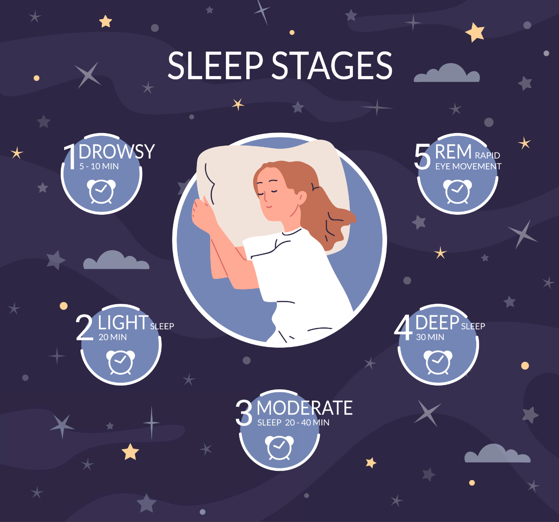
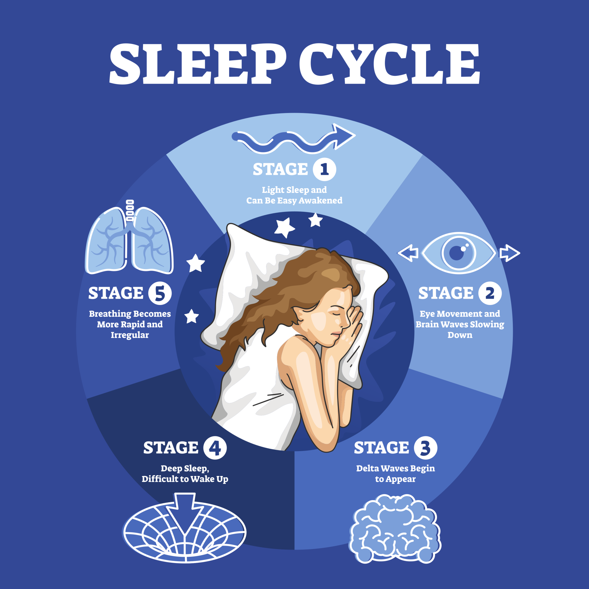
There are five different sleep stages, including REM (rapid eye movement) and NREM (non-rapid eye movement) sleep. The five stages make one sleep cycle, which usually repeats every 90 to 110 minutes.
Sleep itself is a readily reversible state of reduced consciousness, unlike coma or general anaesthesia, in which the brain stays organized and active rather than simply switching off.
-
Stage 1 (NREM): The transition from wakefulness to light sleep. Heart rate and breathing slow; muscles begin to relax.
-
Stage 2 (NREM): A deeper light sleep where body temperature drops and brain waves show specific bursts of activity (spindles). You spend the most time here.
-
Stage 3 (NREM): The beginning of Deep Sleep. It is difficult to wake someone from this stage; the body repairs tissues and builds bone/muscle.
-
Stage 4 (NREM): The deepest stage of sleep, characterized by slow delta waves. This is crucial for physical recovery and immune health.
-
REM (Rapid Eye Movement): Brain activity increases to near-awake levels. This is where dreaming occurs, while skeletal muscles are temporarily paralyzed to prevent acting them out.
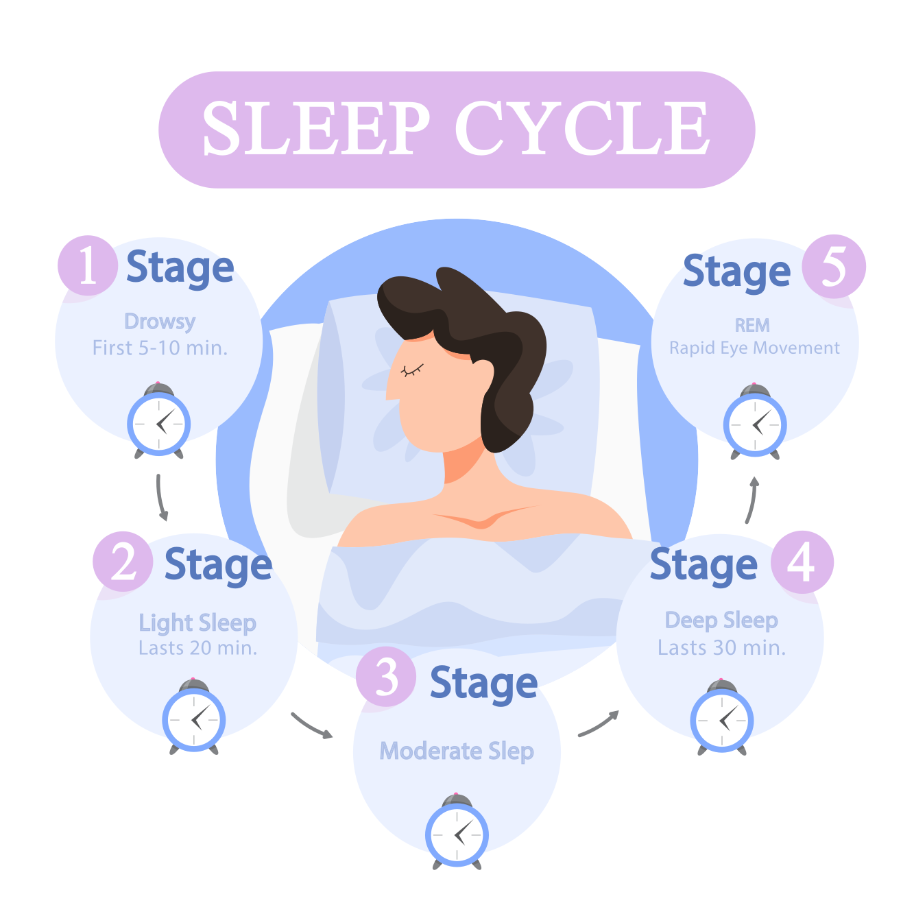

This five-stage numbering follows the classic Rechtschaffen and Kales (1968) scheme. Modern sleep medicine uses a newer system instead. It groups deep sleep differently, as N1, N2, and N3.
The AASM merged the old Stages 3 and 4 into one: N3. The original 3/4 cutoff was unreliable between raters. Stages 1, 2, and 3/4 below map onto N1, N2, and N3.
Stage 1: lightest sleep
Stage 1 sleep is the initial phase of the sleep cycle, functioning as a transitional period between wakefulness and sleep.
It is the first of the non-rapid eye movement (NREM) stages and is characterized as a “light sleep” during which an individual drifts off. This stage typically lasts only a few minutes.
Neural Activity and Brain Waves
During Stage 1, brain activity shifts distinctly from the patterns observed during wakefulness.
This transition is visualized via electroencephalography (EEG) through changes in the frequency and amplitude of brain waves:
- Alpha Waves: Low-frequency (8-12 Hz), high-amplitude waves dominate the early part of Stage 1, resembling the pattern of someone awake but deeply relaxed.
- Theta Waves: As Stage 1 continues, slower, higher-amplitude theta waves (4-7 Hz) take over from alpha activity.
- Overall Pattern: Generally, this stage is defined by relatively rapid, low-amplitude brain waves compared to the deeper stages that follow.
Physiological Changes
As the body enters Stage 1 sleep, several marked physical changes occur to facilitate the transition into deeper rest:
- Respiration and Heart Rate: Both the rate of breathing and the heartbeat begin to slowdown.
- Muscle Tension: There is a marked decrease in overall muscle tension.
- Body Temperature: Core body temperature begins to drop.
- Eye Movement: While rapid eye movements are absent (as this is an NREM stage), slow, rolling eye movements may occur during this period.
Subjective Experience and Wakefulness
Because Stage 1 is such a light phase of sleep, it is very easy to awaken someone during this time.
- Perception of Sleep: People awoken during Stage 1 often report that they have not been asleep at all.
- Mental Imagery: Images resembling still photos may appear to the sleeper during this stage. This differs from “true dreaming,” which occurs later, during REM sleep.
Clinical Context
Stage 1 sleep plays a role in the broader architecture of sleep health and pathology:
- Sleep Cycles: Sleep progresses through stages in cycles lasting roughly 90 minutes. Stage 1 opens the cycle, followed by deeper NREM stages and then REM sleep.
- Depression: Individuals suffering from depression often exhibit altered sleep architecture, the way sleep stages are composed across the night. This includes more Stage 1 sleep and less slow-wave sleep.
- Disorders: In conditions like narcolepsy, individuals may skip NREM stages, including Stage 1, and go directly from wakefulness into REM sleep.
Stage 2: light sleep
Following the fleeting transition of Stage 1, the body enters Stage 2 sleep, a state of deep relaxation.
This phase represents a deepening of sleep where it becomes increasingly difficult to awaken the individual compared to the lighter initial stage.
In this stage, your body temperature drops, and your eye movements stop completely.
For young adults, Stage 2 accounts for approximately half of their total sleep time.
Neural Activity and Brain Waves
While theta waves continue to dominate brain activity during this stage, the electroencephalogram (EEG) reveals distinct patterns that differentiate Stage 2 from the wakefulness–sleep transition:
- Sleep Spindles: Short bursts of about 11-16 Hz activity, generated by a circuit looping between the thalamus and cortex. Spindle density is linked to overnight memory improvement.
- K-complexes: Large, high-amplitude waves that can occur spontaneously or be evoked by an external stimulus, such as a sound. They may help both maintain sleep and monitor the environment.
Progression of Sleep Architecture
Stage 2 plays a significant role in the structure of a night’s rest:
- NREM Cycle: It is one of the distinct non-rapid eye movement (NREM) stages that people progress through in cycles lasting approximately 90 minutes.
- Shift Over Time: As the night progresses, sleep architecture shifts toward less deep Stage 4 sleep and more REM, which lengthens toward morning.
Stage 3: deep sleep
Stage 3 sleep, often referred to as deep sleep or slow-wave sleep, is a period marked by greater muscle relaxation and a dramatic slowing of physiological functions.
As the body progresses from Stage 2 into Stage 3, it becomes much more difficult to awaken the sleeper compared to the earlier stages.
Neural Activity and Brain Waves
In this stage, the brain’s electrical activity shifts significantly:
- Delta Waves: Delta waves, the slowest and highest-amplitude brain-wave pattern in sleep (below 4 Hz), appear in Stage 3.
- Alpha Intrusion: Some people show alpha activity, usually linked to wakefulness, intruding into Stage 3. They often report feeling unrefreshed on waking, however long they slept.
Physiological Functions and Benefits
Stage 3 sleep serves critical restorative and maintenance functions for the brain and body:
- Physical Restoration: Heart rate and respiration slow dramatically during Stage 3. Raising brain temperature, through head heating or vigorous exercise, increases subsequent slow-wave sleep, supporting a restorative function.
- Neurotoxin Clearance: This stage is critical for clearing neurotoxic proteins linked to Alzheimer’s disease, such as beta-amyloid and tau, which build up during wakefulness.
- Memory and Learning: Slow-wave sleep appears to be essential for effective memory formation. Engaging in slow-wave sleep after learning a new task can improve subsequent performance on that task.
Sleep Architecture and Parasomnias
Stage 3 plays a distinct role in the timing and phenomena of the sleep cycle:
- Timing: Deep sleep (Stages 3 and 4) dominates the first third of the night. As the night progresses, less time is spent in these deep stages and REM lengthens instead.
- Parasomnias: Parasomnias, such as sleepwalking (somnambulism) and night terrors, typically arise from slow-wave sleep. Sleepwalking is not dream enactment: narrative dreams are least likely during this phase.
Stage 4: deep sleep
Stage 4 sleep is the deepest phase of the sleep cycle, characterized as a period where the individual is least responsive to outside stimulation.
It is the final stage of non-rapid eye movement (NREM) sleep before the cycle typically loops back or transitions toward REM sleep.
Because of their physiological similarities, Stage 3 and Stage 4 are often grouped together and referred to collectively as slow-wave sleep or deep sleep.
Neural Activity and Brain Waves
In this stage, the brain’s electrical activity slows further than in the preceding stages. The electroencephalogram (EEG) displays a specific pattern:
- Delta Waves: Stage 4 is marked by the predominance of delta waves. These are high-amplitude, low-frequency waves (less than 4 Hz).
- Comparison to Stage 3: Stage 3 also features delta waves, but the activity is more continuous in Stage 4, with an even slower, more regular pattern.
Physiological Changes and Functions
During Stage 4, the body is in a state of profound relaxation, making it very difficult to awaken the sleeper.
- Vital Signs: Heart rate and respiration slow dramatically during this phase.
- Growth Hormone: Growth hormone is released in a large pulse during the first few hours of sleep, when Stage 4 dominates, supporting physical development and repair.
- Restoration: Deep, uninterrupted Stage 4 sleep is vital for feeling rested. Alpha-wave intrusions during this stage often leave people feeling unrefreshed, however long they slept.
Sleep Architecture
The presence of Stage 4 sleep changes as the night – and life – progresses:
- Early Night Dominance: Stage 4 dominates the first third of the night. Across the ~90-minute cycles that follow, sleepers spend less time in Stage 4 and progressively more time in REM.
- Age-Related Changes: The proportion of slow-wave sleep declines with age. In later life, Stage 4 sleep decreases significantly and may eventually disappear altogether.
Parasomnias
Specific sleep disturbances are closely linked to Stage 4 sleep. These phenomena are distinct from the narrative dreams that typically occur during REM sleep:
- Night Terrors: Occurring most frequently in children between ages 3 and 8, night terrors are sudden awakenings from Stage 4 sleep accompanied by extreme fear, panic, and strong physiological arousal.
- Sleepwalking and Sleeptalking: Both disturbances usually occur during Stage 4 sleep. Sleepwalkers may have a vague awareness of their surroundings and can manoeuvre around obstacles despite being deep in NREM sleep.
Stage 5: REM sleep
Stage 5 sleep, most commonly known as REM (Rapid Eye Movement) sleep, is the final phase of the sleep cycle.
It is a unique state of consciousness characterized by rapid movements of the eyes under closed eyelids and brain activity that closely resembles wakefulness.
While the earlier stages (1 through 4) are collectively termed non-REM (NREM) sleep, REM sleep occupies a little more than 20% of an adult’s total sleeping time.
Neural Activity and “Paradoxical Sleep”
REM sleep is often referred to as paradoxical sleep because the brain appears to be awake while the body remains deeply asleep.
- Brain Waves: Unlike the slow delta waves of Stage 4, Stage 5 EEG patterns are low-voltage, mixed-frequency beta waves. These resemble a waking brain.
- PGO Waves: In animal studies, REM onset brings bursts of spike discharges called PGO waves, travelling from the pons through the lateral geniculate nucleus to the occipital cortex.
- Hippocampal Activity: Rhythmic theta activity (4-7 Hz) recorded from the hippocampus during REM may be linked to memory consolidation, the stabilizing of new memories for long-term storage.
Physiological Changes
Physiologically, Stage 5 is a time of high internal arousal contrasted with external immobility:
- Muscle Atonia: While the brain is active, the major voluntary muscle groups are paralyzed (atonia), likely to stop the sleeper acting out dreams.
- Autonomic Storms: Despite muscle paralysis, the heart rate increases and becomes irregular, blood pressure rises, and breathing becomes faster.
- Sexual Arousal: This stage is associated with increased genital blood flow, resulting in erections in males.
- Thermoregulation: The body’s homeostatic control of temperature is poor or reduced during REM sleep.
Dreaming and Function
Stage 5 is the period of sleep in which dreaming occurs most vividly.
- Dream Content: While some dreaming can occur in NREM stages, REM dreams are more likely to be vivid, emotional, bizarre, and easily remembered upon waking.
- Memory and Learning: REM sleep is linked to procedural memory (skills) and emotional processing: the dampening of a memory’s emotional charge while its content is retained.
- Development: REM may play a key role in brain development: neonates spend up to 50% of sleep in REM, far more than adults, suggesting it aids neural maturation.
Regulation and REM Rebound
REM sleep is actively controlled by centers in the brain stem:
- Neurochemistry: Cholinergic neurons in the pons trigger REM onset. Noradrenergic (locus coeruleus) and serotonergic (raphé) neurons inhibit REM; REM begins once these neurons fall silent.
- REM Rebound: The body appears to need a specific amount of REM sleep. Depriving someone of it makes them spend significantly more time in REM on later nights, a phenomenon called REM rebound.
Disorders and Pathology
- Narcolepsy: People with narcolepsy may skip NREM stages, passing directly from wakefulness into REM. They may also experience cataplexy: a sudden loss of muscle tone, like REM paralysis, while awake.
- REM Sleep Behavior Disorder (RBD): The normal muscle paralysis of Stage 5 fails to occur, allowing people to physically act out their dreams, which can cause injury.
- Depression: Interestingly, individuals suffering from major depression often exhibit more REM sleep and less slow-wave sleep. Deprivation of REM sleep can sometimes temporarily improve depressive symptoms.
The Sleep Cycle
Your body cycles through these stages four to five times each night.
Cycles earlier in the night tend to have more NREM sleep, while later cycles have a higher proportion of REM.
By the final cycle, NREM deep sleep may vanish entirely. This pattern repeats nightly.
The sleep cycle is the orchestrator that binds the five stages of sleep into a cohesive biological rhythm.
Sleep is not a uniform state of quiescence. It is active and dynamic instead. The brain cycles through distinct phases of electrical and physiological activity all night.

The 90-Minute Ultradian Rhythm
The alternation between non-REM (NREM) and REM sleep occurs in a regular, cyclical pattern called an ultradian rhythm. An ultradian rhythm is a biological rhythm that repeats more often than once every 24 hours.
- Cycle Duration: Individuals progress through the sleep stages in cycles that last approximately 90 minutes.
- The Sequence: A typical cycle descends from wakefulness through Stages 1-2 into the slow-wave sleep of Stages 3-4, then ascends back through the lighter stages into Stage 5 (REM sleep).
- Cycle Frequency: During a typical night, a sleeper will experience approximately four to six of these cycles.
Shifting Architecture Throughout the Night
A critical feature of the sleep cycle is that it does not repeat identically throughout the night.
The composition of the 90-minute loops changes as the night progresses:
- First Half of the Night: Early cycles are dominated by slow-wave sleep (Stages 3 and 4), suggesting the body prioritises physical restoration and growth-hormone release right after falling asleep.
- Second Half of the Night: Deep slow-wave sleep shortens through the night and may vanish, while REM lengthens toward morning, so dreaming is reported more often on natural morning waking.
Regulation: The Two-Process Model
The timing and structure of the sleep cycle are governed by the interplay of two major biological mechanisms.
Homeostatic Drive (Process S): The internal “pressure” to sleep that builds up during wakefulness.
- Sleep Debt: If an individual goes without sleep, they accrue a sleep debt, which results in decreased alertness and mental efficiency.
- Chemical Buildup: This pressure is partly regulated by neurochemicals such as adenosine, which accumulates in the basal forebrain during waking hours and inhibits neurons that promote wakefulness.
- Rebound Effect: Depriving someone of a stage such as REM triggers compensation: once allowed to sleep undisturbed, they spend significantly more time in that stage, a REM rebound, to “catch up.”
Circadian Rhythm (Process C): The internal biological clock that regulates the timing of sleep and wakefulness over a 24-hour period.
- The Master Clock: This rhythm is controlled by the suprachiasmatic nucleus (SCN) located in the hypothalamus.
- Light Entrainment: Light synchronises the SCN with the outside world: light-sensitive retinal neurons send signals that align the body’s internal clock with the solar day.
- Melatonin: The SCN regulates the pineal gland’s secretion of melatonin, a hormone that rises during darkness to promote sleep and is inhibited by light.
Factors Influencing the Cycle
The architecture of the sleep cycle is not static and changes significantly across the lifespan and in response to environmental factors:
- Ageing:
- Infants: Newborns exhibit a polyphasic sleep pattern (sleeping multiple times a day) and spend approximately 50% of their sleep in REM.
- Adults: By adulthood, the pattern becomes monophasic (one major sleep period), and REM constitutes about 20–25% of total sleep.
- Elderly: In later life, total sleep may decrease and deep sleep (Stages 3-4) declines or disappears, leading to more frequent awakenings.
- Disruptions:
- Jet Lag: Travelling across time zones desynchronises the internal circadian rhythm from the external environment (zeitgebers), causing sleep disturbances.
- Shift Work: Working rotating shifts forces individuals to attempt sleep when their biological clock is promoting wakefulness, often resulting in insomnia and fatigue.
The Function of the Cycle
Why does the brain enforce this complex cycling? While fully understood, current theories suggest distinct functions for different phases:
- Memory Consolidation: Slow-wave sleep replays newly learned facts in the hippocampus and transfers them to the neocortex for storage (Diekelmann & Born, 2010); REM instead favours procedural and emotional memory.
- Restoration: Ian Oswald (1966) proposed that NREM restores the body while REM restores the brain, partly through protein synthesis. The clearest evidence is the growth-hormone pulse tied to early deep sleep.
- Brain Development: The high proportion of REM sleep in infants suggests it plays a key role in the maturation of neural circuits.
Key Study: Does Consolidation Require Sleep?
- Aim: Stickgold, James, and Hobson (2000) tested whether overnight improvement on a learned skill needs sleep itself, or simply time.
- Method: Participants trained on a visual texture discrimination task and were retested after different delays. One group stayed awake the first night, then had two recovery nights before retesting.
- Results: Time alone brought no improvement, however long the delay before retesting; sleep-deprived participants showed no gain even after two recovery nights.
- Conclusion: Consolidating this skill requires sleep on the first night after learning, and a lost night cannot be recovered later. Both slow-wave sleep and REM appear to contribute.

Overall, your body spends more time in the NREM phases of sleep

REM and Non-REM Sleep
Sleep has been traditionally divided into two categories: Non-rapid eye movement (NREM) and rapid eye movement (REM).
Non-REM Sleep
Non-REM sleep is marked by a reduction of physiological activity as bodily functions slow down. There are three phases: N1, N2, and N3.
Each stage has unique characteristics. They differ in depth of sleep and in how disconnected the senses and muscles become.
During non-REM sleep, electrical activity in the brain slows, growth hormone secretion occurs, and muscle activity, heart rate, respiration, and oxygen consumption all decrease (Purves et al., 2001).
Non-REM sleep is regulated by many brain structures, especially the thalamus and the cerebral cortex (De Andrés, Garzón, & Reinoso-Suárez, 2011).
REM Sleep
REM sleep, on the other hand, is marked by intense brain activity and is a much more active period of sleep than non-REM.
This stage is heavily regulated by the brainstem (McCarley et al., 1995), which is the region of the brain that connects the cerebrum with the spinal cord. It consists of the midbrain, medulla oblongata, and pons.
REM sleep occurs after the brain passes through stages one, two, and three and typically occurs approximately every 90 minutes (McCarley et al., 1995).
During REM sleep, brain activity increases, voluntary muscles are inhibited, and rapid eye movements and dreams occur (McCarley et al., 1995).
Sleep Cycles in Other Animals
Humans are not the only species that sleeps. Sleep supports recovery, memory, and growth in humans, so it makes sense that most other animals need it too.
However, the length of sleep, the brain’s state of consciousness, and whether dreaming occurs differ among species.
Which Animals Actually Sleep?
Not every animal loses consciousness the way humans do. Research shows that birds and mammals sleep (Siegel, 2008). They become unconscious for a time.
Reptiles sleep too, but it is unclear whether they enter a REM-like state.
Fish and amphibians only reduce their awareness. They never fully lose consciousness (Siegel, 2008).
Insects appear not to sleep at all, and have never been shown to enter REM sleep. They do, however, have periods of inactivity (McCarley et al., 1995).
How Sleep Cycles Differ by Species
Sleep cycle length varies widely between species. Birds and mammals share non-REM and REM sleep, but bird cycles are far shorter than ours.
A bird’s non-REM phase averages roughly two and a half minutes. Its REM phase lasts only nine seconds (Ogden, 2015).
Mammal cycles vary too. REM sleep lasts 24 minutes in a cat but only 12 minutes in a rat (McCarley et al., 1995).
Reptile data is less complete. A 2016 study of the Australian dragon lizard’s brain found slow-wave and REM-like patterns oscillating continuously, each phase lasting around 80 seconds (Shein-Idelson et al., 2016).
Do Other Animals Dream?
Since many animals enter REM sleep, researchers have asked whether they dream too.
Brainwave studies support the claim that both mammals and birds dream, because both groups enter REM sleep, the state most linked to dreaming in humans.
Reptiles may dream as well. Some studies suggest they show a form of REM sleep (Libourel et al., 2018; Shein-Idelson et al., 2016), but researchers remain uncertain.
Knowing whether an animal dreams is hard enough. Knowing what it dreams about is harder still, and the field needs more research to answer it.
When Should I Set My Alarm?
A sleep cycle typically lasts ninety minutes to two hours as the brain moves from slow-wave sleep to REM. But the cycle does not repeat identically all night.
The practical answer follows from where in the cycle the alarm lands.
Waking from deep N3 sleep, most common in the first third of the night, produces heavy sleep inertia because the brain must climb out of its most slow-wave state.
An alarm timed to the end of a ~90-minute cycle, or set during the lighter N2 and REM sleep that dominate later cycles, tends to feel far less jarring.
How Long Is Each Sleep Cycle?
The length and order of the stages change as the night goes on.
For example, the average length of the first non-REM-REM sleep cycle is 70 to 100 minutes.
The second and later cycles run longer, about 90 to 120 minutes (Carskadon & Rechtschaffen, 2011). REM sleep grows across the night. It peaks in the final third.
N2 comes to dominate non-REM sleep as the night goes on, and N3 can disappear altogether (Altevogt & Colten, 2006).
Adults typically need four or five cycles a night, or 7 to 9 hours of sleep (Hirshkowitz et al., 2015).
Needs vary widely from person to person and night to night (Carskadon & Rechtschaffen, 2011).
Babies cycle faster. Their cycles last only about 50 minutes for the first nine months of life, and newborns typically sleep 14 to 18 hours a day (Hirshkowitz et al., 2015).
How Long Should You Nap?
Shorter naps generally leave people feeling more refreshed and alert. Yet one 2019 study found that even 25-, 35-, and 45-minute naps cut stress and fatigue in physically active men, and improved their attention and performance (Hsouna, 2019).
The National Sleep Foundation warns that longer naps can leave you groggy, because you wake from deeper sleep.
Aim for a nap that is neither too short to feel refreshing nor too long to disrupt REM sleep. At night, aim for at least 7 hours of sleep. Sleep matters, so make it a daily priority.
William Dement: The Father of Sleep
How do we know anything about sleep if we are, well, asleep? We cannot hand someone a questionnaire mid-nap. We cannot ask them to talk while unconscious. One man solved most of this puzzle: Dr. William Dement.
Dement became known as the “Father of Sleep Medicine.” He was a Washington native. He once dreamed of becoming a journalist instead.
The journalism classes at the University of Washington were full that term. So Dement enrolled in an Introduction to Psychology course instead.
He found the class fascinating. He dropped his journalism plans. He decided to become a psychoanalyst instead (Stanford, 2008).
After Washington, Dement moved on. He joined the University of Chicago School of Medicine, where the only person studying sleep was faculty member Nathaniel Kleitman (Stanford, 2008).
The two began working together in the 1950s, making some of the greatest sleep discoveries in the field. Their first came in 1953: the discovery of rapid eye movement (REM) sleep (Aserinsky & Kleitman, 1953).
Key Study: Discovering REM Sleep
Before this study, sleep was seen as one single, passive state.
- Aim: Aserinsky and Kleitman (1953) set out to characterize eye movements during sleep and test whether bursts of eye motility related to sleep depth and dreaming.
- Method: Volunteers slept in the lab wired to an EOG (eye movement) and EEG (brain activity). Researchers woke some during bursts of rapid eye movement, and others during still periods, for a dream report.
- Results: Rapid eye movement recurred several times a night with a fast, low-voltage EEG and irregular heart rate and breathing. Waking during these periods produced a detailed dream report about three-quarters of the time; still periods rarely did.
- Conclusion: Sleep is not one uniform state. A recurring, active phase, later named REM sleep, is strongly linked to dreaming, opening up sleep research.
Critical Evaluation
The five-stage model is a useful summary, not a complete account of sleep. Five issues are worth weighing before taking it as the final word.
- Staging Is Partly Conventional: the neat N1-N3 and REM labels simplify a more continuous, graded process; the old stage-3/4 boundary was dropped for poor reliability.
- Function Evidence Is Correlational: much support for sleep’s functions comes from correlations or deprivation studies, both confounded by stress and rebound effects.
- Restoration Has Real Limits: tissue repair continues around the clock without sleep, and REM’s high energy use fits awkwardly with a pure energy-saving account.
- Individual Differences Are Large: normal sleep need ranges from about five to ten hours, and REM/deep-sleep proportions vary hugely with age and species.
- Glymphatic Evidence Is Still Indirect: the clearance mechanism is best proven in rodents; human studies so far show coupling and association, not direct waste removal.
Staging Is a Human Classification
The shift from four NREM stages to three was not a change in the brain. Scorers disagreed constantly. Those using the Rechtschaffen and Kales (1968) system often could not agree where Stage 3 ended and Stage 4 began.
The American Academy of Sleep Medicine fixed this by merging Stage 3 and Stage 4 into one, N3. Reliability improved right away. The fix also revealed something: sleep depth is graded, not boxed.
Different regions of the cortex can be more asleep than others at the same time. A single, whole-brain label oversimplifies that.
The N1 to N3 system stays clinically useful. It is a convention, not a natural category.
This matters for accuracy. A textbook label is not the same as a boundary drawn by the brain itself.
Function Evidence Leans on Correlation and Deprivation
Spindle density, for instance, correlates with overnight memory gains. That link alone cannot prove spindles cause the improvement, since a third factor could drive both.
This pattern turns up across many sleep-and-memory findings, not just spindles.
The other main method is deprivation: keep someone awake, then measure the damage. Both approaches share the same weakness.
Depriving someone of sleep also creates stress, and stress alone can hurt memory and mood. Rebound sleep afterward is hard to separate from the original deficit.
Ethics compound the problem. Researchers cannot deprive humans of sleep for long periods, so the strongest, most damaging findings come from rats (Rechtschaffen et al., 1989).
Even those extreme results may partly reflect the stress of forced wakefulness, rather than sleep loss itself.
Restoration Has Real Limits
If sleep exists mainly to repair the body, the pattern of repair is puzzling. Repair does not stop for sleep. It proceeds around the clock, whether a person is asleep or awake, so sleep is not strictly required for it.
Quiet rest is not a substitute either. Lying still and relaxed does not equal sleep. Something more than inactivity must be at work.
REM sleep is the clearest problem for a pure restoration account. It is high-energy and active. Neural firing rises toward waking levels.
That fits awkwardly with an account built around resting and conserving energy.
Some species can sharply cut their sleep time without obvious harm. That variability implies sleep often runs longer than any pure restorative need would demand.
Individual and Species Differences
Normal adult sleep need is not one number, and the variation runs from individuals right up to whole species.
- Adult range: Normal sleep need spans roughly five to ten hours a night, which alone challenges any single theory of why we sleep.
- Age: REM makes up roughly half of a newborn’s sleep. That share roughly halves by young adulthood, while deep slow-wave sleep also declines steeply into old age.
- Species: Sleep time ranges from around three hours in some grazing prey animals to eighteen or twenty hours in some predators. That gap tracks how safe or exposed each animal is.
Any single, universal function of sleep has to explain why a three-hour sleeper and a twenty-hour sleeper both thrive. No current theory fully manages that; the variation itself is a clue worth explaining.
Glymphatic Clearance Is Still Indirect in Humans
Sleep’s newest candidate function is cleaning the brain. The glymphatic system flushes metabolic waste out through channels that open wider during sleep, including proteins linked to Alzheimer’s disease.
The clearance mechanism itself is best proven in rodents. Sleep visibly speeds the removal of injected waste protein there.
Human evidence is newer, and so far more indirect.
Human studies show that brain fluid moves in coordinated waves locked to deep-sleep brain activity. Disrupting deep sleep also raises waste-protein levels.
Neither result directly shows a night’s sleep clearing toxic protein from a living human brain.
The theory deserves real weight. It unifies restoration and memory findings under one physiology. But direct human clearance evidence is not yet complete, so the claim stays probable rather than proven.
Contemporary Research
Recent research has moved sleep’s functions from plausible ideas toward mechanisms backed by large-scale evidence and direct human physiology.
Does Targeted Memory Reactivation Work?
The strongest recent evidence comes from a meta-analysis, not a single experiment. It targets sleep-dependent memory consolidation directly.
- Aim: Hu, Cheng, Chiu, and Paller (2020) set out to establish, across the whole published literature, whether cueing memories during sleep reliably improves them.
- Method: The team pooled 91 experiments and 212 effect sizes, covering roughly 2,000 participants, all testing targeted memory reactivation (TMR). In TMR, a cue linked to earlier learning is replayed during sleep to bias which memories get consolidated.
- Results: TMR reliably improved later memory, with a small-to-moderate effect overall. Crucially, the benefit appeared during N2 and slow-wave sleep, but not during REM sleep or wakefulness.
- Conclusion: The effect appears only in specific NREM stages, and only when memories are deliberately cued. That points to an actively orchestrated, stage-specific process, beyond earlier correlational work.
Does Sleep Clear Waste From the Brain?
A second research thread has followed the brain’s glymphatic system, the network that flushes metabolic waste from brain tissue using cerebrospinal fluid.
The foundational evidence is still animal work. Xie et al. (2013) showed that natural sleep expands the space between brain cells in mice by around 60%, roughly doubling how fast the brain clears injected waste protein.
Fultz et al. (2019) then showed a human counterpart. Scanning sleeping adults inside an MRI machine, the team found large, coherent waves of cerebrospinal fluid tightly time-locked to the slow brain waves of deep sleep.
Supporting evidence points the same way in people. Disrupting slow-wave sleep raises amyloid-beta levels in spinal fluid (Ju et al., 2017).
Even one night of sleep deprivation raises amyloid-beta levels in the living human brain on brain scans (Shokri-Kojori et al., 2018).
Taken together, this is a promising but still-maturing picture. Direct proof that sleep clears toxic waste exists mainly in rodents, while the human evidence remains coupling and association rather than clearance itself.
Is Sleep Really Made of Discrete Stages?
A newer research theme questions the neat N1-N3-and-REM picture itself. Sleep depth is not one whole-brain value.
Some cortical regions can show sleep-like slow waves while a person is behaviourally awake after sleep loss, a state called local sleep. Waking and sleep, in other words, can overlap.
Tononi and Cirelli (2014) tracked how the slope and amplitude of slow waves change across the night, the signature their synaptic homeostasis theory predicts for desaturated synapses. The pattern held locally as well as globally, across the whole brain.
This reframes the five-stage model above as a useful summary of a more graded, regional process.
FAQs
How long is a sleep cycle?
A typical sleep cycle lasts about 90 to 110 minutes, consisting of different stages: non-REM sleep stages 1, 2, 3, and the REM (Rapid Eye Movement) stage. Most adults experience 4 to 6 of these cycles per night.
How long is a REM cycle?
A REM (Rapid Eye Movement) cycle, in which most dreaming occurs, varies in length throughout the night. The first REM cycle is typically short, about 10 minutes, and occurs 90 minutes after falling asleep. Subsequent REM cycles lengthen, with the final one lasting up to an hour.
How do brain waves change as a sleeper progresses from stage 1 sleep to rem sleep?
As a sleeper progresses from stage 1 sleep to REM (Rapid Eye Movement) sleep, brain waves undergo distinct changes. In stage 1 sleep, brain waves slow down with irregular patterns known as theta waves.
In contrast, during REM sleep, brain waves become faster and more similar to the waves observed during wakefulness. This is when vivid dreaming occurs, and the brain shows increased activity and resembles the alert state. These transitions between sleep stages reflect different aspects of sleep and the corresponding brain activity.
Key Takeaways
- Five Stages: Sleep cycles through N1, N2, N3 (often still called Stages 1-4) and REM roughly every 90 minutes, four to six times a night.
- Staging Is Conventional: Modern AASM staging merged the old Stage 3 and Stage 4 into one, N3, because the boundary between them was unreliable.
- REM Physiology: REM shows a fast, awake-like brain but near-total muscle paralysis, which stops us acting out dreams.
- Consolidation: Sleep actively stabilizes memory; a 2020 meta-analysis found that cueing memories during NREM sleep reliably strengthens them (Hu et al., 2020).
- Glymphatic Clearance: Deep sleep helps flush metabolic waste from the brain, though direct proof in humans is still limited (Fultz et al., 2019).
- Function Is Debated: No single theory fully explains why we sleep; restoration, memory, and waste-clearance accounts each capture only part of the picture.
References
Abeln, V., Kleinert, J., Strüder, H. K., & Schneider, S. (2014). Brainwave entrainment for better sleep and post-sleep state of young elite soccer players–A pilot study. European Journal of Sport Science, 14 (5), 393-402.
Abhang, P. A., Gawali, B. W., & Mehrotra, S. C. (2016). Introduction to EEG-and speech-based emotion recognition. Academic Press.
Altevogt, B. M., & Colten, H. R. (Eds.). (2006). Sleep disorders and sleep deprivation: an unmet public health problem. National Academies Press.
Aserinsky, E., & Kleitman, N. (1953). Regularly occurring periods of eye motility, and concomitant phenomena, during sleep. Science, 118 (3062), 273-274.
Brown, R. E., & McCarley, R. W. (2008). Neuroanatomical and neurochemical basis of wakefulness and REM sleep systems. In Neurochemistry of sleep and wakefulness/Eds.: JM Monty, SR Pandi-Perumal, CM Sinton (p. 23).
Carskadon, M. A., & Rechtschaffen, A. (2011). Monitoring and staging human sleep. Principles and practice of sleep medicine, 5, 16-26.
De Andrés, I. T., Garzón, M., & Reinoso-Suárez, F. (2011). Functional anatomy of non-REM sleep. Frontiers in neurology, 2, 70.
Dement, W., & Kleitman, N. (1957). Cyclic variations in EEG during sleep and their relation to eye movements, body motility, and dreaming. Electroencephalography and clinical neurophysiology, 9 (4), 673-690.
Diekelmann, S., & Born, J. (2010). The memory function of sleep. Nature Reviews Neuroscience, 11(2), 114-126. https://doi.org/10.1038/nrn2762
Dubocovich, M. L. (2007). Melatonin receptors: role on sleep and circadian rhythm regulation. Sleep medicine, 8, 34-42.
Eiser, A.S. (2005). Physiology and psychology of dreams. Seminars in Neurology 25, 97–105.
Fogel, S. M., & Smith, C. T. (2011). The function of the sleep spindle: a physiological index of intelligence and a mechanism for sleep-dependent memory consolidation. Neuroscience & Biobehavioral Reviews, 35 (5), 1154-1165.
Forget, D., Morin, C. M., & Bastien, C. H. (2011). The role of the spontaneous and evoked k-complex in good-sleeper controls and in individuals with insomnia. Sleep, 34 (9), 1251-1260.
Freud, S., & Strachey, J. (1984). On Metapsychology: the theory of psychoanalysis: ‘Beyond the pleasure principle,’ The ego and the id and other works. Penguin.
Fultz, N. E., Bonmassar, G., Setsompop, K., Stickgold, R. A., Rosen, B. R., Polimeni, J. R., & Lewis, L. D. (2019). Coupled electrophysiological, hemodynamic, and cerebrospinal fluid oscillations in human sleep. Science, 366(6465), 628-631. https://doi.org/10.1126/science.aax5440
Hirshkowitz, M., Whiton, K., Albert, S. M., Alessi, C., Bruni, O., DonCarlos, L., … & Neubauer, D. N. (2015). National Sleep Foundation’s sleep time duration recommendations: methodology and results summary. Sleep health, 1 (1), 40-43.
Hsouna, H., Boukhris, O., Abdessalem, R., Trabelsi, K., Ammar, A., Shephard, R. J., & Chtourou, H. (2019). Effect of different nap opportunity durations on short-term maximal performance, attention, feelings, muscle soreness, fatigue, stress and sleep. Physiology & behavior, 211, 112673.
Hu, X., Cheng, L. Y., Chiu, M. H., & Paller, K. A. (2020). Promoting memory consolidation during sleep: A meta-analysis of targeted memory reactivation. Psychological Bulletin, 146(3), 218-244. https://doi.org/10.1037/bul0000223
Iyo, J. (2020). How Sleep Affects Your Human Growth Hormone (HGH) Levels. Retrieved from https://www.tuck.com/sleep-hgh/
Ju, Y. S., Ooms, S. J., Sutphen, C., Macauley, S. L., Zangrilli, M. A., Jerome, G., Fagan, A. M., Mignot, E., Zempel, J. M., Claassen, J. A. H. R., & Holtzman, D. M. (2017). Slow wave sleep disruption increases cerebrospinal fluid amyloid-beta levels. Brain, 140(8), 2104-2111. https://doi.org/10.1093/brain/awx148
Kales, A., Soldatos, C. R., Bixler, E. O., Ladda, R. L., Charney, D. S., Weber, G., & Schweitzer, P. K. (1980). Hereditary factors in sleepwalking and night terrors. Br J Psychiatry, 137 (2), 111-118.
Libourel, P. A., Barrillot, B., Arthaud, S., Massot, B., Morel, A. L., Beuf, O., … & Luppi, P. H. (2018). Partial homologies between sleep states in lizards, mammals, and birds suggest a complex evolution of sleep states in amniotes. PLoS biology, 16 (10), e2005982.
Lockett, E. (2020). The Stages of Sleep: What Happens During Each. Retrieved from https://www.healthline.com/health/healthy-sleep/stages-of-sleep
Massimini, M., Boly, M., Casali, A., Rosanova, M., & Tononi, G. (2009). A perturbational approach for evaluating the brain’s capacity for consciousness. Progress in brain research, 177, 201-214.
McCarley, R. W., Greene, R. W., Rainnie, D., & Portas, C. M. (1995). Brainstem neuromodulation and REM sleep. In Seminars in neuroscience (Vol. 7, No. 5, pp. 341-354). Academic Press.
Medrano-Martinez, P., & Ramos-Platón, M. J. (2014). Generation and functions of dreams. Revista de neurologia, 59 (8), 359-370.
Niedermeyer, E., & da Silva, F. L. (Eds.). (2005). Electroencephalography: basic principles, clinical applications, and related fields.
Lippincott Williams & Wilkins.
Ogden, D. (2015). How do birds sleep? Retrieved from https://ca.audubon.org/news/how-do-birds-sleep
Oswald, I. (1966). Sleep. Penguin.
Pace-Schott, E. F. (2013). Dreaming as a story-telling instinct. Frontiers in Psychology, 4, 159.
Purves, D., Augustine, G. J., Fitzpatrick, D., Katz, L. C., LaMantia, A. S., McNamara, J. O., & Williams, S. M. (2001). Physiological changes in sleep states. Neuroscience . 2nd edn. Sunderland, MA: Sinauer Associates.
Schwartz, J. R., & Roth, T. (2008). Neurophysiology of sleep and wakefulness: basic science and clinical implications. Current neuropharmacology, 6 4), 367-378.
Shein-Idelson, M., Ondracek, J. M., Liaw, H. P., Reiter, S., & Laurent, G. (2016). Slow waves, sharp waves, ripples, and REM in sleeping dragons. Science, 352 (6285), 590-595.
Shokri-Kojori, E., Wang, G.-J., Wiers, C. E., Demiral, S. B., Guo, M., Kim, S. W., Lindgren, E., Ramirez, V., Zehra, A., Freeman, C., Miller, G., Manza, P., Srivastava, T., De Santi, S., Tomasi, D., Benveniste, H., & Volkow, N. D. (2018). Beta-amyloid accumulation in the human brain after one night of sleep deprivation. Proceedings of the National Academy of Sciences, 115(17), 4483-4488. https://doi.org/10.1073/pnas.1721694115
Siegel, J. M. (2008). Do all animals sleep? Trends in neurosciences, 31 (4), 208-213.
[Stanford]. (2008). A Tribute for William C. Dement [Video file]. Retrieved from https://www.youtube.com/watch?v=UWkeI45kbNU
Stickgold, R., James, L., & Hobson, J. A. (2000). Visual discrimination learning requires sleep after training. Nature Neuroscience, 3(12), 1237-1238. https://doi.org/10.1038/81756
Tirapu-Ustarroz, J. (2012). Neuropsychology of dreams. Revista de Neurologia, 55 (2), 101-110.
Tononi, G., & Cirelli, C. (2006). Sleep function and synaptic homeostasis. Sleep medicine reviews, 10 (1), 49-62.
Tononi, G., & Cirelli, C. (2014). Sleep and the price of plasticity: From synaptic and cellular homeostasis to memory consolidation and integration. Neuron, 81(1), 12-34. https://doi.org/10.1016/j.neuron.2013.12.025
Valli, K., Revonsuo, A., Pälkäs, O., Ismail, K. H., Ali, K. J., & Punamäki, R. L. (2005). The threat simulation theory of the evolutionary function of dreaming: Evidence from dreams of traumatized children. Consciousness and cognition, 14 (1), 188-218.
Verma, A., Radtke, R. A., VanLandingham, K. E., King, J. H., & Husain, A. M. (2001). Slow wave sleep rebound and REM rebound following the first night of treatment with CPAP for sleep apnea: correlation with subjective improvement in sleep quality. Sleep medicine, 2 (3), 215-223.
Xie, L., Kang, H., Xu, Q., Chen, M. J., Liao, Y., Thiyagarajan, M., O’Donnell, J., Christensen, D. J., Nicholson, C., Iliff, J. J., Takano, T., Deane, R., & Nedergaard, M. (2013). Sleep drives metabolite clearance from the adult brain. Science, 342(6156), 373-377. https://doi.org/10.1126/science.1241224

BSc (Hons) Psychology, MRes, PhD, University of Manchester
Chartered Psychologist (CPsychol)
Saul McLeod, PhD, is a qualified psychology teacher with over 18 years of experience in further and higher education. He has been published in peer-reviewed journals, including the Journal of Clinical Psychology.

BSc (Hons) Psychology, MSc Psychology of Education
Associate Editor for Simply Psychology
Olivia Guy-Evans is a writer and associate editor for Simply Psychology, where she contributes accessible content on psychological topics. She is also an autistic PhD student at the University of Birmingham, researching autistic camouflaging in higher education.

Charlotte Ruhl graduated from Harvard University with a degree in Psychology and African American Studies. During her studies she worked at Harvard's Implicit Social Cognition Lab under Dr. Mahzarin Banaji, researching implicit racial bias and outgroup exposure, and contributed to the Decision Science Lab administering studies in behavioural economics and social psychology.