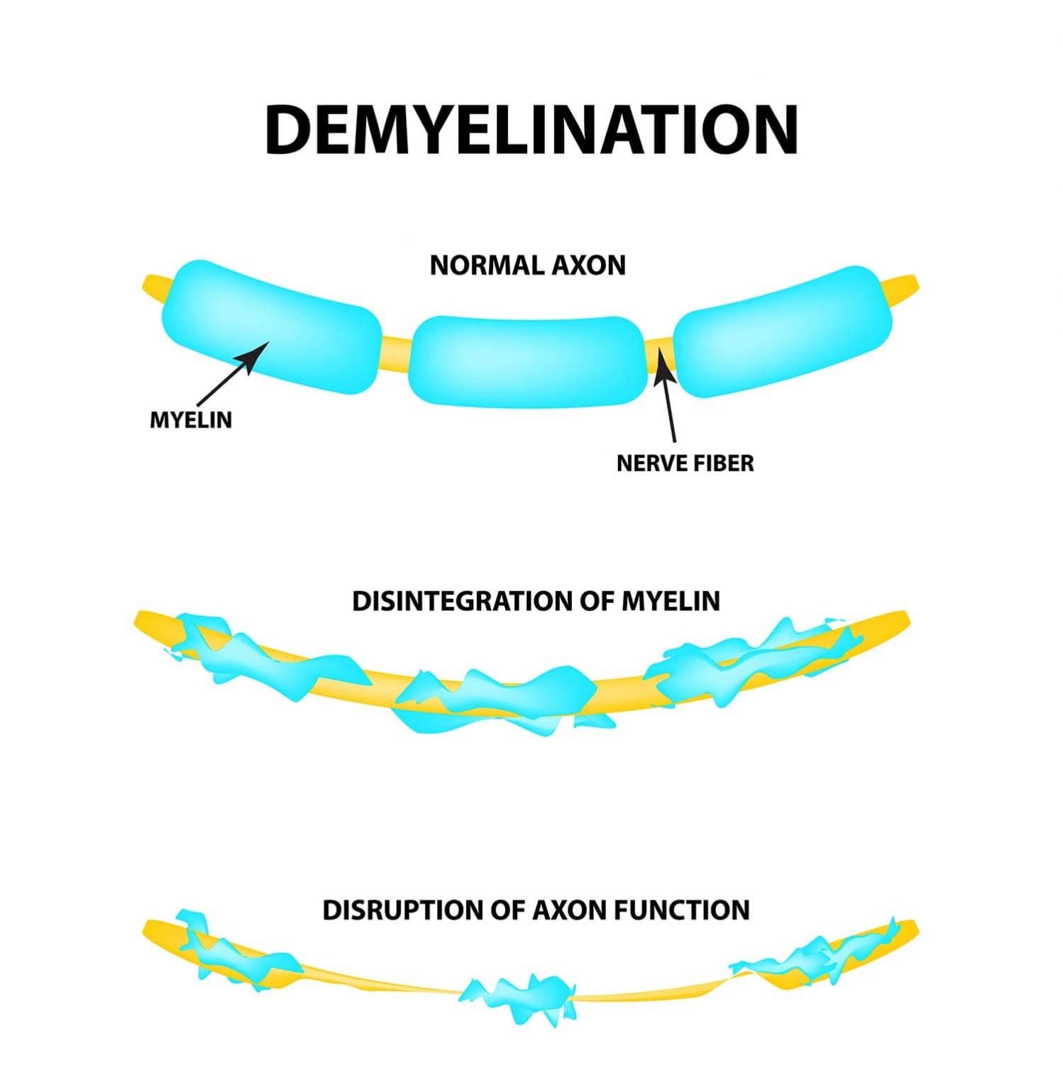
Demyelination is the loss or damage to the myelin sheath, the protective covering that surrounds nerve fibers and allows signals to be quickly and efficiently conducted along neurons.
When myelin is damaged or stripped away, neuronal signaling slows, leading to impaired sensation, movement, cognition, or other nervous system functions.
Signals arrive late, out of step, or not at all.
Disorders such as multiple sclerosis are characterized by demyelination, causing symptoms ranging from muscle weakness to impaired coordination or vision.
Disclaimer: This article is for informational purposes only and is not a substitute for professional medical advice, diagnosis, or treatment. Always seek the guidance of a qualified healthcare provider with any questions you may have about a medical condition.
Why is Myelin Important?
Myelin allows for fast, reliable communication between the brain, spinal cord, and body.
Imagine trying to stream a video with a frayed internet cable. The signal cuts in and out. Damaged myelin does the same to nerves.
Damage to this layer causes delays or complete blockages in nerve signaling, which can lead to physical and mental symptoms such as muscle weakness, numbness, or cognitive difficulties.

How Myelin Speeds Up Nerve Signals
Myelin works like the plastic coating around an electrical wire. This fatty insulation stops the electrical signal leaking out of the nerve fibre (the axon) as it travels.
The sheath is not continuous. It comes in segments, separated by tiny bare gaps called nodes of Ranvier. These gaps occur roughly every 0.2 to 2 millimetres along the axon.
Inside each insulated segment, current flows almost instantly. Only at the nodes is the action potential (the nerve impulse) regenerated at full strength. The impulse seems to leap from node to node. This is called saltatory conduction.
The payoff is large.
In big myelinated fibres, signals can reach around 100 to 120 metres per second. That is up to 50 to 100 times faster than along a comparable bare fibre.
Myelin also saves energy. Sodium enters the fibre only at the nodes, so far less pumping is needed to restore its resting state.
Why Losing Myelin Slows or Blocks Signals
Without its insulation, current leaks out across the exposed membrane and the signal weakens. Demyelination can also disrupt the seals at the edges of each node. The sodium channels that regenerate the impulse then spread out.
Conduction then slows. It can fail altogether, even while the fibre itself is intact.
Partly demyelinated fibres can still carry slow, unreliable signals. That is one reason early relapses in multiple sclerosis can partly recover.
Long-term loss does lasting harm because myelin does more than insulate. Support cells also feed the axons they wrap (Nave, 2010). In mice, oligodendrocytes were shown to pass the fuel lactate to the axon (Fünfschilling et al., 2012).
Stripping myelin therefore removes that fuel supply too. This helps explain why chronic demyelination can end in permanent loss of the nerve fibre itself.
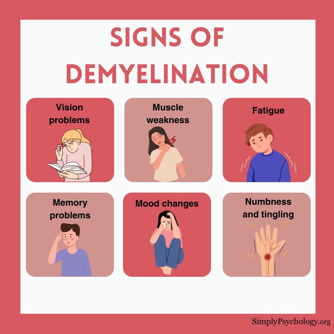
Signs of demyelination
Symptoms depend on the condition and on where in the nervous system the damage has occurred. Peripheral neuropathy, for instance, affects the hands and feet.
Symptoms vary from person to person, although some are common. Early signs include:
- Sudden vision loss or blurred vision
- Tingling or numbness in limbs
- Fatigue that doesn’t improve with rest
- Muscle weakness or poor coordination
- Unusual nerve pain
- Bladder or bowel dysfunction

Below are some of the common areas where demyelination can cause impairments and how they affect different parts of the nervous system:
Vision
Demyelinating conditions can lead to blurry or double vision. They can also result in a loss of vision that can be either temporary or permanent, depending on the severity of the demyelination.
Reflexes and movement
Demyelination can cause changes to the motor system which can lead to muscle weakness, stiffness, uncoordinated movements, and problems with balance.
This can ultimately affect how someone moves, meaning that they may not be able to perform some motor tasks or find it harder to walk, speak, or swallow.
Sensations
Those with demyelinating conditions may experience numbness and tingling, burning, or prickling sensations in their arms, legs, or feet.
In some cases, they may even feel pain when touched lightly, which can make it extremely difficult to navigate life as normal.
Cerebellum
The cerebellum is a part of the brain that plays a role in controlling balance and coordination.
Demyelination in this area can lead to tremors or incoordination such as finding it harder to walk without losing balance.
Genitourinary system
Some people with demyelinating conditions may experience problems with urination and bowel movements.
This can also affect sexual health and make someone more likely to develop a urinary tract infection (UTI).
Mood and thinking
Demyelination can also change mood. Many people experience depression, anxiety, and irritability.
There may also be problems with thinking, such as memory issues and a loss of focus, with some taking longer to process their thoughts.
Blood pressure and heart
In some cases, demyelination may result in people having poorer control over their blood pressure, as well as having a racing heartbeat or palpitations with no apparent cause.
What causes demyelination?
Demyelination can result from various factors, including immune system attacks, infections, or environmental and genetic influences.
Primary causes include:
- Autoimmune reactions: Conditions like multiple sclerosis (MS) occur when the immune system mistakenly attacks myelin.
- Viral infections: Viruses such as the JC virus may trigger demyelination, especially in those with weakened immune systems.
- Genetics: Some inherited disorders, like leukodystrophies (conditions in which faulty genes stop myelin forming or being maintained normally), affect myelin production.
Other contributing factors:
- Reduced oxygen to the brain
- Vitamin deficiencies (e.g., B12)
- Exposure to toxins or heavy metals
- Chronic alcoholism or liver damage
- Vascular diseases and inflammation
Demyelinating Disorders
Several neurological disorders are caused by or associated with demyelination:
Multiple Sclerosis (MS)
MS is the most well-known demyelinating condition. It affects the brain, spinal cord, and optic nerves. In MS, immune cells damage myelin, leading to scarring and disrupted nerve signaling.
People with MS may experience fluctuating symptoms such as numbness, vision problems, fatigue, or difficulty walking. It typically follows a relapsing-remitting or progressive course.
In MS, the immune attack leaves patches of demyelination called plaques in the brain, spinal cord and optic nerves (Compston & Coles, 2008). Early on, some myelin regrows and symptoms often partly recover. This repair is incomplete and does not last.
Over time, lasting loss of nerve fibres takes over. That is why disability tends to build up in a largely irreversible way.

Guillain-Barré Syndrome (GBS)
GBS involves rapid-onset muscle weakness due to immune attacks on peripheral nerves. It’s often triggered by infections and may progress quickly but can be treatable with early intervention.
GBS is the most common acute paralytic neuropathy, a sudden illness of the nerves that causes weakness. It is also the most severe. It affects roughly 100,000 people worldwide each year (Willison et al., 2016).
Infection with the bacterium Campylobacter jejuni is a frequent trigger. Germ molecules resemble nerve molecules, so the immune system attacks both. This mix-up is called molecular mimicry.
Weakness climbs up the body, and reflexes are lost. In the most severe cases, around 20 to 30%, the breathing muscles fail and the person needs a ventilator.
Most people improve substantially. Peripheral myelin can be rebuilt. Intravenous immunoglobulin or plasma exchange, plus supportive care, improves outcomes.
Optic Neuritis
This condition causes inflammation of the optic nerve, often leading to pain and temporary vision loss in one eye.
It can be an early sign of MS.
Chronic Inflammatory Demyelinating Polyneuropathy (CIDP)
CIDP is a rare, long-term condition where immune cells attack peripheral nerve myelin.
It causes gradual weakness, numbness, and sensory loss, especially in the limbs.
Leukodystrophies and Charcot-Marie-Tooth Disease
Some demyelinating conditions are built into the body from the start. In leukodystrophies, a genetic fault means the brain’s white matter never develops normally or slowly breaks down.
They are individually rare. They usually appear in childhood, as a loss of movement and thinking skills the child had already gained.
Charcot-Marie-Tooth disease is the most common inherited demyelinating nerve disease. The classic form is caused by an extra copy of the PMP22 gene, which helps hold peripheral myelin together. Other forms involve the gene for protein zero, another key myelin protein.
Nerve signals slow, and nerve damage slowly worsens in the feet, legs and hands. These conditions prove that myelin’s structural proteins matter.
Why Central and Peripheral Demyelination Differ
Two kinds of support cell (glial cells) make myelin. Oligodendrocytes do it in the brain and spinal cord. Schwann cells do it in the nerves running to muscles and skin.
One oligodendrocyte can wrap segments of up to about 50 different axons at once. If that cell is damaged or dies, many fibres lose myelin together. A Schwann cell wraps just one segment of one axon.
That difference changes outcomes.
Peripheral myelin is comparatively easy to rebuild, so Guillain-Barré syndrome often recovers well. Central myelin repairs poorly, so multiple sclerosis tends toward lasting disability over time.
Central myelin also blocks repair.
Two teams working independently identified Nogo-A. This protein is made by oligodendrocytes, strongly inhibits nerve-fibre growth and is essentially absent from Schwann cells (Chen et al., 2000; GrandPré et al., 2000).
A damaged central axon therefore struggles to regrow. A peripheral one can be guided back along a chain of Schwann cells.
How Is Demyelination Diagnosed?
Diagnosing demyelination involves a combination of clinical evaluation and diagnostic tests:
- Neurological exam: Doctors assess reflexes, strength, coordination, and sensory function.
- MRI scans: Used to detect lesions or inflammation in the brain and spinal cord.
- Lumbar puncture: Analyzes cerebrospinal fluid for markers of inflammation or infection.
- Blood tests: Rule out vitamin deficiencies or autoimmune markers.
- Evoked potentials: Measure how quickly electrical signals move through the nervous system.
Early diagnosis is crucial for managing symptoms and preventing long-term damage.
Treatment
While myelin damage can’t always be reversed, treatments can slow progression, reduce inflammation, and improve symptoms.
Treatment strategies include:
- Medications: Corticosteroids reduce inflammation; disease-modifying therapies (DMTs) are used in MS.
- Plasma exchange (plasmapheresis): Removes harmful antibodies from the blood, often used in GBS or severe relapses.
- Immunoglobulin therapy: Provides antibodies that modulate the immune response.
- Physical therapy: Helps maintain strength, flexibility, and function.
- Vitamin supplementation: Especially important for deficiencies such as B12.
Remyelination Research
Current treatments mainly calm the immune attack. Researchers are now testing whether drugs can rebuild myelin itself.
In the ReBUILD trial, Green et al. (2017) tested the antihistamine clemastine. The 50 participants had relapsing MS and long-standing damage to the optic nerve.
Each person took clemastine for 90 days and a placebo for 60 days, in either order. Neither they nor the researchers knew which was which.
The main outcome was how fast signals crossed the optic nerve, measured with evoked potentials. Clemastine shortened the delay by 1.7 milliseconds per eye (95% CI 0.5 to 2.9).
It was the first randomised evidence that a drug can repair chronic myelin damage in a person. The effect was modest. Fatigue was a common side effect.
Scientists are also learning to see myelin in living people. Stikov et al. (2015) built an MRI method that estimates the g-ratio. This is the width of a nerve fibre divided by its width including myelin.
In one person with MS, the ratio was higher inside lesions. That suggests thinner myelin where the disease had struck.
Living with Demyelination
A diagnosis of a demyelinating condition can be overwhelming, but many people manage symptoms successfully with treatment and support.
Regular follow-ups, symptom tracking, and healthy lifestyle choices can make a significant difference.
Support groups, counseling, and patient education also play vital roles in managing the emotional and psychological impact of chronic neurological conditions.
Key Takeaways
- Definition: Demyelination is damage to or loss of myelin, the fatty insulation around nerve fibres, which slows or blocks nerve signals.
- Symptoms: Effects depend on which nerves are affected, and can include vision loss, numbness, weakness, poor coordination and fatigue.
- Causes: Autoimmune attacks, infections, inherited faults, toxins and vitamin deficiencies can all damage myelin.
- CNS vs PNS: Peripheral myelin rebuilds more easily than central myelin, so Guillain-Barré syndrome often recovers better than multiple sclerosis.
- Diagnosis: Doctors combine a neurological exam, MRI, lumbar puncture, blood tests and evoked potentials.
- Treatment: Treatments calm the immune attack and ease symptoms. A 2017 trial showed a drug can modestly repair myelin.
References
Railton, D. (2021, November 28). What is demyelination, and what causes it? Medical News Today. https://www.medicalnewstoday.com/articles/318750
Apetauerova, D. (2021, September 20). An Overview of Demyelination. Very Well Health. https://www.verywellhealth.com/demyelination-4691934#toc-causes
Healthline Editorial Team. (2019, October 1). Demyelination: What Is It and Why Does It Happen? Healthline. https://www.healthline.com/health/multiple-sclerosis/demyelination
Reiff Ellis, R. (2020, August 9). What Are Demyelinating Diseases? WebMD. https://www.webmd.com/multiple-sclerosis/what-are-demyelinating-disorders
National Multiple Sclerosis Society. (n.d.) Understanding MS. Retrieved 2022, January 10, from https://www.nationalmssociety.org/What-is-MS/MS-FAQ-s
Centers for Disease Control and Prevention. (2019, December 20). Guillain-Barré Syndrome. https://www.cdc.gov/campylobacter/guillain-barre.html
Chen, M. S., Huber, A. B., van der Haar, M. E., Frank, M., Schnell, L., Spillmann, A. A., Christ, F., & Schwab, M. E. (2000). Nogo-A is a myelin-associated neurite outgrowth inhibitor and an antigen for monoclonal antibody IN-1. Nature, 403(6768), 434–439. https://doi.org/10.1038/35000219
Compston, A., & Coles, A. (2008). Multiple sclerosis. The Lancet, 372(9648), 1502–1517. https://doi.org/10.1016/S0140-6736(08)61620-7
Fünfschilling, U., Supplie, L. M., Mahad, D., Boretius, S., Saab, A. S., Edgar, J., Brinkmann, B. G., Kassmann, C. M., Tzvetanova, I. D., Möbius, W., Diaz, F., Meijer, D., Suter, U., Hamprecht, B., Sereda, M. W., Moraes, C. T., Frahm, J., Goebbels, S., & Nave, K.-A. (2012). Glycolytic oligodendrocytes maintain myelin and long-term axonal integrity. Nature, 485(7399), 517–521. https://doi.org/10.1038/nature11007
GrandPré, T., Nakamura, F., Vartanian, T., & Strittmatter, S. M. (2000). Identification of the Nogo inhibitor of axon regeneration as a Reticulon protein. Nature, 403(6768), 439–444. https://doi.org/10.1038/35000226
Green, A. J., Gelfand, J. M., Cree, B. A., Bevan, C., Boscardin, W. J., Mei, F., Inman, J., Arnow, S., Devereux, M., Abounasr, A., Nobuta, H., Zhu, A., Friessen, M., Gerona, R., von Büdingen, H. C., Henry, R. G., Hauser, S. L., & Chan, J. R. (2017). Clemastine fumarate as a remyelinating therapy for multiple sclerosis (ReBUILD): A randomised, controlled, double-blind, crossover trial. The Lancet, 390(10111), 2481–2489. https://doi.org/10.1016/S0140-6736(17)32346-2
Nave, K.-A. (2010). Myelination and support of axonal integrity by glia. Nature, 468(7321), 244–252. https://doi.org/10.1038/nature09614
Stikov, N., Campbell, J. S. W., Stroh, T., Lavelée, M., Frey, S., Novek, J., Nuara, S., Ho, M.-K., Bedell, B. J., Dougherty, R. F., Leppert, I. R., Boudreau, M., Narayanan, S., Duval, T., Cohen-Adad, J., Picard, P.-A., Gasecka, A., Côté, D., & Pike, G. B. (2015). In vivo histology of the myelin g-ratio with magnetic resonance imaging. NeuroImage, 118, 397–405. https://doi.org/10.1016/j.neuroimage.2015.05.023
Willison, H. J., Jacobs, B. C., & van Doorn, P. A. (2016). Guillain-Barré syndrome. The Lancet, 388(10045), 717–727. https://doi.org/10.1016/S0140-6736(16)00339-1

