Neuroimaging, or brain scanning, refers to techniques that produce images of the brain’s structure or activity. These tools allow researchers and clinicians to observe the brain in action and identify abnormalities without surgery.
Used in both clinical and research settings, neuroimaging helps us better understand how different parts of the brain work, how disorders affect brain function, and how treatments may influence brain activity.
Why Do We Use Neuroimaging in Psychology?
Neuroimaging is especially valuable in psychology and neuroscience for exploring how thoughts, emotions, and behaviors link to brain regions. It can:
- Show differences in brain structure and activity between individuals.
- Help diagnose brain injuries, tumors, or disorders like Alzheimer’s.
- Map brain function in real time (e.g., watching how the brain responds during memory tasks).
This non-invasive window into the brain allows scientists to test theories about cognition, behavior, and mental illness with unprecedented detail.
Types of Brain Imaging Techniques
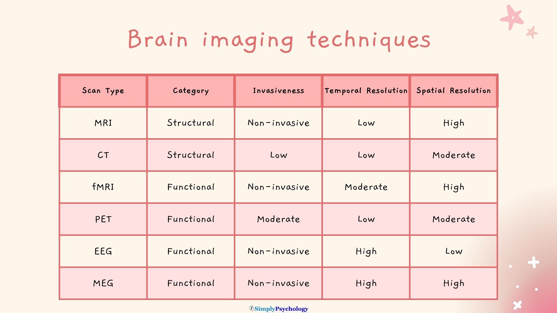
Most neuroimaging techniques fall into two main categories:
- Structural imaging reveals the brain’s anatomy (e.g., MRI, CT).
- Functional imaging tracks brain activity in real time (e.g., fMRI, PET, EEG).
Below are some of the brain imaging techniques:
Structural Neuroimaging
These scans provide detailed images of brain anatomy and are often used to detect abnormalities like tumors, bleeding, or injury.
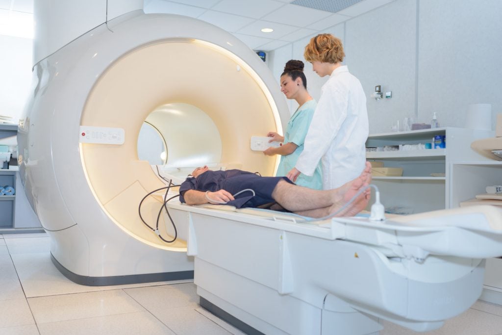
MRI (Magnetic Resonance Imaging)
- What it does: Uses strong magnetic fields and radio waves to create high-resolution images of brain tissue.
- Used for: Identifying brain tumors, multiple sclerosis, stroke damage, or brain malformations.
- What to expect: You’ll lie still inside a long tube. The scan is painless but noisy; you may be given earplugs. No radiation is involved. It typically takes 30–60 minutes.
CT Scan (Computed Tomography)
- What it does: Uses X-ray images to create cross-sectional views of the brain.
- Used for: Detecting bleeding, skull fractures, and brain swelling — especially in emergencies.
- What to expect: You’ll lie on a table that moves through a large ring-shaped scanner. The process is fast (5–10 minutes), and the machine is quiet. Some scans may require a contrast dye injection.
Functional Neuroimaging
These scans show brain activity and are useful in research and diagnosis of cognitive or neurological disorders.
fMRI (Functional Magnetic Resonance Imaging)
- What it does: Measures blood oxygen levels to map active areas of the brain during tasks or rest.
- Used for: Studying memory, emotion, attention, decision-making, and brain disorders like depression.
- What to expect: Similar setup to an MRI. You may be asked to complete simple tasks (like viewing images or tapping fingers) while the machine tracks brain responses.
PET Scan (Positron Emission Tomography)
- What it does: Uses a small amount of radioactive tracer to measure metabolic activity in brain tissue.
- Used for: Diagnosing Alzheimer’s disease, epilepsy, cancer, and some psychiatric conditions.
- What to expect: A radioactive tracer is injected into your bloodstream. After a short wait, you’ll lie on a table while the scanner captures images. The scan itself takes around 30 minutes and is painless.

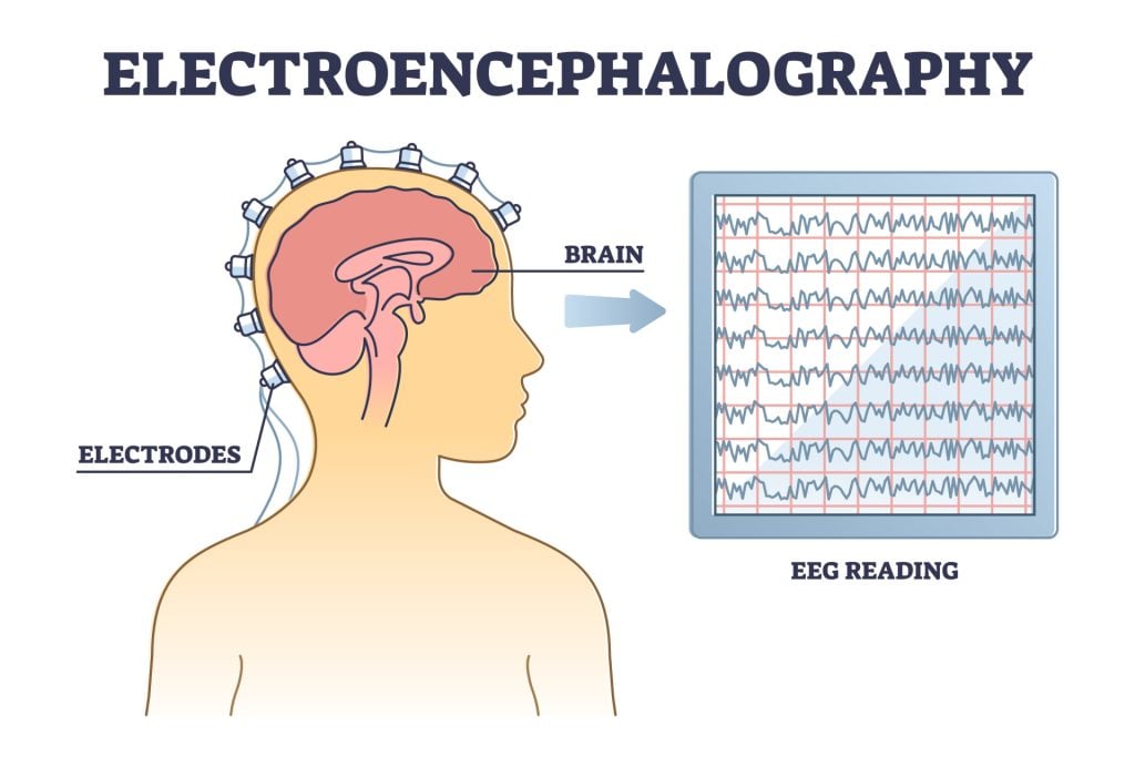
EEG (Electroencephalography)
- What it does: Records electrical activity in the brain using sensors placed on the scalp.
- Used for: Monitoring brain waves, diagnosing epilepsy, sleep disorders, or attention-related conditions like ADHD.
- What to expect: Electrodes are gently placed on your scalp using a cap or adhesive. The test is quiet, non-invasive, and typically lasts 20–60 minutes.
MEG (Magnetoencephalography)
- What it does: Measures the magnetic fields produced by neuronal activity to pinpoint where and when brain processes occur.
- Used for: Mapping sensory functions, diagnosing epilepsy, and guiding brain surgery.
- What to expect: You’ll sit or lie with your head positioned near a helmet-like device. It’s silent, non-invasive, and often used alongside MRI for detailed brain mapping.
History of Early Neuroimaging Techniques
Before the invention of MRI or fMRI, scientists used much cruder methods to study the brain.
While some early approaches are now outdated or discredited, they helped shape the evolution of modern neuroimaging techniques.
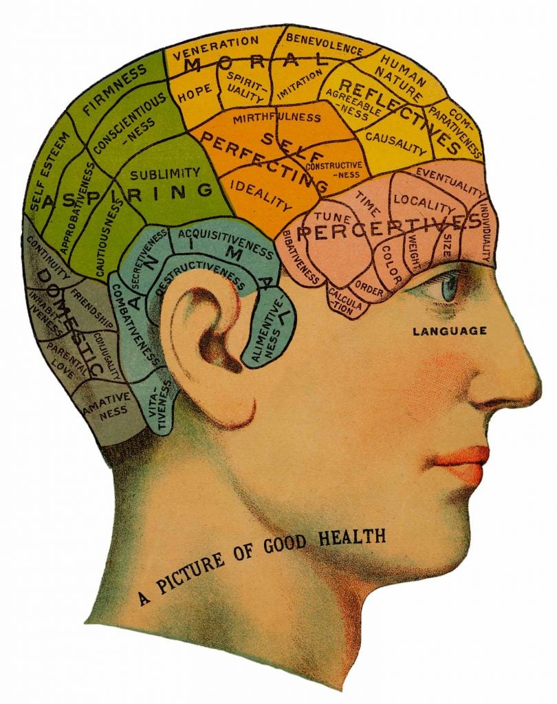
Phrenology (1800s)
One of the earliest (and most controversial) attempts to understand the brain was phrenology. This pseudoscientific theory claimed that bumps and indentations on the skull could reveal a person’s character traits and mental abilities.
Though inaccurate, phrenology introduced the idea of localization of brain function—the notion that different brain areas serve different purposes—a principle still central in neuroscience today.
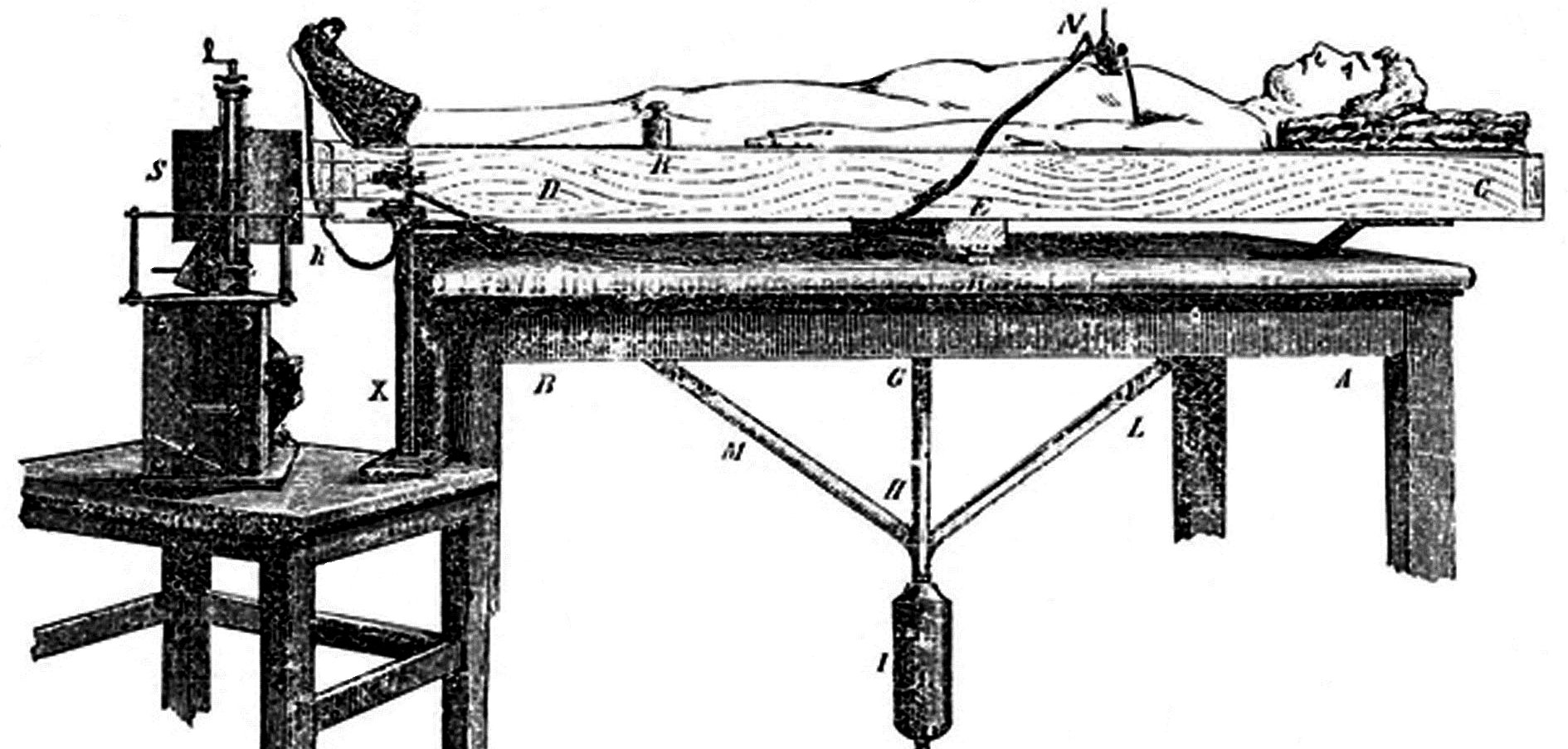
Human Circulation Balance
Italian physiologist Angelo Mosso developed the first device to measure changes in brain blood flow.
His “balance bed” showed that blood shifted toward the brain during mental effort. This idea—linking brain activity with blood flow—is the basis for modern fMRI scans
Pneumoencephalography (1918)
This brutal-sounding method involved draining cerebrospinal fluid and replacing it with air to make brain structures visible on X-rays.
While it helped visualize the brain’s ventricles, it caused severe side effects like headaches, nausea, and discomfort. Still, it marked a major step toward visualizing internal brain anatomy.
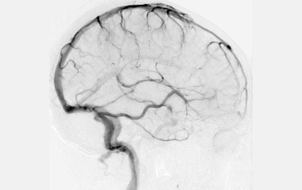
Cerebral Angiography (1927)
Developed by Egas Moniz, this method used contrast dyes injected into the bloodstream to make brain blood vessels visible on X-rays.
Though invasive, it was revolutionary for diagnosing brain tumors and vascular conditions. This technique is still used today in a safer, refined form.
The Rise of Modern Imaging (1970s)
The invention of Computed Tomography (CT) in the 1970s brought non-invasive imaging into clinical use.
Shortly after, Magnetic Resonance Imaging (MRI) was developed, offering clear, detailed images of brain tissue without radiation. These advances ushered in a new era of safe, high-resolution brain scanning.
What Can Neuroimaging Reveal?
Neuroimaging can help identify:
- Brain structure issues: tumors, strokes, trauma.
- Mental health patterns: activity differences in depression, anxiety, or schizophrenia.
- Brain function: which areas light up during reading, math, or emotion regulation.
It’s a powerful tool for both clinical diagnosis and understanding the biology behind behavior.
Can Brain Scans Read Thoughts?
Not quite. While functional scans show which areas of the brain are active, they can’t reveal exact thoughts or feelings.
For example, seeing activity in the amygdala may suggest emotional arousal, but it doesn’t tell us what the person is feeling or why.
How Neuroimaging Helps in Psychology
Researchers use brain scans to:
- Test theories of behavior and emotion.
- Compare brain differences in mental health conditions.
- Track changes after therapy or medication.
For instance, studies show people with ADHD often have different patterns of activation in attention-related brain regions. Neuroimaging can help tailor treatments based on individual brain responses.
Limitations of Neuroimaging
While revolutionary, brain imaging has important limitations:
- Correlational, not causal: Seeing activity doesn’t prove cause.
- Costly: Some techniques like fMRI and PET are expensive.
- Interpretation required: Data must be analyzed carefully by experts.
- Ethical concerns: Misuse could lead to overgeneralization (e.g., using scans to predict behavior).
What’s Next for Neuroimaging?
Advancements on the horizon include:
- Real-time brain-computer interfaces.
- Portable neuroimaging devices.
- AI-enhanced image analysis.
- Personalized treatments using brain scan profiles.
As technology evolves, neuroimaging could help us better predict, prevent, and treat brain-based conditions.
FAQs
Can neuroimaging diagnose mental illness?
It can support a diagnosis, but it’s not a standalone tool. Diagnosis still relies on clinical interviews and behavioral assessments.
Is neuroimaging safe for children?
Yes—non-invasive techniques like MRI and EEG are safe and commonly used with children.
What’s the difference between MRI and CT?
MRI uses magnets and shows more detail. CT uses X-rays and is faster but exposes you to radiation.
Does neuroimaging show emotions?
It shows the brain regions involved in emotion but not the specific feeling itself.
References
American Psychological Association. (2014, August). Scanning the Brain. https://www.apa.org/action/resources/research-in-action/scan
Artico, M., Spoletini, M., Fumagalli, L., Biagioni, F., Ryskalin, L., Fornai, F., Salvati, M., Frati, A., Pastore, F. S., & Taurone, S. (2017). Egas Moniz: 90 Years (1927-2017) from Cerebral Angiography. Frontiers in neuroanatomy, 11, 81.
Britannica, T. Editors of Encyclopaedia (2009, February 9). Pneumoencephalography. Encyclopedia Britannica. https://www.britannica.com/science/pneumoencephalography
Carter, R. (2019). The Human Brain Book: An Illustrated Guide to its Structure, Function, and Disorders (3rd ed). DK.
Grant, F. C. (1927). Indications for and technic of ventriculography. Radiology, 9 (5), 388-395.
Lumen (n.d.). Brain Imaging Techniques. Retrieved June 8, 2021, from https://courses.lumenlearning.com/boundless-psychology/chapter/brain-imaging-techniques/
Sandrone, S., Bacigaluppi, M., Galloni, M. R., Cappa, S. F., Moro, A., Catani, M., Filippi, M., Monti, M. M., Perani, D. & Martino, G. (2014). Weighing brain activity with the balance: Angelo Mosso’s original manuscripts come to light. Brain, 137 (2), 621-633.
Van Wyhe, J. (2000). The History of Phrenology. Victorian Web. https://victorianweb.org/science/phrenology/intro.html
Further Reading
- Kniermin J. Neuroscience online: an electronic textbook for the neurosciences. Chapter 5: Cerebellum. University of Texas Health Science Center at Houston.
- Electroencephalography (EEG)
- Casson, A. J., Abdulaal, M., Dulabh, M., Kohli, S., Krachunov, S., & Trimble, E. (2018). Electroencephalogram. In Seamless Healthcare Monitoring (pp. 45-81). Springer, Cham.

