The default mode network (DMN) is a set of connected brain regions. It is most active when a person is resting and thinking inwardly, and it quiets down when attention turns to a demanding external task.
Raichle et al. (2001) named it the “default mode.” Researchers now see it as the brain’s ‘intrinsic’ activity, studied mainly through resting-state brain imaging.
It supports self-referential thought, reminiscing, and future planning (Davey, Pujol and Harrison, 2016; Smallwood et al., 2021).
Where is the Default Mode Network in the Brain?
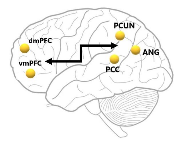
The DMN is distributed across three major subdivisions (Raichle, 2015):
- Ventromedial prefrontal cortex (vmPFC),
- Dorsomedial prefrontal cortex (dmPFC),
- Posterior hub including the posterior cingulate cortex (PCC), adjacent precuneus and angular gyrus (Figure 1).
In resting functional magnetic resonance (fMRI) studies, these regions show coordinated temporal activity. This is a known feature of large-scale brain networks (Greicius et al., 2003).
Greicius et al. (2003) tested this directly. They studied healthy adults at rest.
They tracked spontaneous fluctuations in the blood-oxygen-level-dependent (BOLD) signal, the blood-flow measure that fMRI uses as an indirect index of brain activity. They mapped the network two ways.
One map started from a seed region in the PCC. The other used independent component analysis, a data-driven method that separates the signal into networks without a starting region.
Both converged on the same network, with the PCC as its most widely connected node. The DMN is therefore a genuine network, not just a set of areas that happen to switch off together.
The vmPFC is an integration area.
It is involved in the generation and regulation of emotions and the attribution of reward value to stimuli. It also supports value-based decision-making and various aspects of social cognition (Mayeli et al., 2020).
Overall, it plays an important modulatory role when coping with adverse conditions, and it regulates motivational drive in favor of goal-oriented behaviors.
The dmPFC has often been linked to self-referential judgments in the present. It is activated when a person judges themselves or reflects on their own psychological state, or on someone else’s (Denny et al., 2012).
The PCC and medial precuneus, along with the angular gyrus, support the recollection of past experiences. This includes autobiographical information and previously studied items.
This process is mediated by functional coupling with the medial temporal lobe and hippocampus.
Both structures are sensitive to the daily accumulation of experience and support the memory-based construction of future scenarios (Andrews-Hanna et al., 2010; Shannon et al., 2013).
The DMN is not one uniform block. Using resting-state connectivity in healthy adults, Andrews-Hanna et al. (2010) found a midline core and two interacting subsystems:
- Midline core: Anterior medial prefrontal cortex and PCC, engaged across many kinds of self-relevant thought.
- Dorsal medial prefrontal subsystem: dmPFC, temporoparietal junction, lateral temporal cortex and temporal pole, linked to mentalizing (inferring mental states) in the present.
- Medial temporal subsystem: Hippocampal formation, parahippocampal and retrosplenial cortex, and posterior inferior parietal lobule, linked to memory-based construction and imagining the future.
Both subsystems couple with the core but serve dissociable functions. This helps explain why one network links to so many cognitive processes.
Distance matters here too. The DMN sits at a functional and spatial distance from more extrinsically driven brain networks, such as the sensorimotor system.
This distance supports the idea that the DMN handles higher-order, abstract cognition, often disconnected from events in the immediate environment (Smallwood et al., 2021).
Role of the Default Mode Network
Self-reflection
The DMN was first identified at rest, in contrast with ‘task-positive’ networks, the attention and control systems that switch on when we engage the outside world.
It has therefore often been linked to spontaneous, self-referential thought that contributes to our sense of self and identity (Yeshurun, Nguyen, and Hasson, 2021).
In fMRI studies, subjects made self-referential judgments about trait adjectives. This activated the PCC and medial PFC, compared with control conditions involving resting-state and non-self-referential tasks (Whitfield-Gabrieli et al., 2011; Davey, Pujol and Harrison, 2016).
The picture is not one-to-one. Whitfield-Gabrieli et al. (2011) found both overlaps and differences between the DMN as defined by resting connectivity and the network engaged by explicit self-reference tasks. Self-referential processing is a core but not exclusive DMN function.
In addition, spontaneous electroencephalography (EEG) studies of DMN areas could predict how often people engage in introspective thoughts (Frewen et al., 2020).
Memories
The DMN’s ability to retrieve previously encoded information relates to its connectivity with medial temporal lobe areas that are crucial for learning and memory.
Autobiographical remembering, and interpreting new stimuli using past knowledge, activates midline DMN regions.
This activation is linked to increased connectivity with hippocampal regions, a key area for learning and memory (Chen et al., 2016; Spreng and Grady, 2010).
Spreng and Grady (2010) compared three abilities. All three recruit a common core that overlaps substantially with the DMN:
- Autobiographical memory: remembering personal events from the past.
- Prospection: imagining the future.
- Theory of mind: inferring what other people think and feel.
One constructive system, in other words, handles all three. It lets people project the self across time and into other minds.
The medial temporal part of the network adds scene construction: assembling the sensory and spatial details that make remembered and imagined episodes feel vivid.
Memory retrieval also matters for decision-making.
Feedback and learning from past experience, accumulated through the DMN’s retrieval role, can guide future behaviour (Buckner and Wheeler, 2001).
Daydreaming
People spend a large share of their waking lives in stimulus-independent thought: thinking not triggered by their surroundings. This includes daydreaming and mind-wandering (thoughts drifting from the task at hand) about plans, everyday concerns, and past experience. It reliably engages the DMN (Fox et al., 2015).
DMN connectivity shifts as daydreaming fluctuates. Moment-to-moment changes in DMN coupling track these fluctuating states, and people differ in how often they daydream day to day (Kucyi and Davis, 2014).
Daydreaming usually serves a purpose. It supports everyday autobiographical planning: greater DMN engagement, together with medial temporal areas, is tied to more frequent future-oriented thought (Andrews-Hanna et al., 2010).
Daydreaming has a cost when attention matters. In a reading study using fMRI, Zhang et al. (2019) linked individual differences in DMN connectivity to off-task thought and to memory for the text.
A DMN region was more decoupled from medial visual regions in people who mind-wandered more often. It was more strongly connected to the medial prefrontal cortex in people who remembered the text better.
Social evaluations
Many studies show DMN regions activate when people try to understand or interact with others. This includes interpreting others’ mental states, inferring intentions and beliefs, and evaluating their behavior.
The vmPFC and its connections with affective regions support general emotional engagement in social settings. In turn, vmPFC-PCC connectivity helps distinguish self from other.
For example, it is active when people attribute qualities and feelings to themselves or to others (Frewen et al., 2020).
The dmPFC’s connections with the temporoparietal junction (TPJ) play a crucial role in theory of mind. It means attributing beliefs and desires to ourselves and others.
People use this to interpret others’ mental states and predict their desires and behavior (Li, Mai, and Liu, 2014).
How Does Meditation Affect the Default Mode Network?

As opposed to states of mind-wandering, meditation involves maintaining attention to the present moment, on purpose and non-judgementally (Bishop et al., 2004).
How Meditation Changes DMN Activity
During meditation, people learn to notice self-related thoughts, emotions, and body sensations. They learn to separate feeling from self-identifying.
This builds emotion regulation, focused attention, and a shift in self-perspective. Greater activation of prefrontal areas linked to executive control and self-monitoring is expected as a result.
This is not passive relaxation. Meditation trains sustained, effortful attention, which is why it recruits brain systems for executive control rather than switching them off.
Over time, this loosens mind-wandering’s grip on attention. Spontaneous, self-generated thought is itself closely tied to DMN activity.
This distinguishes meditation from ordinary rest, where the mind is free to wander without any such training. That is why the practice itself, not rest alone, draws such interest from DMN researchers.
Evidence From Brain-Imaging Studies
This is exactly what fMRI studies of experienced meditators have found. A stronger coupling of prefrontal regions appears alongside deactivation of DMN nodes, probably due to reduced mind-wandering (Brewer et al., 2011).
Three findings stand out:
- Across styles: Brewer et al. (2011) tested concentration, loving-kindness, and choiceless-awareness meditation (observing whatever arises without choosing a focus). The DMN’s main nodes were relatively deactivated in all three.
- Moment to moment: DMN activity fluctuates with how focused a person is. When meditators pressed a button on noticing a distraction, those moments meant greater DMN activity (Hasenkamp and Barsalou, 2012).
- Over time: Meditation weakens connectivity between DMN regions involved in self-referential processing and emotional appraisal (Taylor et al., 2013; Simon and Engström, 2015).
Clinicians can track this shift to monitor meditation’s therapeutic effects over time.
Role of the Default Mode Network in Disorders
Some disorders share a common thread. They involve poor coordination between the DMN, a control network, and a salience network that flags what matters and shifts attention inward or outward.
This “triple-network” framework helps explain why DMN changes appear across such different conditions.
The main finding for each condition is summarized below, with detail in the sections that follow.
| Condition | DMN pattern | Key evidence |
|---|---|---|
| Alzheimer’s disease | PCC-hippocampus connectivity breaks down early; DMN topography overlaps where amyloid builds up | Mevel et al. (2011); Buckner et al. (2008) |
| Schizophrenia | Increased connectivity within the task-negative (DMN) network, shared by patients and their unaffected siblings | Liu et al. (2012) |
| ADHD | Delayed maturation; late separation of the DMN from task-positive networks | Sripada et al. (2014) |
| Depression | Heightened DMN engagement tied to brooding rumination; stronger coupling with the subgenual cortex | Hamilton et al. (2015); Zhou et al. (2020) |
Alzheimer’s Disease
Alzheimer’s disease (AD) is a progressive condition primarily linked to memory loss. It has a distinct molecular signature.
AD involves a buildup of amyloid-β plaques and tau tangles, mainly in the medial cortical regions and hippocampus (Braak and Braak, 1991). These protein clumps disrupt normal neuron function.
These same regions support normal DMN function. AD-related changes there are thought to alter DMN activity too.
Buckner et al. (2008) noted a striking overlap. The DMN’s topography matches where amyloid accumulates.
This suggests that chronic activity in the network confers vulnerability to degeneration. It is a hypothesis, not proof.
Functional connectivity between the PCC and hippocampus breaks down early in AD (Mevel et al., 2011). Resting-state DMN measures can also help distinguish AD from healthy aging (Greicius et al., 2004).
Studies of amnestic mild cognitive impairment (aMCI), a stage that can precede AD, found similar though milder PCC-hippocampus desynchronization compared with normal aging (Mevel et al., 2011). Task-switching is affected too.
Delayed switching between resting-state and task-related brain function has been linked to inefficient synchronization of DMN areas in both AD and aMCI (Rombouts et al., 2005).
Schizophrenia
Schizophrenia is a complex psychiatric disorder.
It involves:
- Altered perception
- Delusions
- Cognitive deficits
- Abnormal emotion regulation
Many fMRI studies have found altered DMN connectivity with other brain areas in people with schizophrenia. The changes are linked to both positive and negative symptoms (Hu et al., 2017).
Liu et al. (2012) studied patients and their unaffected siblings.
Both groups showed increased connectivity within the DMN, but not within the anticorrelated task-positive network.
The pattern points to an inherited vulnerability. DMN hyper-connectivity may be an endophenotype of the illness (Liu et al., 2012): a heritable trait that sits between genetic risk and the full disorder.
Preliminary findings point to DMN connectivity as a potential target for future antipsychotic drugs. More research on the DMN’s response to medication is needed (Hu et al., 2017).
ADHD
Characteristics of ADHD, including inattention and impulsivity, have been linked to delayed maturation of the DMN. Findings on connectivity within the DMN itself are more mixed across studies (Harikumar et al., 2021).
The clearer finding concerns separation. Connectivity between the DMN and task-positive networks (systems for externally directed attention), such as the ventral attentional and frontoparietal networks, matures late (Sripada, Kessler, and Angstadt, 2014). Weak DMN suppression during tasks may let internal thought intrude.
Abnormal DMN connectivity with attentional-control areas has also been found in adults with ADHD, supporting the maturational-delay hypothesis.
However, the contribution of other intellectual impairments, often present in co-morbidity with ADHD, still needs to be clarified (Harikumar et al., 2021).
Depression
One defining feature of depression is brooding rumination: a passive, recurrent focus on depressed mood and its consequences (Treynor, Gonzalez, and Nolen-Hoeksema, 2003).
Several fMRI studies have identified DMN areas critically involved in ruminative processes.
A coordinate-based meta-analysis found that rumination consistently maps onto the medial prefrontal cortex, posterior cingulate cortex and precuneus, and angular gyrus (Zhou et al., 2020).
Connectivity changes go further. In major depressive disorder, connectivity also increases between the DMN and the subgenual prefrontal cortex (sgPFC). This area is involved in appraising negative emotion and behavioral withdrawal (Hamilton et al., 2015).
This has shaped new treatments. Transcranial magnetic stimulation (TMS) of the dorsolateral prefrontal cortex appears to work partly by normalizing DMN connectivity, particularly with the subgenual cingulate (Liston et al., 2014).
The next step is personalization.
Cash et al. (2021) developed computational methods to find each patient’s own best stimulation site: the point whose connectivity with the subgenual cingulate best matches the therapeutic target.
In scans from 1,000 healthy adults, the method located the same target to within about 2 mm across separate days. This could replace a fixed scalp location with individualized, connectivity-guided coil placement.
Critical Evaluation
The DMN has reshaped how psychologists think about the resting brain, but it is not without limitations. Several points are worth weighing carefully.
How the DMN Was Discovered
- Aim: Raichle et al. (2001) had a puzzle. Certain brain regions reliably reduced activity during goal-directed tasks, and no one knew why.
- Method: Using PET scans, the team measured blood flow and oxygen use across the brain. They pooled data from many tasks to find regions that reliably reduced activity compared with quiet rest.
- Findings: Oxygen use was uniform across the resting brain, confirming rest as a valid baseline. Medial regions, including the PCC and medial prefrontal cortex, consistently deactivated whenever attention turned outward.
- Conclusion: The brain has a default mode: an organised baseline that supports internal thought and is suspended during external tasks. It gave the field a name and a physiological basis for the DMN.
The study was transformative.
But it has limits: PET has poor time resolution, and it shows only that regions deactivate together, not that they form one network.
Proof of a real network came later, from resting-state connectivity studies.
Built on Correlational Evidence
A second finding deepened the picture. Fox et al. (2005) showed that the DMN and “task-positive” attention networks are anticorrelated. When one rises in activity, the other tends to fall, even at rest.
This built the opposition between outward attention and inward thought into the brain’s intrinsic organisation.
The finding is influential, but contested. Removing the brain’s average “global” signal during preprocessing can artificially push correlations negative. Critics argued some of the original anticorrelation was an artefact of this choice.
Later work found the anticorrelation survives without that step, so the effect is now considered real, though its exact size depends on analysis choices.
More broadly, the DMN is defined mainly through correlation. That makes its precise causal role in any single function hard to pin down.
One Network, an Expanding List of Functions
A third concern is scope. The DMN has been linked to self-reference, memory, mind-wandering, and theory of mind, among other functions. That breadth risks turning it into a catch-all explanation.
Buckner et al. (2008) proposed that a single principle, constructive simulation, ties these functions together. But an account broad enough to unify daydreaming, memory, and social cognition is also very broad. It is difficult to falsify against narrower, competing explanations (Smallwood et al., 2021).
Rival Accounts of Resting-State Organisation
Beyond Task-Positive Versus Task-Negative
The task-positive versus task-negative split is a useful first approximation, not a fixed rivalry. The DMN itself divides into a core and subsystems (Andrews-Hanna et al., 2010).
The “task-positive” side is not one camp either. Dorsal attention and frontoparietal control networks do different jobs: orienting attention and flexible task control.
Multi-network maps have largely replaced the binary as the field’s working model.
Global Workspace and Predictive Processing
Other theories ask why the brain is organized this way at all. Global neuronal workspace theory places flexible, effortful thought in a fronto-parietal system that broadcasts information across the brain (Dehaene and Changeux, 2011).
On that view, DMN activity at rest is one configuration of a general-purpose workspace.
Friston’s (2010) free-energy principle goes further. It treats all spontaneous cortical activity, the DMN’s included, as the brain minimizing prediction error by testing internal models of the world.
Neither account overturns the DMN’s findings. The field has not settled which framing is closer to the truth.
Is Mind-Wandering Just the DMN?
Christoff et al. (2016) argue that spontaneous thought is best defined by how freely it moves, meaning how weakly deliberate or automatic control constrains it.
The DMN supplies much of the content of that thought. Executive-control regions determine whether it is experienced as mind-wandering, rumination, daydreaming, or creative ideation.
Contemporary Research
Depression evidence stands out most.
- Aim: Zhou et al. (2020) set out to quantify, across many brain-imaging studies, how strongly rumination relates to the DMN.
- Method: The team ran a coordinate-based meta-analysis, pooling the brain coordinates reported across many independent studies of rumination to find regions of convergent evidence.
- Findings: Rumination consistently mapped onto the core DMN, especially the medial prefrontal cortex, posterior cingulate cortex, and angular gyrus. This DMN-linked circuitry overlapped with the regions typically implicated in depression.
- Conclusion: DMN abnormality is a robust, cross-study marker of rumination and a plausible mechanism linking self-focused thought to depression.
Every study has limits. As a meta-analysis, this one pools peak coordinates rather than raw brain images, and it cannot show that DMN engagement causes rumination.
Its strength is convergence across many independent studies, which is more reliable than any single resting-state experiment.
Clinical Value and Open Questions
Promising, but Not Specific
The DMN has real clinical value, but it is not yet a diagnostic test.
It helps tell Alzheimer’s disease from healthy aging (Greicius et al., 2004). Shared hyperconnectivity in patients and their siblings points to a possible inherited marker in schizophrenia (Liu et al., 2012).
The catch is specificity. The same network is disrupted in broadly similar directions across depression, Alzheimer’s disease, schizophrenia, and ADHD (Hamilton et al., 2015; Hu et al., 2017; Sripada et al., 2014).
DMN disruption alone cannot yet separate these conditions. It works better as a general marker of disrupted internal-external balance.
Direct modulation is earlier still. In a pilot study, 27 healthy adults used live fMRI feedback to try to raise activity in the ventromedial prefrontal cortex.
Training did not significantly raise that region’s activity. The feedback signal tracked the wider DMN, so changing one DMN region without affecting the others looks hard (Mayeli et al., 2020).
Causality Remains Open
Most DMN evidence is correlational. Whether DMN abnormalities cause a disorder, follow from it, or reflect a shared upstream vulnerability is still unresolved (Liu et al., 2012; Mevel et al., 2011).
Longitudinal and genetically informed studies that track DMN change before onset would help more than further comparisons of patients with controls.
Frequently Asked Questions
Is the default mode network active during sleep?
fMRI studies have shown the persistence of DMN connectivity during light sleep, probably reflecting the permanence of self-reflective thoughts that gradually decrease as a person falls asleep (Horovitz et al., 2009).
In later stages of sleep, changes in consciousness produce a reduction in functional correlations between frontal and posterior regions of DMN regions, finally resulting in mPFC decoupling from the rest of the DMN.
Overall, this evidence supports the hypothesis that integrated DMN activity is necessary to promote ongoing mentation and conscious awareness.
What is the role of the default mode network during creative activities?
Creativity is increasingly acknowledged as a process involving both idea generation and idea evaluation.
During the generation phase, subjects were asked to convert conventional mental schemas into alternative ones or to create multiple solutions to a problem. Activation was observed within the DMN and areas supporting novel combinations of associations, such as the insula and hippocampus (Kleinmintz, Ivancovsky, and Shamay-Tsoory, 2019).
In the evaluation phase, the involvement of executive control processes is crucial to support the rejection of inappropriate and non-original ideas.
Specifically, interactions between frontoparietal regions and correlations with DMN activity can support working memory processes that facilitate shifting between different types of thinking modes (Heinonen et al., 2016).
How can the default mode network be deactivated?
Relaxation techniques, including mindfulness meditation and breathing exercises, can help reduce DMN activity, dampening the impact of self-reflective thoughts and resulting in increased present-moment awareness (Brewer et al., 2011).
Engaging in hobbies and novel activities can also induce a shift in thought processes, contributing to an increase in a person’s sense of self-worth and self-efficacy.
Finally, engaging in social interactions can help the person assume alternative points of view on problems and disengage from passive, ruminative problem-solving tendencies (Yeshurun, Nguyen, and Hasson, 2021).
Key Takeaways
- Discovery: The DMN was named by Raichle et al. (2001), who showed that specific medial brain regions quiet down whenever attention turns to an external task.
- Anatomy: Its main hubs are the medial prefrontal cortex, the posterior cingulate cortex and precuneus, and the angular gyrus, working together with the hippocampus.
- Function: The DMN underlies self-referential thought, autobiographical memory, mind-wandering, and understanding other people’s mental states.
- Disorders: DMN changes appear across depression, Alzheimer’s disease, schizophrenia, and ADHD, though the same pattern shows up in strikingly different conditions.
- Meditation: Regular meditation practice reliably lowers DMN activity and strengthens prefrontal control over it.
- Caveat: Most DMN evidence is correlational, so its exact causal role in any one function is still not fully settled.
References
Andrews-Hanna, J. R., Reidler, J. S., Sepulcre, J., Poulin, R., & Buckner, R. L. (2010). Functional-anatomic fractionation of the brain’s default network. Neuron, 65(4), 550–562. https://doi.org/10.1016/j.neuron.2010.02.005
Bishop, S. R., Lau, M., Shapiro, S., Carlson, L., Anderson, N. D., Carmody, J., … & Devins, G. (2004). Mindfulness: A proposed operational definition. Clinical psychology: Science and practice, 11(3), 230.
Braak, H., & Braak, E. (1991). Neuropathological stageing of Alzheimer-related changes. Acta neuropathologica, 82(4), 239-259.
Brewer, J. A., Worhunsky, P. D., Gray, J. R., Tang, Y. Y., Weber, J., & Kober, H. (2011). Meditation experience is associated with differences in default mode network activity and connectivity. Proceedings of the National Academy of Sciences, 108(50), 20254-20259.
Buckner, R. L., Andrews-Hanna, J. R., & Schacter, D. L. (2008). The brain’s default network: Anatomy, function, and relevance to disease. Annals of the New York Academy of Sciences, 1124(1), 1–38. https://doi.org/10.1196/annals.1440.011
Buckner, R. L., & Wheeler, M. E. (2001). The cognitive neuroscience of remembering. Nature Reviews Neuroscience, 2(9), 624-634.
Cash, R. F. H., Cocchi, L., Lv, J., Wu, Y., Fitzgerald, P. B., & Zalesky, A. (2021). Personalized connectivity-guided DLPFC-TMS for depression: Advancing computational feasibility, precision and reproducibility. Human Brain Mapping, 42(13), 4155–4172. https://doi.org/10.1002/hbm.25330
Chen, J., Honey, C. J., Simony, E., Arcaro, M. J., Norman, K. A., & Hasson, U. (2016). Accessing real-life episodic information from minutes versus hours earlier modulates hippocampal and high-order cortical dynamics. Cerebral cortex, 26(8), 3428-3441.
Christoff, K., Irving, Z. C., Fox, K. C. R., Spreng, R. N., & Andrews-Hanna, J. R. (2016). Mind-wandering as spontaneous thought: A dynamic framework. Nature Reviews Neuroscience, 17(11), 718–731. https://doi.org/10.1038/nrn.2016.113
Davey, C. G., Pujol, J., & Harrison, B. J. (2016). Mapping the self in the brain’s default mode network. Neuroimage, 132, 390-397.
Dehaene, S., & Changeux, J.-P. (2011). Experimental and theoretical approaches to conscious processing. Neuron, 70(2), 200–227. https://doi.org/10.1016/j.neuron.2011.03.018
Denny, B. T., Kober, H., Wager, T. D., & Ochsner, K. N. (2012). A meta-analysis of functional neuroimaging studies of self-and other judgments reveals a spatial gradient for mentalizing in medial prefrontal cortex. Journal of Cognitive Neuroscience, 24(8), 1742-1752.
Fox, K. C., Spreng, R. N., Ellamil, M., Andrews-Hanna, J. R., & Christoff, K. (2015). The wandering brain: Meta-analysis of functional neuroimaging studies of mind-wandering and related spontaneous thought processes. Neuroimage, 111, 611-621.
Fox, M. D., Snyder, A. Z., Vincent, J. L., Corbetta, M., Van Essen, D. C., & Raichle, M. E. (2005). The human brain is intrinsically organized into dynamic, anticorrelated functional networks. Proceedings of the National Academy of Sciences, 102(27), 9673–9678. https://doi.org/10.1073/pnas.0504136102
Frewen, P., Schroeter, M. L., Riva, G., Cipresso, P., Fairfield, B., Padulo, C., … & Northoff, G. (2020). Neuroimaging the consciousness of self: Review, and conceptual-methodological framework. Neuroscience & Biobehavioral Reviews, 112, 164–212.
Friston, K. (2010). The free-energy principle: A unified brain theory? Nature Reviews Neuroscience, 11(2), 127–138. https://doi.org/10.1038/nrn2787
Greicius, M. D., Krasnow, B., Reiss, A. L., & Menon, V. (2003). Functional connectivity in the resting brain: a network analysis of the default mode hypothesis. Proceedings of the National Academy of Sciences, 100(1), 253–258. https://doi.org/10.1073/pnas.0135058100
Greicius, M. D., Srivastava, G., Reiss, A. L., & Menon, V. (2004). Default-mode network activity distinguishes Alzheimer’s disease from healthy aging: evidence from functional MRI. Proceedings of the National Academy of Sciences, 101(13), 4637-4642.
Hamilton, J. P., Farmer, M., Fogelman, P., & Gotlib, I. H. (2015). Depressive rumination, the default-mode network, and the dark matter of clinical neuroscience. Biological Psychiatry, 78(4), 224–230. https://doi.org/10.1016/j.biopsych.2015.02.020
Harikumar, A., Evans, D. W., Dougherty, C. C., Carpenter, K. L., & Michael, A. M. (2021). A review of the default mode network in autism spectrum disorders and attention deficit hyperactivity disorder. Brain connectivity, 11(4), 253-263.
Hasenkamp, W., & Barsalou, L. W. (2012). Effects of meditation experience on functional connectivity of distributed brain networks. Frontiers in human neuroscience, 6, 38.
Heinonen, J., Numminen, J., Hlushchuk, Y., Antell, H., Taatila, V., & Suomala, J. (2016). Default mode and executive networks areas: Association with the serial order in divergent thinking. PloS one, 11(9), e0162234.
Horovitz, S. G., Braun, A. R., Carr, W. S., Picchioni, D., Balkin, T. J., Fukunaga, M., & Duyn, J. H. (2009). Decoupling of the brain’s default mode network during deep sleep. Proceedings of the National Academy of Sciences, 106(27), 11376-11381.
Hu, M. L., Zong, X. F., Mann, J. J., Zheng, J. J., Liao, Y. H., Li, Z. C., … & Tang, J. S. (2017). A review of the functional and anatomical default mode network in schizophrenia. Neuroscience Bulletin, 33, 73-84.
Kleinmintz, O. M., Ivancovsky, T., & Shamay-Tsoory, S. G. (2019). The two-fold model of creativity: The neural underpinnings of the generation and evaluation of creative ideas. Current Opinion in Behavioral Sciences, 27, 131–138.
Kucyi, A., & Davis, K. D. (2014). Dynamic functional connectivity of the default mode network tracks daydreaming. Neuroimage, 100, 471-480.
Li, W., Mai, X., & Liu, C. (2014). The default mode network and social understanding of others: what do brain connectivity studies tell us. Frontiers in human neuroscience, 8, 74.
Liston, C., Chen, A. C., Zebley, B. D., Drysdale, A. T., Gordon, R., Leuchter, B., … & Dubin, M. J. (2014). Default mode network mechanisms of transcranial magnetic stimulation in depression. Biological psychiatry, 76(7), 517-526.
Liu, H., Kaneko, Y., Ouyang, X., Li, L., Hao, Y., Chen, E. Y., … & Liu, Z. (2012). Schizophrenic patients and their unaffected siblings share increased resting-state connectivity in the task-negative network but not its anticorrelated task-positive network. Schizophrenia Bulletin, 38(2), 285-294.
Mayeli, A., Misaki, M., Zotev, V., Tsuchiyagaito, A., Al Zoubi, O., Phillips, R., … & Bodurka, J. (2020). Self‐regulation of ventromedial prefrontal cortex activation using real‐time fMRI neurofeedback—Influence of default mode network. Human Brain Mapping, 41(2), 342-352.
Mevel, K., Chételat, G., Eustache, F., & Desgranges, B. (2011). The default mode network in healthy aging and Alzheimer’s disease. International Journal of Alzheimer’s disease, 2011.
Raichle, M. E. (2015). The brain’s default mode network. Annual Review of Neuroscience, 38, 433–447. https://doi.org/10.1146/annurev-neuro-071013-014030
Raichle, M. E., MacLeod, A. M., Snyder, A. Z., Powers, W. J., Gusnard, D. A., & Shulman, G. L. (2001). A default mode of brain function. Proceedings of the National Academy of Sciences, 98(2), 676–682. https://doi.org/10.1073/pnas.98.2.676
Rombouts, S. A., Barkhof, F., Goekoop, R., Stam, C. J., & Scheltens, P. (2005). Altered resting state networks in mild cognitive impairment and mild Alzheimer’s disease: An fMRI study. Human Brain Mapping, 26(4), 231–239.
Shannon, B. J., Dosenbach, R. A., Su, Y., Vlassenko, A. G., Larson-Prior, L. J., Nolan, T. S., … & Raichle, M. E. (2013). Morning-evening variation in human brain metabolism and memory circuits. Journal of Neurophysiology, 109(5), 1444-1456.
Simon, R., & Engström, M. (2015). The default mode network as a biomarker for monitoring the therapeutic effects of meditation. Frontiers in Psychology, 6, Article 776.
Smallwood, J., Bernhardt, B. C., Leech, R., Bzdok, D., Jefferies, E., & Margulies, D. S. (2021). The default mode network in cognition: a topographical perspective. Nature reviews neuroscience, 22(8), 503-513.
Spreng, R. N., & Grady, C. L. (2010). Patterns of brain activity supporting autobiographical memory, prospection, and theory of mind, and their relationship to the default mode network. Journal of cognitive neuroscience, 22(6), 1112-1123.
Sripada, C. S., Kessler, D., & Angstadt, M. (2014). Lag in maturation of the brain’s intrinsic functional architecture in attention-deficit/hyperactivity disorder. Proceedings of the National Academy of Sciences, 111(39), 14259-14264.
Taylor, V. A., Daneault, V., Grant, J., Scavone, G., Breton, E., Roffe-Vidal, S., … & Beauregard, M. (2013). Impact of meditation training on the default mode network during a restful state. Social cognitive and affective neuroscience, 8(1), 4-14.
Treynor, W., Gonzalez, R., & Nolen-Hoeksema, S. (2003). Rumination reconsidered: A psychometric analysis. Cognitive Therapy and Research, 27, 247–259.
Whitfield-Gabrieli, S., Moran, J. M., Nieto-Castañón, A., Triantafyllou, C., Saxe, R., & Gabrieli, J. D. (2011). Associations and dissociations between default and self-reference networks in the human brain. NeuroImage, 55(1), 225–232.
Yeshurun, Y., Nguyen, M., & Hasson, U. (2021). The default mode network: where the idiosyncratic self meets the shared social world. Nature Reviews Neuroscience, 22(3), 181-192.
Zhang, M., Savill, N., Margulies, D. S., Smallwood, J., & Jefferies, E. (2019). Distinct individual differences in default mode network connectivity relate to off-task thought and text memory during reading. Scientific Reports, 9, Article 16220. https://doi.org/10.1038/s41598-019-52674-9
Zhou, H. X., Chen, X., Shen, Y. Q., Li, L., Chen, N. X., Zhu, Z. C., … & Yan, C. G. (2020). Rumination and the default mode network: Meta-analysis of brain imaging studies and implications for depression. NeuroImage, 206, Article 116287. https://doi.org/10.1016/j.neuroimage.2019.116287


