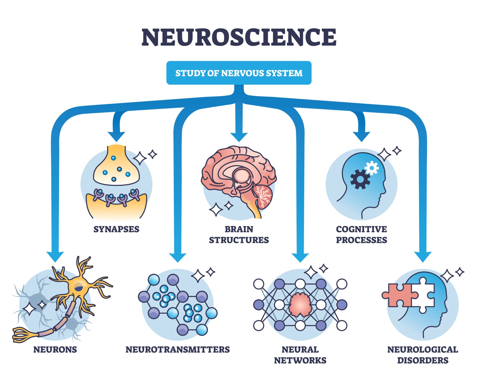
Neuroscience is the branch of science concerned with studying the nervous system.
It is a multidisciplinary field integrating numerous perspectives from biology, psychology, and medicine. It consists of several sub-fields ranging from the study of neurochemicals to behavior and thought.
Key Takeaways
- Field: Neuroscience studies the nervous system, and cognitive neuroscience is the branch that links brain structures to mental processes like memory and language.
- Method: Researchers combine brain scans such as fMRI with behavioral testing to connect neural activity to specific mental functions.
- Neurons: The brain runs on roughly 100 billion neurons that fire electrical signals called action potentials and communicate at synapses.
- Brain Divisions: The nervous system splits into the central nervous system (brain and spinal cord) and the peripheral nervous system (nerves throughout the body).
- Key Studies: Landmark cases such as Phineas Gage and Patient HM showed that damage to one brain area can impair one function while sparing others.
- A Real Limit: Reverse inference, assuming a mental process occurred just because its usual brain region was active, is a widely criticized weakness of neuroimaging.
For example, cognitive neuroscience is the scientific study of the influence of brain structures on mental processes, done using brain scanning techniques such as fMRI.

What is Cognitive Neuroscience?
Cognitive neuroscience aims to discover how brain structures influence how we process information and map mental cognitive functions to specific areas of the brain.
Researchers do this using brain imaging techniques such as fMRI and PET scans.
The field has old roots. The ancient Egyptians practiced trephination, drilling a hole into the skull to treat brain and mental disorders, and already recognized some symptoms of brain damage (Mohamed, 2008).
Centuries later, the microscope changed everything. New staining procedures let Santiago Ramón y Cajal identify individual neurons in the late 1890s, setting the stage for the modern study of the brain (Guillery, 2004).
Neuroscience itself only became a distinct field in the 20th century, pioneered by David Rioch, Francis O. Schmitt, and Stephen Kuffler (Cowan et al., 2000).
Its branches are defined by scale. That is, the level at which each one examines the nervous system.
Molecules form the basis for neuronal function and communication. This is the focus of molecular neuroscience.
Those molecular processes give rise to larger-scale cellular functions such as neural signaling, the focus of cellular neuroscience. Such functions in turn enable complex networks of communication between neurons, the focus of systems neuroscience.
Finally, these systems underlie thought and behavior. That is the focus of cognitive and behavioral neuroscience.

The scientific study of the nervous system provides crucial insights into the workings of the mind and brain, and is thus indispensable to psychology.
Neuroscience allows us to understand the many workings of the mind as operating through networks of neural connections, just as computers operate through electrical connections.
By studying how these neural connections work, we can better comprehend normal human cognition and disease—i.e., when these neural connections go awry.
The Naming of the Field
The term “cognitive neuroscience” has an unusually well-documented birth.
Neuroscientist Michael S. Gazzaniga and cognitive psychologist George A. Miller coined it together, in the back seat of a New York taxi in the late 1970s. They were on their way to a dinner meeting about how the brain enables the mind (Cognitive Neuroscience Society, n.d.).
Neither man says which of them spoke the phrase first. It stuck anyway.
Turning a coined term into a working field took another decade and a half.
Gazzaniga secured Sloan Foundation funding in the early 1980s to found a dedicated cognitive neuroscience institute (Cognitive Neuroscience Society, n.d.). He launched the Journal of Cognitive Neuroscience in 1989.
In 1994 he helped found the Cognitive Neuroscience Society, now more than 2,000 members strong (Cognitive Neuroscience Society, n.d.).
Four Related Approaches to Studying Cognition
Researchers who study cognition don’t all use the same evidence. Four related approaches make different trade-offs between behavior and the brain.
- Experimental cognitive psychology: studies cognition using behavioral evidence alone, such as reaction times and accuracy, without measuring the brain directly.
- Cognitive neuroscience: uses evidence from both behavior and the brain, measuring neural activity or structure alongside performance.
- Cognitive neuropsychology: studies the cognitive effects of naturally occurring brain damage, such as stroke or tumor, to reveal normal brain organization.
- Computational cognitive science: builds formal, runnable computer models of cognitive processes that generate precise, testable predictions.
A researcher might use cognitive neuropsychology to show a stroke patient’s word problem is selective for verbs, then use cognitive neuroscience to find which networks distinguish that from nouns.
These are not rival camps. They are complementary lenses that different research questions call for.
The Methodological Toolkit
Cognitive neuroscience’s methods trade off spatial precision, timing, and whether they can show a brain region is truly necessary for a function, not just active alongside it.
Most tools are correlational. fMRI and PET track blood flow as an indirect proxy for neural activity; EEG and MEG record electrical or magnetic signals from large groups of neurons. All of them show that a region was active, not that it was needed.
Two methods can test necessity directly. Single-unit recording measures the firing of individual neurons, usually in animals, at a level of detail no scanner can match.
Transcranial magnetic stimulation (TMS) does something scanners cannot: it briefly disrupts a small patch of cortex using a pulsed magnetic field. That creates a temporary “virtual lesion” in a healthy volunteer (Barker, Jalinous, & Freeston, 1985).
If disrupting a region impairs a task, that matters. It is far stronger evidence of a causal role than any scan alone can offer.
The Society for Neuroscience (2015) lists the following as the field’s “core concepts”:
- The brain is the body’s most complex organ.
- Neurons communicate using both electrical and chemical signals.
- Genetically determined circuits are the foundation of the nervous system.
- Life experiences change the nervous system.
- Intelligence arises as the brain reasons, plans, and solves problems.
- The brain makes it possible to communicate knowledge through language.
- The human brain endows us with a natural curiosity to understand how the world works.
- Fundamental discoveries promote healthy living and the treatment of disease.
Neuron Structure
Neurons are the basic cellular units that constitute the nervous system. Humans possess approximately 100 billion neurons. An individual neuron generally consists of a soma (cell body), dendrites, and axons.
Each part has a job.
The soma contains the cell’s nucleus (where its DNA is stored) and produces proteins necessary for the neuron’s function.

Dendrites listen; axons speak.
Extending out from the soma are dendrites, which are branch-like structures that form connections with other neurons from which they receive and process electrical signals. Finally, an axon projects out from the other end of the soma, producing and carrying an electrical signal to other neurons.
Each neuron usually only contains one axon, although the structure may be branched following the initial projection from the soma (Woodruff, 2019).
The electrical signals carried by axons and transmitted to dendrites are called action potentials. Neurons are electrical devices — they contain channels that allow positive and negative ions to pass from outside to inside the cell or vice versa, which gives rise to an electrical potential concerning a cell’s membrane (the barrier around the outside of a cell).

The Action Potential
By default, when neurons are “at rest,” there is a more negative charge on the inside of the cell than outside. This creates a resting potential of -70 millivolts.
However, this electrical potential constantly changes in response to inputs from other cells, which cause ions to flow in or out of the cell.
Some of these inputs are “excitatory,” meaning they make the cell’s membrane potential less negative, for example by letting positive ions flow into the cell. Others are “inhibitory,” meaning they make the membrane more negative.
If a neuron receives enough excitatory input, and not too much inhibitory input, its membrane potential rises above a threshold. Neuroscientists call this the “action potential threshold,” roughly -50 millivolts, and an action potential fires.
Electrically, action potentials are brief but dramatic spikes in a neuron’s membrane potential. Neuroscientists often refer to action potentials simply as “spikes.”
When a neuron’s membrane potential passes the action potential threshold, it opens voltage-gated sodium channels. These let positively charged sodium ions rush into the cell.
This makes the membrane rapidly more positive: a spike.
This signal then rapidly travels down the length of the neuron’s axon. The spike itself causes voltage-gated sodium channels farther down the axon to open too, and so on.
Finally, the action potential reaches the end of the axon, and the neuron passes this signal along to other neurons.
Synaptic Transmission
Neurons communicate with one another through structures called synapses. A single synapse consists of a presynaptic terminal, a synaptic cleft, and a postsynaptic terminal.
Release happens fast.
Once an action potential reaches the end of a neuron’s axon, it reaches the presynaptic terminal, which causes neurotransmitters to be released from the cell. These neurotransmitters are released into the synaptic cleft, a small (20-40nm) gap between the pre-and postsynaptic terminals.
Then diffusion takes over.
The neurotransmitters then travel across the synaptic cleft and activate neurotransmitter receptors on the postsynaptic terminal. When these receptors are activated, they let positive or negative ions flow into the postsynaptic neuron. That triggers excitation or inhibition, respectively.

When neurotransmitters cause positive ions to flow into the postsynaptic neuron, that is called excitation. It brings the neuron closer to its action potential threshold, making it more likely to fire.
Inhibition works the opposite way.
Conversely, when neurotransmitters cause negative ions to flow in, that is called inhibition. It moves the neuron further from its action potential threshold, making it less likely to fire.
As a result, some neurotransmitters are referred to as excitatory neurotransmitters (since their action on receptors causes excitation), while others are referred to as inhibitory neurotransmitters.
Glutamate and dopamine are common excitatory neurotransmitters. GABA and glycine are common inhibitory ones. Some neurotransmitters, such as serotonin, can be either excitatory or inhibitory depending on the type of receptor it acts upon.
The Nervous System
Our nervous system comprises billions of neurons, all firing action potentials and communicating with each other through synapses.
These networks of neurons ultimately give rise to larger structures that perform specialized functions. By studying the anatomy of the nervous system, we can begin to understand how it divides up its many tasks.
Anatomy reveals the division of labor.
The most important anatomical division of the nervous system is between the central nervous system and the peripheral nervous system.
The central nervous system consists of the brain and the spinal cord. The peripheral nervous system consists of the nerves throughout the body that communicate with the central nervous system.

The central and peripheral nervous systems act together to interpret sense data and initiate movement (Sukel, 2019).
Sensory information is sent from the peripheral nerves to the spinal cord and then relayed to the brain; motor information travels from the brain down to the spinal cord and then ultimately to the muscles via the peripheral nerves.
The brain itself consists of three parts: the brainstem, the cerebellum, and the cerebral cortex.
The brainstem primarily controls so-called “autonomic” functions, meaning unconsciously regulated bodily functions, such as heart rate and breathing. The cerebellum is next to the brainstem and controls balance and movement coordination.
Finally, the cerebral cortex lies above the brainstem and cerebellum and is what most people think of when they think of the brain — it is responsible for the perceptual and cognitive functions that make up our mental lives (Sukel, 2019).
The cerebral cortex, in turn, is divided into two hemispheres and four lobes. A bridge of neural fibers connects the right and left hemispheres called the corpus callosum.
Contrary to popular belief, most cognitive processes are associated with both hemispheres of the cerebral cortex (i.e., the right side is not more “creative” and the left side more “analytical”).
However, one exception is that most neural structures related to language reside in the left hemisphere (Sukel, 2019).
In addition to the two hemispheres, the cerebral cortex is also divided into four lobes: the occipital lobe, the temporal lobe, the parietal lobe, and the frontal lobe. The occipital lobe is located toward the back of the brain and is mostly responsible for processing visual information.
The temporal lobe is located behind the forehead temples and largely deals with sound information (including language) and some aspects of memory.
The parietal lobe is located above the ear and mainly processes sensory, touch, and spatial information. Lastly, the frontal lobe (the largest lobe) is located above the eyes in the front of the cortex. It is responsible for higher-level cognitive functions such as reasoning, decision-making, and planning.
It is thought that our highly developed frontal cortex separates humans from primate ancestors (Sukel, 2019).
Each lobe contains two distinct types of neural tissue: gray matter and white matter. Gray matter appears gray in color and comprises neurons’ somas, dendrites, and non-neuron supporting cells. White matter is white in color and comprises neurons’ axons, which serve to form connections between areas in the brain.
The white color results from myelin, a fatty substance wrapped around axons to enable them to send signals more efficiently (Sukel, 2019).
The brain also contains numerous smaller regions with more specific functions. Important regions include:
- Hypothalamus — the control center of autonomic functions such as body temperature and blood pressure, as well as behaviors like hunger, thirst, and sex drive.
- Pituitary gland — connected to the hypothalamus, regulates the endocrine system, secreting hormones involved in sexual development, bone and muscle growth, and stress.
- Thalamus — major “relay station” that regulates information coming and going from the cerebral cortex.
- Basal ganglia — in conjunction with the cerebellum, helps coordinate fine motor movements.
- Amygdala — plays a significant role in emotional response to stimuli.
- Hippocampus — responsible for long-term memory (“Anatomy,” 2018).
Landmark Case Studies
Long before brain scanners existed, three patients did more than any single experiment to show that specific brain structures make specific, separable contributions to behavior.
Phineas Gage
Aim: In 1848, railway foreman Phineas Gage survived an accident in which an iron rod was driven through his skull, destroying much of his left frontal lobe.
Method: His physician, John Martyn Harlow, documented the injury at the time (Harlow, 1848).
Twenty years later, after Gage’s death, Harlow published a fuller account of his personality changes (Harlow, 1868).
Results: Gage’s memory, language, and movement were untouched. But Harlow’s later account described a profound change in temperament, self-control, and social judgment.
Conclusion: Gage’s case became the first widely cited evidence that the frontal lobe contributes to personality and social conduct, not just movement or sensation. Modern historians note the popular story outruns the thin nineteenth-century record, but the case still opened the question of localized personality function.
Patient HM
Aim: In 1953, surgeon William Scoville removed tissue from both sides of a young patient’s brain, including most of the hippocampus, to treat severe epilepsy. The patient became known as HM.
Method: Scoville and neuropsychologist Brenda Milner tested HM’s memory and other cognitive functions in detail over the following years (Scoville & Milner, 1957).
Results: HM’s epilepsy improved. But he could no longer form new long-term memories. His short-term memory, intelligence, language, and even his ability to learn new motor skills stayed intact (Scoville & Milner, 1957).
Conclusion: HM’s case showed that long-term declarative memory depends on the medial temporal lobe and hippocampus, while short-term memory and motor-skill learning rely on separate systems.
This double dissociation became foundational to every later model of human memory.
Split-Brain Patients
Aim: From the 1960s, neuroscientist Roger Sperry, joined by Michael Gazzaniga, studied patients who had undergone a rare surgery. It severed the corpus callosum, the band of fibers connecting the brain’s two hemispheres, as a last-resort epilepsy treatment.
Method: Because the surgery disconnects the hemispheres, researchers could show information to just one hemisphere at a time and test what each side, independently, appeared to know (Gazzaniga, 1967).
Results: The left hemisphere led language and sequential tasks. The right hemisphere could not produce speech, but excelled at certain spatial and facial-recognition tasks, and each side could act on information the other had no access to (Sperry, 1968).
Conclusion: Split-brain research showed that localization of function operates between the two hemispheres, not just within one, a pattern called lateralization. Because the disconnection was surgical and precise, it offered an unusually clean demonstration of functional specialization.
What These Cases Show Together
Together, these three cases established the logic the field still leans on.
If damage to one structure produces a selective deficit while sparing other functions, a double dissociation, that is strong evidence the structure makes a distinct contribution.
HM’s case is the clearest example: procedural learning, such as motor skills, stayed fully intact while declarative memory was devastated, showing the two depend on entirely separate brain systems.
Each case has the same weakness.
A single natural case is never a controlled experiment.
Its lesion is rarely as clean or as precisely mapped as later, imaging-informed reconstructions suggest.
And its behavioral record, especially Gage’s, which rests on retrospective, non-systematic clinical description, can be read through whichever theory the observer already holds.
That vulnerability is precisely why later, better-controlled methods such as TMS became so valuable to the field.
Neuroimaging Methods
Brain and mind are linked.
The scientific study of the brain is indispensable to the scientific study of the mind.
Although neuroscience and psychology focus on different domains, neuroscience deals with the realm of physical properties, while psychology deals with the more abstract realm of the mental.
Our ever-evolving ability to correlate brain states with mental states means that the two disciplines can engage in meaningful dialogue.
That dialogue is new.
Scientists have sought to understand the relationship between the brain and mind in normal and abnormal human cognition. This is the main goal of cognitive neuroscience.
Much research in cognitive neuroscience is done through the use of neuroimaging (which refers to any technology that aids in the visualization of the brain) because it allows us to “look inside” living people’s skulls.
The most common form of neuroimaging used in cognitive neuroscience studies is magnetic resonance imaging (MRI). MRI uses responses from hydrogen ions in different settings to obtain information about the brain.
That signal reveals brain structure and activity.
MRI can provide structural information about the brain, information about someone’s brain anatomy, such as the sizes of different regions. It does this by differentiating different types of tissue in the skull, creating a physical brain map.
It can also provide functional information—i.e., information about the activity of different areas of the brain—by detecting regions with high levels of oxygenated blood, which is correlated with brain activity.
Function and form, both visible.
Through neuroimaging studies, cognitive neuroscientists can build models of human cognition using structural and functional information. That helps them understand the roles different brain systems and regions play in thought and behavior (Kalra, 2012).
Depression and Neurogenesis
In addition to shedding light on the neural processes underlying the human mind in general, neuroscience has revolutionized clinical psychology by generating significant advances in our understanding of psychiatric illness.
By comparing the brains of healthy subjects to those of individuals with psychiatric disorders, neuroscientists have improved our knowledge of the causes of these illnesses.
They have also improved knowledge of the most effective treatments.
For example, neuroimaging studies have suggested that some depressed people may have a smaller hippocampus. This may be related to stress, which is thought to decrease neurogenesis (the production of new neurons) in the hippocampus.
Depression may shrink the hippocampus.
This finding is also consistent with evidence that antidepressant medications serve to promote neurogenesis in the hippocampus. That process takes a long time, which may explain why patients typically do not notice the effects of antidepressants for several weeks.
Time is the key variable.
Antidepressants do produce direct effects on neurotransmitter levels thought to be involved in mood. But these findings suggest neurogenesis is ultimately the more significant mechanism, and that drugs should be developed to specifically target it (“What causes,” 2019).
Schizophrenia and Dopamine
Another example is schizophrenia.
Various neurochemical, neuroimaging, and animal model studies point to dopamine playing a prominent role in this disorder. In particular, patients often show abnormally high dopamine levels in a part of the brain called the striatum.
It is thought that one of the roles of dopamine is to signal the salience of external stimuli.
Salience is the key concept.
For example, food might be signaled as salient because it is necessary for survival.
Scientists have thus hypothesized that abnormal dopamine activity in the striatum may cause innocuous stimuli to seem falsely salient in people with schizophrenia. That can cause delusions and hallucinations.
As a result, a key mechanism of antipsychotic drugs is to block dopamine receptors (Winton-Brown et al., 2014).
Addiction and the Reward Circuit
Dopamine is also implicated in addiction.
The neurotransmitter plays a key role in motivation and reward, a certain kind of salience. When drugs that increase dopamine levels in the reward circuit are taken, a conditioned response is evoked.
Typically, this reward circuit is kept in check by circuits in the prefrontal cortex that control executive functioning: the ability to resist short-term cravings in service of a longer-term goal.
Addiction breaks that balance.
However, in individuals with addiction, the drug’s conditioned response is so strong that reward circuits override prefrontal circuits. The result is compulsive drug-seeking, even in the face of negative consequences (Volkow and Boyle, 2018).
Contemporary research has shown that neuroscience and psychology can work together for mutual benefit. By learning about the mental and physical relationship, we can better understand both.
Critical Evaluation
Cognitive neuroscience’s tools carry real limits. Several issues shape how much weight a single finding deserves.
Reverse Inference
Aim: Poldrack (2006) asked a hard question. How much can a single brain scan really prove about a mental process?
Method: He ran a Bayesian analysis of BrainMap, a large database of published neuroimaging results. This let him calculate how much a region’s activity actually raises the odds that a process occurred, based on how selectively that region responds.
Results: Reverse inference is not logically valid on its own. Most regions respond to many different tasks, so activity in one usually gives only weak evidence for a single process, unless that region’s selectivity is proven high.
Conclusion: Researchers should stay cautious about reverse inference. This matters most for regions like the amygdala, where selectivity for one process is weak or untested.
A brain scan showing “the amygdala lit up” is not proof, by itself, that fear occurred. That headline-friendly shortcut is exactly what Poldrack’s analysis was built to discipline.
Correlation, Causation, and Causal Methods
Most of the field’s core toolkit, fMRI, PET, EEG, MEG, is correlational: it shows a region was active while a process occurred, not that the region was necessary for it.
That is precisely why methods that can manipulate the brain rather than just observe it, the lesion tradition, and modern TMS, hold special evidential weight. They come closest, in humans, to a genuinely causal experiment.
The Multiple-Comparisons Problem
Aim: Bennett, Baird, Miller, and Wolford (2009) set out to show something memorable.
Failing to correct for the number of statistical tests in an fMRI scan can manufacture false brain activity.
It can do this even in a subject incapable of any cognitive process at all.
Method: The target was absurd on purpose. The researchers placed a single dead Atlantic salmon, chosen because it could not be having a cognitive experience, in an fMRI scanner and had it “complete” a social perspective-taking task.
They analyzed the images exactly as they would a live human dataset, first without correcting for the very large number of statistical tests involved, then with standard correction applied.
Results: The uncorrected analysis found 16 voxels, clustered implausibly in the salmon’s brain and spinal cord, showing statistically “significant” activity. Once corrected, every result vanished.
Conclusion: Uncorrected whole-brain fMRI analysis does not just lack precision. It can manufacture entirely spurious findings in a subject with no brain function to detect. Proper statistical correction is therefore a precondition for trusting any fMRI result (Bennett, Baird, Miller, & Wolford, 2009).
Contemporary Research
Two studies define where cognitive neuroscience stands today. They pull in different directions.
Aim: Glasser et al. (2016) set out to build a single, reproducible map of how the human cerebral cortex is organized. It is the modern descendant of the same localization question Broca and Wernicke asked over a century earlier.
Method: The team combined four independent types of MRI data. These covered architecture, task activity, resting connectivity, and topography, from 210 healthy adults in the Human Connectome Project.
A machine-learning algorithm then mapped cortical areas from sharp changes across all four measures at once.
Results: The atlas identified 180 distinct areas per hemisphere. Ninety-seven had never been formally described before; the rest matched areas already known from decades of earlier mapping.
Conclusion: Combining several independent neuroimaging methods produced a far more precise, reproducible map than any single measure could achieve alone. The atlas has since become a standard reference used across much of human neuroimaging research.
That achievement has a flip side. A second strand of research asked something more basic.
When an fMRI study finds a brain region “activates” during a task, how much can that specific finding be trusted to replicate?
Aim: Elliott et al. (2020) set out to measure this directly. They wanted to know the test-retest reliability of task-based fMRI, how consistently it reproduces when the same people are tested again.
Method: They combined two sources of evidence. One was a meta-analysis of 90 published test-retest fMRI studies, pooling 1,008 participants.
The other was new data from 11 widely used tasks, given to two large, independent cohorts.
Results: Both the meta-analysis and the new data agreed: task-fMRI reliability is poor. The number was low: .397 on average.
That is well below what is considered adequate for studying differences between individuals.
Conclusion: Task-fMRI activation is valuable at the group level (Elliott et al., 2020). But not for one individual.
It remains useful for showing which regions are involved in a task across a group, just not for a single person.
Read together, the two studies frame the field’s current position with unusual clarity. One maps; the other measures trust.
Cognitive neuroscience can now map the group-average brain with striking precision, but its most widely used tool for studying any one person has been over-trusted.
Generalisability and Sample Bias
Cognitive neuroscience shares a further limit with psychology more broadly.
Most neuroimaging participants are drawn from WEIRD populations, Western, Educated, Industrialized, Rich, and Democratic (Henrich, Heine, & Norenzayan, 2010). Because scanners, funding, and recruitment concentrate in a few wealthy countries, claims about “the human brain” are often claims about a narrow slice of humanity.
Henrich and colleagues’ wider analysis found that WEIRD populations are often outliers on the very dimensions being measured, not a neutral baseline. That caution applies to brain scans, not just behavior.
A finding that holds up in a lab in the United States or United Kingdom is not guaranteed to hold up worldwide.
Yet most of what cognitive neuroscience currently claims about “the human brain” comes from exactly those kinds of samples.
The Reductionism Debate
A neural correlate is not an explanation. Not by itself.
Even a perfect one leaves a question open: why does this pattern of brain activity happen at all?
Knowing which brain regions activate when someone feels grief or makes a moral judgment is genuinely informative. But something gets lost in translation.
Turning a rich psychological experience into a list of active voxels can flatten real complexity. Social context, personal history, and meaning are exactly what a purely biological description struggles to capture.
A colorful brain scan can also make a claim feel more scientifically settled than the evidence actually warrants. Pictures persuade.
This wider levels-of-explanation debate, whether psychology should ultimately reduce to biology, is explored in full in Simply Psychology’s reductionism guide.
Ecological Validity
Finally, the scanning environment itself is artificial: lying immobile in a loud, cramped machine, doing a simplified task under time pressure.
Does this generalize to cognition as it actually happens, in open, social, moving, real-world settings? The question remains open.
Naturalistic-stimulus studies push back on this. Hasson, Nir, Levy, Fuhrmann, and Malach (2004) showed that different people watching the same film clip show tightly synchronized cortical activity.
That happens across a wide swath of the brain, even for a richly naturalistic stimulus.
Rich, real-world-like stimulation can still be analyzed rigorously. Mobile EEG, and wireless recording in freely moving animals, extend the same logic outside the scanner altogether.
The field treats this as a real, unresolved limitation, not a solved problem, and keeps developing new naturalistic paradigms to close the gap.
References
Cowan, W. M., Harter, D. H., & Kandel, E. R. (2000). The Emergence of Modern Neuroscience: Some Implications for Neurology and Psychiatry . Annual Review of Neuroscience, 23 (1), 343–391.
Dorland, W. A. N. (2011). Dorland’s Illustrated Medical Dictionary E-Book. Elsevier Health Sciences.
Guillery, R. W. (2004). Observations of synaptic structures: origins of the neuron doctrine and its current status. Philosophical Transactions of the Royal Society B: Biological Sciences, 360 (1458), 1281–1307.
Harvard University. (2019, June 24). What causes depression? Harvard Health Publishing. https://www.health.harvard.edu/mind-and-mood/what-causes-depression.
Kalra, P. (2012, July 1). Cognitive neuroscience: Connecting neuroimaging and neural nets. Science in the News. http://sitn.hms.harvard.edu/flash/2012/cognitive-neuroscience/
Mayfield Brain & Spine. (2018, April). Anatomy of the Brain. Mayfield Brain & Spine. https://mayfieldclinic.com/pe-anatbrain.htm
Mohamed, W. (2008). The Edwin Smith Surgical Papyrus: Neuroscience in Ancient Egypt. IBRO History of Neuroscience. https://archive.vn/20140706060915/http://www.ibro1.info/Pub/Pub_Main_Display.asp?LC_Docs_ID=3199#selection-437.0-437.28
Scandola, M., Esposito, M., Guidotti, R., & Romano, D. (2025). How artificial intelligence is shaping neuropsychology: A focus on cognitive assessment of neurodegenerative disorders. Journal of Neuropsychology. https://doi.org/10.1111/jnp.70009
Society for Neuroscience. (2015). Neuroscience Core Concepts: The Essential Principles of Neuroscience . Washington, DC.
Sukel, K. (2019, August 25). Neuroanatomy: The Basics. Dana Foundation. https://www.dana.org/article/neuroanatomy-the-basics
Sussex Publishers. What Is Neuroscience? Psychology Today. https://www.psychologytoday.com/gb/basics/neuroscience
The University of Queensland. (2017, November 9). Action potentials and synapses. Queensland Brain Institute. https://qbi.uq.edu.au/brain-basics/brain/brain-physiology/action-potentials-and-synapses
Volkow, N. D., & Boyle, M. (2018). Neuroscience of Addiction: Relevance to Prevention and Treatment. American Journal of Psychiatry, 175 (8), 729–740.
Winton-Brown, T. T., Fusar-Poli, P., Ungless, M. A., & Howes, O. D. (2014). Dopaminergic basis of salience dysregulation in psychosis. Trends in Neurosciences, 37 (2), 85–94.
Woodruff, A. (2019, August 13). What is a neuron? Queensland Brain Institute. https://qbi.uq.edu.au/brain/brain-anatomy/what-neuron.